INTRODUCTION
Fencing is one of the oldest known sports and is one of only four sports that has been included in every modern Olympic Games.1 Rapid growth in participation in the United States (US) in recent years has been driven, in part, by international success and the resultant media attention garnered by US fencers over the past two decades.2,3 A unique sport that involves both aerobic and anaerobic exertion, fencing is inherently asymmetrical, as fencers stand with their dominant arm and leg pointed toward their opponent, with the rear leg at a ninety-degree angle.4,5 The dominant arm is the weapon-holding arm, and the non-dominant arm plays a minor role in providing balance during explosive movements. The motions involved in a fencing “bout” are a combination of fine repetitive motions and rapid explosive movements. Multiple studies have shown that these factors lead to significant physiologic and biomechanical demands, and can lead to asymmetries in both structure (e.g. muscle bulk) and function (e.g. reaction time).1,6
Comparative data from recent Olympic Games indicate that fencing is a relatively safe sport. In the 2016 Rio Olympic Games, 9% of fencers experienced an injury, which was relatively low, especially when compared to higher-risk sports such as BMX where 38% of athletes were injured.7 Additionally, the vast majority of the fencing injuries reported involved no time-loss, suggesting that they were predominantly minor injuries.7 Catastrophic or penetrating injuries are rare, contrary to what might be expected given the weapons used.2 This safety profile is likely due, at least in part, to significant improvements in protective equipment.1,8
Multiple studies have looked at the biomechanics of fencing, however the data on fencing injuries are sparse, with few published studies over the past few decades.6,9–15 Rare case studies of atypical injuries in fencers have been published, as well as a few larger studies investigating injury rates and types in different environments, which have generally found low injury rates and a predominance of mild injuries.2,3,16–18 Several smaller, mostly survey-based studies have been done investigating injury characteristics in fencers in a variety of populations.19–24 Research has consistently shown the lower extremity to be the most common area of injury; however, results vary in terms of the most frequently injured joints and the effects of sex, age, and hand dominance on injury rates and types.2,3,8,18–24 To the authors’ knowledge, no published studies have been based at medical centers, and therefore generally lack clinical data including specific diagnoses (especially those requiring confirmation by diagnostic imaging) and medical management, except when self-reported on athlete questionnaires.
There is a need for better understanding and additional data on injuries in fencing athletes to aid in the care of the growing fencing population. This is especially true of pediatric injuries given the rise in youth fencing, youth sports in general, and early sports specialization.25–28 Therefore, the purpose of this retrospective study was to examine the types of injuries incurred by fencing athletes and to analyze the associations between age, sex, and handedness on the type and location of injury. It was hypothesized that injuries would be more common in the lower extremity and that overuse injuries would be more common than acute/traumatic injuries. Additionally, it was anticipated that dominant-sided injuries would be more common, and that female fencers would have higher rates of knee injuries.
METHODS
Institutional Review Board approval was obtained. A medical record review was conducted using an intradepartmental database, Hound Dog, with keywords “fencing” or “fencer” over a 10-year period from January 1, 2011 to December 31, 2020. “Injury” was defined as any fencing-related pain or disorder that led to evaluation in the sports medicine or general orthopedic clinic at the investigators’ institution. Only injuries documented in patients actively engaged in the sport of fencing at the time of injury were included. Injuries that were unrelated to fencing (e.g., fencer injured during another activity, or fencer with pain that does not affect fencing) were excluded. Additionally, patients with underlying medical diagnoses that predisposed them to the injury for which they were evaluated (e.g., fencer with osteogenesis imperfecta presenting with a fracture) were excluded. Demographic data and descriptive information were collected, including age at time of presentation, sex, dominant hand, and number of separate fencing-related injuries in that patient during the study timeframe, body part involved, side of injury (right, left, or bilateral), and whether injury was acute/traumatic or chronic/overuse. Injury classification (i.e., the type of injury) was recorded; of note the “joint” classification indicates joint dysfunction (e.g., patellofemoral pain, shoulder instability). “Acute/traumatic” injuries were defined as any injury that occurred at a clearly identifiable time point per patient report. “Chronic/overuse” injuries were defined as occurring with gradual or unknown time of onset.
Patient and injury characteristics were summarized for the cohort by frequency and percent or mean and standard deviation, as appropriate. Injury characteristics were further stratified and compared by age, sex, and handedness. Comparisons across stratification variables were conducted using chi-square tests for categorical characteristics and Student’s t-test for continuous characteristics. Pearson’s correlation analysis was used to assess the degree of association between continuous characteristics. Pearson’s correlation coefficients were reported along with 95% confidence intervals. All tests were two-sided and p-values less than 0.05 were considered significant.
RESULTS
Patient Characteristics
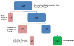
A total of 452 patient records containing the word “fencing” or “fencer” with clinic visits in the Orthopedics and/or Sports Medicine Departments from January 1, 2011 to December 31, 2020 were found. Of these, 49 patients were not fencing athletes and were excluded. Of the remaining 403 records, 200 were injuries in fencers that did not occur during fencing, so these were also excluded. An additional 17 charts of the remaining 203 were excluded due to underlying medical diagnoses that predisposed the patient to their specific injury (e.g. patients with Ehlers-Danlos Syndrome presenting with a joint subluxation event during fencing). A total of 186 patients (98 male, 88 female) were included in the study (Figure 1), with a total of 313 fencing-related injuries. Average age at time of injury was 14.6 years (range of 9 to 32 years); the majority of patients were 21 years of age or younger. Distribution of injuries by age is shown in Figure 2. The patient’s dominant hand was documented for 102 of the 186 patients. The majority (62%) of patients had one fencing-related injury during the 10-year study period; only seven (4%) had five separate injuries, and none presented with more than five. (Table 1)
Injury Characteristics
The characteristics, management, and outcomes of injuries are summarized in Tables 2 and 3. The majority of injuries (73%) were in the lower extremity (227/313), while 16% (49/313) occurred in the upper extremity and 10% (32/313) were in the back. Within the lower extremity, the most commonly affected joint was the knee (49%), followed by the ankle (16%) and the hip (11%). Of the upper extremity injuries, the hand was most commonly affected (35%), followed by the shoulder (31%) and the wrist (24%). The most common injury classification was joint pathology (e.g. patellofemoral pain or hip femoroacetabular impingement, 31%), followed by tendon injuries (27%). The majority of injuries were evaluated by plain film radiograph (64%) with a smaller percentage requiring advanced imaging modalities such as MRI (34%), CT (1%), and ultrasound (3%). The majority of injuries were treated with physical therapy (PT, 80%) and only a small percentage required surgical intervention (5%). The majority of injuries (51%) did not involve time loss from sport, and very few (8%) had time loss of 3 months or greater, although it should be noted that this information was only available for 140/313 (45%) of injuries. The ten most common diagnoses seen in this study population are summarized in Table 4.
Effect of dominant side
Both patient hand dominance and injury laterality were documented in the chart for 202 injuries. Upper extremity injuries in this subset were seen almost exclusively in the dominant side (46/48, 96%, p =0.003). Lower extremity injuries were also seen more often in the dominant side (99/141, 70%). Within the lower extremity, there was a significant difference in location of injury between dominant and non-dominant sided injuries (p=0.007), with the majority of joints more frequently injured on the dominant side (Table 5). Within the most common diagnoses of the lower extremity, the distributions of extensor and FAI injuries were similar between dominant and non-dominant injuries; however, hamstring injuries were more frequent on the dominant side, whereas non-dominant injuries were more commonly seen in the ankle and Achilles (p = 0.02).
Effect of age
There were 240 injuries (77%) in patients 13 years of age or older. Injuries occurring in younger patients (under 13 years) were more likely to be in the lower extremity (90%) compared to older patients (67%), with a p value of 0.003. Younger patients were only slightly more likely to have apophysitis injuries (14%) compared to older patients (6%) (p=0.05). It was found that age was mildly correlated with RTP (r=0.20; 95% CI, 0.04-0.36; p=0.02), such that younger subjects returned to participation earlier than older subjects. There appears to be a difference in the distribution of return to participation across age groups; however, this did not reach a statistically significant difference (p=0.07). Within the most common diagnoses in the lower extremity, extensor injuries were the most common in both age groups. However, the younger group had a higher occurrence of Achilles injuries whereas the older group had a higher occurrence of hamstring injuries (p= 0.003).
Effect of sex
Males comprised 53% of the study patients and 51% of injuries (160/313). Females had a slightly higher proportion of chronic/overuse injuries (85%) compared to males (74%) (p=0.04). There was no significant difference between the sexes for frequency of the most common lower extremity diagnoses.
DISCUSSION
This study is the only recent English-language study to look at primarily youth fencing-related injuries evaluated at a medical institution, thus providing greater information on specific diagnoses made, imaging utilized, and treatment modalities prescribed. Consistent with expectations, lower extremity injuries were most common, and just over half of all injuries required no time out of sports. Interestingly, several cases of spondylolysis were found, which has not been commonly described in fencers. The effect of handedness was demonstrated, especially in upper extremity injuries, which occurred almost exclusively in the dominant side. Younger patients had fewer injuries overall and were more likely to return to play earlier than their older counterparts. Surprisingly, there was no effect of sex on injury location or specific diagnosis, although male athletes had higher rates of acute/traumatic injuries, compared to female athletes.
As anticipated, this study found that the lower extremity was the most commonly injured area of the body, affected in almost 75% of all injuries; these results are similar to previously published studies.18 Also noted were very low rates of knee intraarticular derangement, with only two ACL ruptures and three meniscal injuries during the study period; this was consistent with other studies which showed ACL and meniscal tears, especially those requiring surgery, were infrequent.2,19,24 The most common diagnosis involving the lower extremity was extensor mechanism dysfunction (including patellofemoral pain, patellar tendinitis, and Osgood-Schlatter), which matches the findings reported in previous studies.19,22
In addition, this study found that the upper extremity was almost exclusively injured on the dominant side in comparison to the non-dominant side. Also, not surprising given the asymmetrical biomechanical demands on fencers’ legs, there were significant differences in injury rates between the dominant and non-dominant sides. For hamstring injuries, the dominant side was more likely to be injured, which is consistent with what is known about the significant acceleration/deceleration forces on the dominant hamstring during a lunge.1
While back pain has previously been reported as a common fencing injury, this study was able to report on specific diagnoses.8 Interestingly, several cases of spondylolysis were found, which has not been previously reported in fencing athletes. The mechanism of spondylolysis is presumed to be repetitive loading of the pars through hyperextension or rotation.29 While there is no emphasis on these motions in typical fencing maneuver, one could hypothesize that repetitive extension of the back during the recovery from a lunge or rotation of the torso in order to avoid a hit may predispose athletes to this type of injury.
Previously reported data indicated increased rates of injury in older fencers in comparison to younger ones.3,21 The data from this study found a similar trend, with 77% of injuries occurring in patients ages 13 years or older. Age also played a role in injury characteristics; those fencers 13 years or older were more likely to have upper extremity injuries, compared to those fencers under age 13. Additionally, younger fencers had a significantly shorter time to return to play in comparison to the older age group. As might be expected, there was a slight increase in diagnosis of apophysitis injuries in the younger age group, although this did not reach statistical significance (p = 0.05).
Previous studies have been mixed in terms of comparing injury rates between female and male fencers.2,3,18,19,30 In this study, there was a similar number of injuries in males and females, but there was a higher rate of acute/traumatic injuries in male fencers (p = 0.04). Interestingly, despite reports of biomechanical differences between the sexes in fencers, the data did not show any difference in injury classification, specifically there was no increase extensor mechanism dysfunction, despite the known higher incidence of these diagnoses in female athletes as a whole.1,6,31–33
While time to return to play was only recorded in a subset of patients, within that group, the majority of injuries required no time-loss. Additionally, only five percent of injuries in this study population required surgery, which is especially notable given that patients were seen in subspecialty clinics, where one might anticipate a higher number of severe injuries requiring surgical intervention. Generally, these findings match previous data suggesting that fencing is a relatively low risk sport from an injury perspective.
This study has several limitations. As a retrospective study, data were limited to information documented in the charts. Several points of interest, such as hand dominance and time to return to play were not available for all study subjects, which may have limited the authors’ ability to detect differences between groups. This study included only those patients who were evaluated in Sports Medicine and Orthopedics clinics, and therefore may not represent the entire population of fencers. Patients with injuries that did not require medical attention or that were managed by athletic trainers or primary care providers would not be included in this study. Additionally, those patients with severe injuries, who presented to the emergency department were also not represented in this study. This did not likely exclude a large proportion of patients since prior research indicates that most injuries in fencers are not severe to the point of requiring time away from sports and catastrophic injuries are rare. Future directions for research include investigating fencing injuries seen in other medical settings such as the emergency department or primary care offices, as well as prospective studies to collect data on other sports-specific factors of interest, including level of training, cross training activities, and weapon fenced.
CONCLUSION
This study provides better understanding of youth fencing injuries. Consistent with prior literature, injuries of the lower extremity, particularly the knee, were the most common, and the majority of injuries were relatively minor, requiring no time out of sports. Interestingly, handedness, age and sex of the athlete affected different aspects of injury location, diagnosis, and mechanism. Ongoing investigation of injury patterns in fencing will be crucial to informing appropriate medical care of and injury prevention strategies for athletes in this rapidly growing sport.
Conflicts of interest
The authors report no conflicts of interest.


