INTRODUCTION
Female and male elite gymnasts begin competing at approximately six and nine years of age, respectively.1 The peak performance age for female and male gymnasts is 16–18 years and early 20s, respectively.2 Gymnastics includes a large population of young athletes, and many injuries occur in gymnasts in elementary and junior high school. Previous studies on gymnastics injuries have covered various ages, and few studies have focused on male gymnasts.3 Furthermore, most previous studies used lost practice and competition time as a reflection of injury severity. However, because gymnasts tend to modify their events and practice content to be able to continue practicing despite having injuries,1 conventional survey methods may not accurately identify chronic disabilities that are not severe enough to cause them to miss the practice sessions.
Height, weight, body fat, age, rapid growth spurts, life stress, and high levels of practice and competition have been cited as risk factors for injury in gymnasts.1 Among female gymnasts aged 10–20 years, those with greater age, weight, and body mass index, smaller shoulder flexion angles, and greater lower back extension were reported to have a higher risk of injury than their counterparts.4 History and hypermobility have also been shown to potentially contribute to injury risk estimates. In contrast, Wright et al.5 identified age, weight gain, greater height, and lack of flexibility as injury risks in female athletes aged 8–18 years. Flexibility is particularly difficult to define, and its association with injury remains speculative.1
Previous studies have not prospectively investigated injury occurrence and physical factors in gymnasts, and only a few have examined physical assessment. Flexibility, an essential element for gymnasts, has also not been associated with injury. In addition, the relationship between physical function and injury occurrence among male and female elementary and junior high school gymnasts was not investigated. Therefore, the purpose of this study was to investigate the physical characteristics that are factors in the injury occurrence in elementary and junior high school gymnasts.
MATERIALS AND METHODS
The study enrolled healthy young gymnasts (at the level of national competition) from one gymnastics club. The inclusion criteria required that participants were healthy and free from injuries that would prevent them from completing the measurements. Exclusion criteria were refusal to cooperate in the study and inability to continue the questionnaire. Questionnaires regarding the participant height, weight, age, and medical history were distributed at the beginning of the study. Prior to the start of the study, the purpose, methods, risks associated with participation in the study, and handling of personal information were explained in writing to the subjects and their guardians, and consent was obtained. In addition, consideration was given to anonymizing the data so that the subjects could not be identified. This study was conducted with the approval of the Research Ethics Committee of the author’s institution.
Incidence of injury
The incidence of injury was investigated using the Oslo Sports Trauma Research Center questionnaire,6 which was modified and translated into Japanese.7 Because the questionnaire was designed for adult athletes, a modified version for elementary and junior high school gymnasts was used. Responses were made once a week for 23 weeks that began in September 2021, and participants completed the questionnaire under the supervision of the researcher. The presence or absence of injury and its severity (0: no injury; 100: injury with maximum severity) were recorded from the responses. When a new injury occurred, the research staff (one identified physical therapist) directly checked the patient’s condition and recorded the distinction between acute trauma and chronic disability, the location, and the name of the diagnosis (if a medical facility was consulted). Acute trauma was defined as an injury that occurred suddenly and had an injury mechanism, whereas chronic disability was defined as an injury in which pain gradually increased or continued without any associated incident.
Physical assessment
Baseline assessments for range of motion (hip flexion and extension, ankle plantar flexion, shoulder flexion, external rotation [upper extremity raising position], and internal rotation [upper extremity raising position], wrist active–passive palmar flexion and dorsiflexion), tightness (Thomas test [iliopsoas muscle], Ely test [quadriceps], straight leg raise (SLR) [hamstrings], triceps surae, combined abduction test (CAT) [latissimus dorsi muscle], horizontal flexion test (HFT) [deltoid muscle and teres minor muscles]), and muscle elasticity [multifidus] were performed. Joint range of motion testing was performed using a goniometer in accordance with the method prescribed by the Japanese Society of Rehabilitation Medicine.8 Only passive range of motion of the joints were measured, except wrist joints. Tightness tests were performed using a ruler (Raymay–Fujii Corporation, APJ188W) to measure the distance at the knee for the Thomas test and the heel-buttock distance for the Ely test.9 A goniometer was used to measure the SLR. The triceps surae were measured using an angle meter (Digi-Pas ® Pocket Digital Leveler DWL-80Pro) to measure the maximum dorsiflexion angle of the ankle joint under a load in the standing position; the CAT10 assessed the shoulder joint abduction angle in the back supine, scapular fixation position (Figure 1); the HFT11 measured the shoulder joint horizontal flexion angle while lying on the back in supine, with scapular fixation, using an angle meter.
Muscle elasticity testing of the multifidus muscle at rest was performed using an ultrasound device with shear wave elastography (SuperSonic Imagine, Aixplorer, France) and a linear probe (50 mm, 4–15 MHz). The intraclass correlation coefficient (ICC) for intra- and interobserver reliability has been shown to be excellent for muscle elasticity testing of the multifidus muscle at rest by ultrasound elastography [ICC = 0.99 and ICC = 0.95, respectively].12 In this study, the authors used the method of Koppenhaver et al.13 Measurements were performed at the 4th–5th intervertebral positions with the probe placed over the muscle belly of the bilaterally multifidus muscle. The probe was rotated counterclockwise approximately 10° from the front and tilted approximately 10° from the sagittal plane to ensure that it was parallel to the muscle fibers of the multifidus muscle. The shear modulus (kPa) was calculated by dividing the Young modulus obtained from the measurement by three. Three pictures were taken on each side, and the average was calculated since the average of the three measurements was considered better with respect to test–retest reliability.13 Three images were taken from each side, and the average was calculated.
Statistical analysis
Injury was defined as a severity of ≥1 on the questionnaire. The injury retention rate was calculated as the 23-week average of the ratio of the number of injured participants divided by the number of respondents per week. The injury retention rate (%) and injury retention rate by location (%) were calculated using the collected data, and 95% confidence intervals (CI) were obtained. Next, the relationships between the top three most frequently injured locations and physical function were examined according to sex. Participants with injuries at each location (wrist, lower back, and foot for males; heel, knee, and lower back for females) were defined as the injury group, and those without injuries were defined as the noninjury group. Differences in physical assessment according to injury location were examined using unpaired t-tests. The significance level was set at 5%. SPSS ver. 22 (IBM Corporation, Chicago, USA) was used for statistical processing.
RESULTS
Thirty-six gymnasts (19 males and 17 females, mean age 12.0 ± 1.8 years) consented to participate. Participant details are presented in Table 1. Of the 19 males, 18 participated in six events (floor exercise, pommel horse, rings, vault, parallel bars, and high bars) and one participated in three events (floor exercise, vault, and high bars). In the female competition, 15 of 17 athletes competed in four events (vault, uneven bars, balance beam, and floor exercise), and two competed in three events (vault, floor exercise, and balance beam).
The average overall injury incidence was 65.9% (95% CI: 62.3–69.5), with averages of 70.0% (65.3–74.8) and 61.7% (57.8–65.6) for males and females, respectively. Overall, 21.2% (19.8–22.7) of the injuries occurred in the lower back, 21.2% (19.2–23.1) in the wrist, and 13.8% (12.4–15.2) in the heel. Among males, common injury locations were the wrist (42.1% [38.1–46.1]), lower back (30.2% [26.9–33.5]), and foot (9.5% [6.5–12.5], whereas for females, injuries occurred most often in the heel (22.2% [19.8–24.6]), knee (16.0% [14.0–18.0]), and lower back (12.8% [10.9–14.7]).
Among male participants, the wrist injury group showed a significantly decreased range of motion in the internal rotation of the left and right shoulder joints (Table 2).
Among male participants, the lower back injury group had significantly lower values for left hip flexion, right hip extension, right and left shoulder external rotation, right and left wrist active palmar flexion, and right and left wrist passive palmar flexion range of motion (Table 3). There was no significant difference in the shear modulus of the multifidus muscle between males with and without lower back injury. No significant differences in physical function were observed between males with and without foot injuries.
Regarding physical function in females with injury, the heel injury group had a significantly higher left SLR and a significantly lower left CAT than the noninjury group (Table 4).
The knee injury group had significantly lower values for left hip flexion and left wrist passive dorsiflexion range of motion, significantly higher values for left shoulder flexion range of motion, and significantly lower values for the right iliopsoas test than the females without knee injury (Table 5).
Although the lower back injury group had significantly higher values for the right Thomas test than the noninjury group (Table 6), there was no significant difference in the shear modulus of the multifidus muscle between the two groups.
DISCUSSION
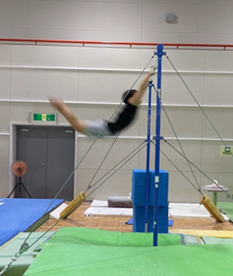
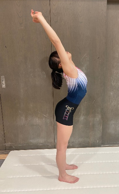
The assessment of incidence of injury in male and female young gymnasts indicates that wrist injuries in males were associated with decreased shoulder joint internal rotation range of motion, and lower back injuries were associated with decreased hip flexion and extension range of motion, shoulder joint external rotation range of motion, and wrist joint palmar flexion range of motion. Male gymnasts often support themselves with their upper extremities; the wrist joints in particular are subjected to excessive physical loading, including compression, rotation, and traction, which have been reported to be twice their body weight and up to 16 times their weight by different authors.14,15 DiFiori et al.14 reported that wrist joint injuries were the most common injury among gymnasts aged 10–14 years, with high weekly practice volume and high skill level as risk factors, but no association with physical function was shown. It is likely that reduced shoulder joint mobility and high levels of support during upper extremity loading places more stress on the wrist joint, and the demonstrated association between reduced shoulder joint range of motion and wrist joint injury may help in intervention for wrist joint injury prevention. The results also suggested that a lack of overall range of motion was associated with the development of lower back injuries in male gymnasts. In this study, the results indicated that lower back injury was associated with decreased hip flexion and extension range of motion, shoulder joint external rotation range of motion. Males have more upper limb supportive techniques and well-developed upper limb muscles, which may be related to the lack of shoulder joint range of motion, which therefore may be relevant. When swinging on the rings and high bar, in order to extend the trunk in during the swinging motion or during shoulder return (from forced shoulder joint flexion to shoulder joint extension), decreased range of motion of the scapulohumeral joint may compensate for the decreased range of motion caused by extension of the thoracolumbar spine. The internal rotation alignment of the humerus characteristic seen in gymnasts may lead to injury,16 and the results of the current study suggest that a decrease in the external rotation range of motion of the shoulder joint during upper limb elevation position may lead to excessive extension of the lumbar spine as a compensation, which may lead to lower back injuries. (Figure 2) Additionally, since hip extension is required during the swinging motion,17 inadequate hip extension range of motion may cause excessive extension stress on the lower back, leading to the onset of the injury.
Heel injuries in female gymnasts were associated with decreased hamstring tightness, knee injuries were associated with increased shoulder joint range of motion and decreased iliopsoas tightness. These findings indicate that the lack of joint range of motion and mobility may not be the only problems contributing to injury. In this study, heel injuries were more common in gymnasts diagnosed with Sever’s disease and were more common in elementary school gymnasts. Mackie et al.18 reported that Sever’s disease was the most common overuse disorder in female gymnasts aged 7–18 years. The primary risk factors include obesity and high-intensity physical activity, with high-impact sports being the primary cause.19 It is necessary to consider age-related characteristics, including muscle strength, due to the age of predilection and athleticism in developing athletes.
Furthermore, lower back injuries were associated with reduced hip extension mobility among female gymnasts. Desai et al.20 reported that a general anterior pelvic tilt in gymnasts influences the development of chronic low back pain and requires improvements in hip flexor group, quadriceps flexibility, trunk muscle strength, and hamstring muscle strength. The results of the current study support the findings of Desai et al.20 that flexibility of the hip flexor group is necessary. Sweeney et al.21 reported decreased iliotibial ligament tightness as a risk factor for lower back pain in female gymnasts, indicating that limited joint flexibility was not associated with lower back pain. However, Sweeney et al.21 also included high school gymnasts, whereas the current study only included elementary and middle school gymnasts. The results of the current study also included participants who reported a history of lower back pain, suggesting that decreased hip extension mobility, which was also associated with a history of lower back pain, may be also related to the occurrence of lower back injuries (Figures 3 and 4).
There was no association between lower back injury and multifidus muscle elasticity in either sex. Masaki et al.22 showed that adults with lower back pain had significantly greater muscle stiffness in the multifidus muscle (L4 level) at rest in the supine position compared to those without the same. These results differed because the age group of the participants in the current study was prone to maturity, and the results may have reflected a variation in muscle elasticity. In addition, Murillo et al.23 found a lower increase of muscular stiffness with contraction of the multifidus muscle in the group with lower back pain than in the group without, suggesting a deficit in multifidus activation. Therefore, there may be a relationship between muscle elasticity during multifidus contraction and lower back injuries in elementary and junior high school athletes, and future studies are warranted.
Limitations
It is difficult to identify physical factors that influence the occurrence of injury based on joint range of motion, tightness, and muscle elasticity alone. Regression analysis should be used to consider the effects of muscle strength, age, individual factors, and left–right differences as well. In this study, there were some items in which left–right differences occurred; the characteristics of gymnasts that tend to cause left–right differences, such as the kicking and pivot leg, as well as the direction of twisting, should be examined in the future.
Moreover, the results of this study were limited by its duration, which did not cover the entire competitive season, and scope, as the injury incidence survey and intervention were conducted in only one club.
CONCLUSIONS
The results of this study indicate that the factors related to range of motion and flexibility differ according to injury location and between males and females. Further studies are required to clarify the physical factors that influence injury occurrence by examining the effects of the gymnasts’ muscle strength, age, individual factors, and left–right differences.
Conflicts of Interest
The authors report no conflicts of interest
_measures_the_shoulder_joint_abduction_angle._the_examiner_is.png)


_measures_the_shoulder_joint_abduction_angle._the_examiner_is.png)


