INTRODUCTION
Dry needling (DN) has emerged as a popular therapeutic intervention for managing musculoskeletal pain, with more than half of physical therapists surveyed using DN in the treatment of their patients.1 Numerous authors have reported DN as an effective tool when used in conjunction with other interventions to manage back pain.2–6 While major adverse events due to DN are reported to be rare,7 those that have been reported to occur from DN around the spine and thorax can be serious, and include pneumothorax, quadriparesis related to acute cervical epidural damage, and hemiplegia related to subdural or epidural hematomas.8–12 In addition, the lack of a standardized method for reporting adverse events likely leads to underreporting, which suggests there may be more adverse events occurring than those currently documented in the literature.1,13
Dry needling of the multifidi is a common intervention for the treatment of back pain and is one that has generally been regarded as a safe technique given that the bony backdrop of the lamina is considered to be protective. While using a bony backdrop, such as the lamina, is thought to decrease the risk of inadvertently entering other unintended structures, this DN strategy for the multifidi presumes the needle tip will not land between adjacent vertebral lamina, where it could potentially pierce through the ligamentum flavum (LF) and enter the spinal canal. Despite this presumption, there is no specific DN technique that has been universally accepted as being the safest approach to ensure the needle successfully reaches the protective bony backdrop of the lamina.
Reported techniques for DN the multifidi suggest that needles should be placed 1-2 cm lateral to the lumbar spinous process and directed medially or inferior-medially until the needle reaches the lamina.14–20 Both the inferior and the medial needle orientation documented among the inferior-medial techniques vary considerably, ranging between 15° and 45°.14,15,17–21 Williams et al. demonstrated that a 0.30 x 50 mm needle can penetrate the LF and enter the spinal canal at the thoracolumbar junction with a needle inserted approximately 1.0 cm lateral to the spinous process, directed in a posterior-anterior orientation, and inclined medially at a 22-degree angle from vertical.22 The purpose of this study was to reproduce the methods employed by Williams et al. but with an inferior-medial multifidus DN technique to determine if a dry needle can penetrate the LF and breach the spinal canal at the thoracolumbar junction.
METHODS
The study was performed on a 77-year-old female, fresh-frozen, lightly-fixed cadaver at Middle Tennessee School of Anesthesia. Ultrasound scans were performed by a certified registered nurse anesthetist with over 20 years of diagnostic ultrasound imaging experience and 15 years of experience performing and teaching regional anesthesia, including ultrasound-guided spinal anesthesia procedures. The fresh/lightly embalmed cadaver was received through the Willed Body Program at The University of North Texas Health Science Center. Exemption from Institutional Review Board approval was granted by Advarra IRB (Pro00070509).
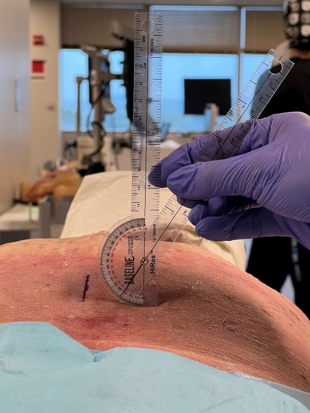
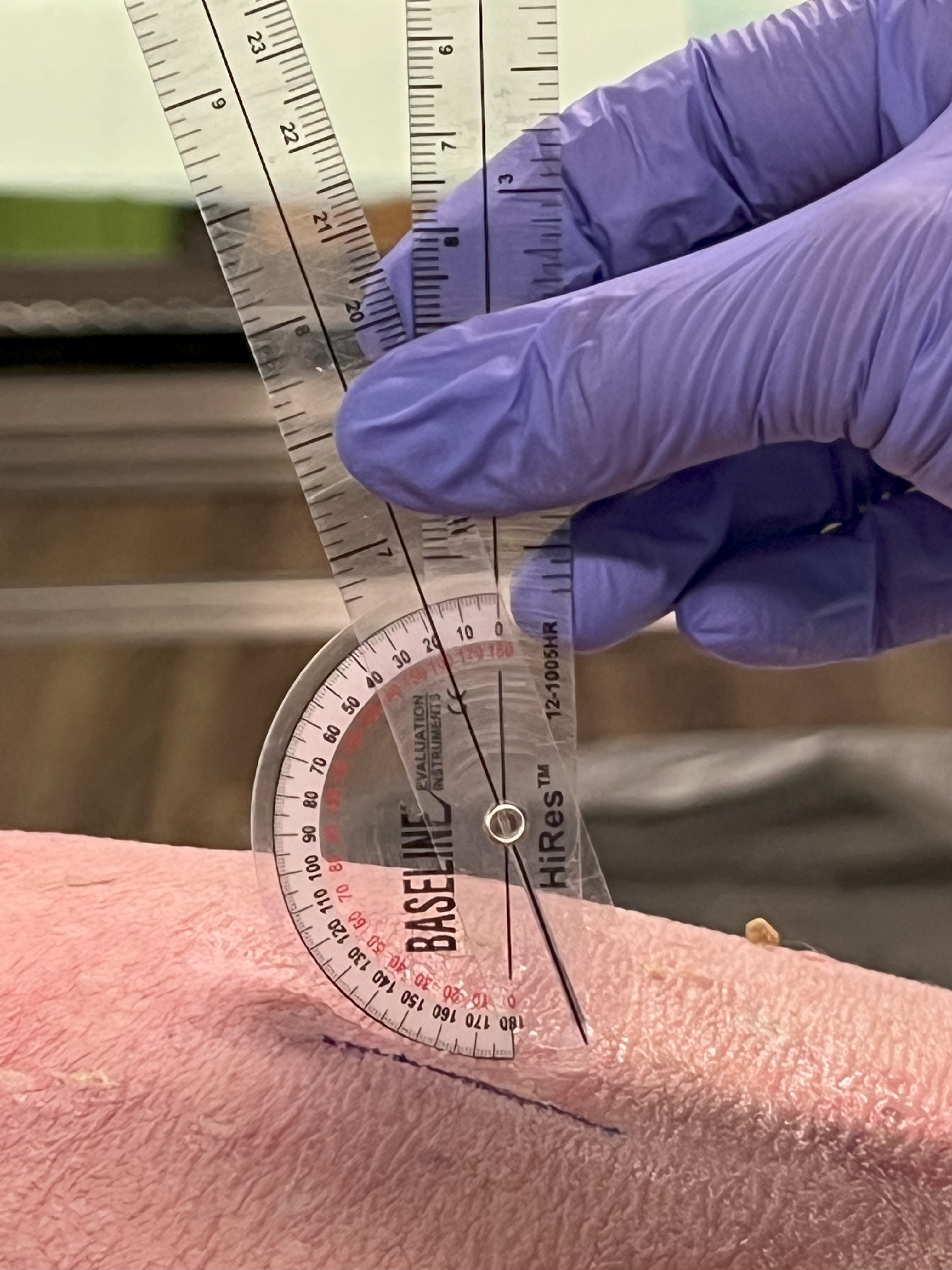
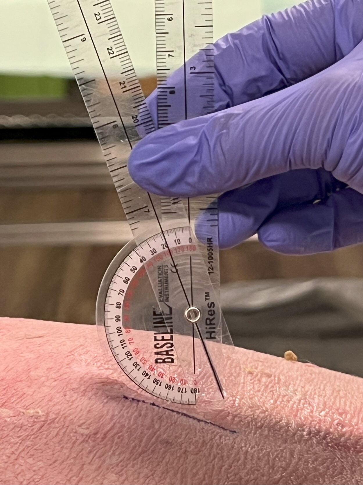
The cadaver was placed in the prone position with the lumbar spine in a relatively neutral position. A Sonosite Edge II ultrasound system with an rC60xi 5-2 MHz curvilinear array transducer (Bothell, WA) was placed in a parasagittal orientation with the orientation indicator facing cephalad. The sacrum, L5-S1 interspace, and L5 spinous process were identified. The transducer was moved cephalad until the interspace between T12 and L1 was identified. A DBCTM ProMaxx 0.30 x 40 mm dry needle was inserted approximately one finger-breadth lateral to the spinous process of T12 and directed in an inferior-medial angulation toward midline while being visualized by ultrasound. If the needle reached the lamina, it was left in place, and another needle was placed in very close proximity. The process was repeated until a needle breached the LF and entered the spinal canal. After the final attempt, measurements were taken to determine the distance along the skin between the needle and the spinous processes and the medial and inferior needle angulations for the needle which entered the spinal canal.
A range of needle lengths between 40 mm and 100 mm have been described when studying DN techniques in the thoracic and lumbar regions, with 50 mm and 60 mm being the most commonly utilized in clinical practice, depending on the spinal location.14,16–21,23 Dry needle length is selected based on the size of the patient’s soft tissue and musculature.14,24 In the previous study by Williams et al., a 50mm needle was used,22 whereas this study utilized a shorter needle (40 mm).
RESULTS
A 0.30 x 40 mm dry needle was able to traverse the space between the vertebral laminae of T12 and L1, penetrate the LF, and enter the spinal canal on the fourth attempt. The needle that entered the spinal canal was located 1.9 cm lateral to the spinous process of T12. More specifically, the insertion point was approximately 4.3 mm inferior to the center of the spinous process of T12, the medial angulation of the needle was 33-degrees from vertical (Figure 1), and the inferior angulation of the needle was 18-degrees (Figure 2).

DISCUSSION
The purpose of this study was to reproduce the methods employed by Williams et al. but with an inferior-medial multifidus DN technique to determine if a dry needle could penetrate the LF and breach the spinal canal at the thoracolumbar junction. The results of this study corroborate a previous finding that a dry needle can enter the spinal canal at the thoracolumbar junction using a medial technique, and also when using an inferior-medial technique.22 The thoracolumbar junction is an area of complex anatomy and an area of presumed increased risk and therefore, it may be important for physical therapists to consider the overall safety of DN techniques when treating this area of the spine.
This study is the second to use ultrasound guidance to document dry needles entering the spinal canal at the thoracolumbar junction when using techniques outlined in the literature for targeting the multifidi. While other healthcare professions utilize ultrasound guidance as a standard tool for training and practicing needle techniques, ultrasound guided DN during training courses for physical therapists is not common practice, nor is ultrasound guided DN routinely used in clinical physical therapist practice in the United States.25 Using ultrasound guidance during DN training may prove valuable, especially in light of the poor reliability and validity of palpation-based methods seen as cornerstone in DN training.26,27 Ultrasound guidance offers a potential solution by providing enhanced visualization and accuracy. Additionally, using ultrasound during DN training may promote trainee’s tactile sensation and spatial awareness by allowing trainees to visualize anatomical structures in real-time to correlate tactile sensations as different tissue layers and structures are engaged. Given the fact that some physical therapists are now being trained in ultrasound imaging techniques in entry-level physical therapy programs,28 as well as dry needling,29 it stands to reason that ultrasound guided DN, especially in areas of increased risk, may be an important consideration in future training and clinical use to improve patient safety.
There are a number of procedural considerations and limitations in this study. First, it is important to recognize that this study did not attempt to determine the likelihood of penetrating the spinal canal during the clinical use of DN, but merely explored the possibility of a dry needle entering the spinal canal when using a technique commonly described in the literature for the multifidi. Therefore, specific needle locations relative to the spinous process and needle angulations are intended to help with replication in future studies, not to be extrapolated as safe/unsafe zones for DN to be performed. Given that the determination of the center of the spinous process was based on palpation and neither imaging nor dissection were used to confirm a true center of the spinous process, generalization of unsafe zones relative to the spinous process would be speculative and inappropriate.
Second, this study was a feasibility study using a 77-year-old, fresh-frozen, lightly-fixed, female cadaver. It is plausible that differences in tissue turgor in cadavers versus living subjects, as well as the age of the donor may have impacted the outcomes. Several authors have documented that the thickness and stiffness of the LF increase linearly with the degree of disc degeneration30 and age31 and can also differ between various lumbar segments.32 The donor in this study was 77-years-old and thus, similar to 90% of individuals 65 years and older, very likely had disc degeneration.33 The age-related changes in the donor used in this study may have actually been protective; however, the dry needle was still able breach the spinal canal. Studies using a larger sample size may have resulted in different outcomes if performed in vivo and with more variability in subjects.
CONCLUSION
The results of this study demonstrate the feasibility of a dry needle entering the spinal canal at the thoracolumbar junction using an inferior-medial technique for the multifidi. These findings support the potential role of ultrasound guidance in the training and clinical practice of DN, especially in regions where safety issues have been documented. Evaluating the safety profiles of DN techniques currently being used, exploring the feasibility and potential impact of ultrasound guidance during training and clinical practice, and examining the clinical outcomes associated with ultrasound-guided dry needling are important next steps.