

INTRODUCTION
Vibration therapy utilizes the human body’s response to vibrations through various mechanoreceptors and is therefore regarded as an effective modality for training and rehabilitation, offering potential health benefits when applied correctly.1 Whole-body vibration (WBV) therapy, which involves standing on a vibrating platform that transmits vibrations through the feet, has been extensively studied.1 In contrast to WBV therapy, local vibration (LV) treatment targets specific muscles or tendons using portable devices such as vibrating foam rollers.2 Percussive therapy (PT) likely integrates LV with elements of conventional massage3,4 and has gained popularity in recent years among athletes, physiotherapists, and other individuals for optimizing post-exercise recovery.5 This is typically achieved through mechanical handheld percussion devices (MPDs), such as massage guns, that combine compression and vibration.4,5
The impact of PT on recovery, physical performance parameters and experiences of pain has been extensively researched. However, variability in MPDs, application protocols (i.e. pre/post exercise, duration of application, targeted muscle group, single/repeated application, frequency/amplitude) and outcomes assessed lead to mixed results. Massage guns, such as the Theragun™ (Therabody, Los Angeles, CA, USA) or the Hypervolt (Hyperice, Irvine, CA, USA) appear to effectively enhance range of motion (ROM) and flexibility, as well as improve short-term recovery-related outcomes6,7 and pain perception.7 However, no significant effects were observed on balance, acceleration, and agility.6 Investigations of the impact of massage guns on muscle strength and explosive activities have produced contradictory results.6,7
Despite the claimed clinical benefits, there is a lack of research explaining the underlying mechanism of PT’s physiological effects.6–9 Recent studies have demonstrated that PT acutely enhances microcirculation blood flow10 and skin temperature.11 Physiological responses of PT have been shown to be more localized and do not involve a general excitation of the cardiovascular system, as there were no significant increases in heart rate or popliteal artery diameter.10 Further investigation is needed to determine whether these PT responses occur at the cutaneous or muscular tissue level.12 The physiological responses of therapy between males and females can differ and there is still an underrepresentation of female participants in biological research, particularly in physiology.13–15
The aim of this study was to investigate the changes and time course of local skin temperature (Tskin), deep tissue perfusion (erythrocyte flow velocity [speed] and deep tissue blood flow [flux]), and muscle oxygenation (SmO2) after a standardized 4-minute treatment with a TheragunTM of the vastus medialis muscle of healthy female volunteers.
METHODS
Participants
The sample size was determined using a power analysis (G*Power Version 3.1; Franz Faul University, Kiel, Germany) with the following parameters: α = 0.05; power = 0.8; effect size = 0.2; statistical test = ANOVA: repeated measures, within-between interaction; number of groups =2; number of measurements = 12. Based on these criteria, the minimum sample size was 20.
The following inclusion criteria were applied: (i) non-smokers; (ii) aged >18 years; (iii) healthy skin condition on the leg on which the TheragunTM was applied; (iv) no existing musculoskeletal injuries to the lower extremities. Participants who met any of the following criteria were excluded from the study: (i) metal implants in the intervention area; (ii) medications like analgesia, muscle relaxants or coagulations; (iii) pregnancy/lactation; (iv) alcohol or drug addiction; (v) diseases such as diabetes mellitus type 1 or 2, polyneuropathies; (vi) inability to follow the procedure of the study due to language barriers, psychological illness, dementia, anxiety; (vii) participation in another intervention study with medication/medical device.
This study protocol was approved by the Swiss Ethical Committee of Zurich (BASEC-No. 2019-01742) and all participants provided written informed consent prior to the experimental sessions being conducted.
Measurements
Before interventions, body height (cm) was measured using a stadiometer (GPM; Zurich, Switzerland). Body weight (kg) and estimated subcutaneous fat percentage (%) were assessed using a digital weighing scale (TBF 611; Tanita, Tokyo, Japan). Body mass index (BMI; kg/m2) and body surface area (BSA; m2) were calculated.
Erythrocytes flow velocity [speed], deep tissue blood flow [flux], and muscle oxygen saturation (SmO2) were assessed unilaterally in the treated area of the intervention leg, while skin temperature (Tskin) was measured bilaterally in both the treated area of the intervention leg and the corresponding area of the control leg. Tskin (°C) was measured using a forward-looking infrared (FLIR) camera (A655 sc series; InfrarotTec Systems, Ranstadt, Germany). Flux (arbitrary units; AU) was measured using a high-power laser doppler monitor (moorVMS-LDF1-HP; Moor Instruments, Devon, UK). SmO2 (%) and speed (arbitrary units; AU) were assessed with a near-infrared spectroscopy monitor (moorVMS-NIRS, Moor Instruments, Devon, UK). Data from moorVMS-LDF1 and moorVMS-NIRS were recorded with the provided software (moor MS PC v4.2.1). All parameters were assessed at 5-minute intervals up to 50 minutes after TheragunTM application.
Experimental Application
Experimental sessions were conducted at the research laboratory under controlled environmental conditions: room temperature at 22.24 ± 0.66 °C and relative humidity at 39.35 ± 1.62%, measured with a digital multimeter (Voltcraft MT-52; Conrad Electronic, Hirschau, Germany). The side of the Theragun™ application was randomly selected by flipping a coin. Participants were instructed to lie supine on a therapeutic plinth, where they remained throughout the preparation and the entire experiment. An area of interest, defined by anatomic structure to account for leg length variations, was marked prior to measurements to ensure standardized procedure. Therefore, the anterior superior iliac spine and middle of the top edge of the patella of each participant were palpated and marked with a skin-friendly highlighter. Another mark was made 2 cm from the middle of the top edge of the patella in the cranial direction. Then, a line was drawn from the anterior superior iliac spine to the middle of the edge of the patella, and a prefabricated template (dimension = 10 cm), was placed so that the lower edge aligned with the mark 2 cm cranial to the upper edge of the patella. The application area was cleaned with an alcohol wipe, marked, and then covered with elastic tape strips to ensure that the FLIR images would be reliable (Figure 1). To support the legs, a foot roller (30 cm x 15 cm) was placed between the lower legs and secured with a light bandage.
_marked_with_elastic_red_tape_.jpeg)
Participants underwent a 20-minute acclimatization period after preparations and were instructed to minimize movement throughout the entire experiment. Baseline measurements (BL) for Tskin, speed, flux and SmO2 were taken before applying the TheragunTM. Following the BL, the TheragunTM was applied using a standardized procedure with the standard ball attachment. The TheragunTM G3Pro offers two speeds (29 and 40 Hz) with an amplitude of 16 mm and a strength of up to approximately 27 kg. For this study, the speed was set to 29 Hz. To simulate a realistic physiotherapeutic setting while maintaining standardization across participants, a 4-minute treatment was applied in a specific pattern within the intervention area: during the first two minutes, the TheragunTM was moved horizontally, and for the remaining two minutes, it was moved vertically, with no external pressure applied.
Following the application, measurements were taken at 5-minute intervals from the initial time point (t0) up to a total duration of 50 minutes (t50). First, skin temperature was assessed using the FLIR camera. Subsequently, the laser sensor of moorVMS-LDF1 and sensor of moorVMS-NIRS were placed on the skin within the targeted area to measure speed, flux, and SmO2 for 30 seconds. To maintain consistent temperatures of the sensors and the skin between measurements, the laser and sensors were positioned on a region of the skin outside the designated measurement area.
Statistical Analyses
Statistical analyses were performed using the Statistical Package for Social Sciences (SPSS), version 29.0 (IBM Corporation, Armonk, NY, USA). Descriptive statistics (mean ± standard deviations (SD)) were computed for Tskin, speed, flux, and SmO2 at each of the 10 time points. The normality of data distribution was verified using the Shapiro–Wilk test. All data, except Tskin on the control leg, was normally distributed and parametric statistical analyses were applied. Repeated-measures analysis of variance (ANOVA) with post hoc Bonferroni test correction was used to assess the effect of speed, flux and SmO2 over time (BL, t0, t5, t10, t15, t20, t25, t30, t35, t40, t45, and t50). A repeated measures analysis of variance (ANOVA) was conducted to assess the effect of time, group, and the interaction between time and group. The within-subject factors included time (BL, t0, t5, t10, t15, t20, t25, t30, t35, t40, t45, and t50) and group (intervention vs control leg). A Greenhouse-Geisser adjustment was applied to adjust the degrees of freedom if the Mauchly test indicated that the sphericity assumption was violated. Post-hoc pairwise comparisons were performed using the Bonferroni correction to control for multiple comparisons, if significant main or interaction effects were observed. The level of significance was set at p < 0.05 for all statistical tests. The effect size was calculated to assess the magnitude of the observed effects and expressed as partial eta-squared (η2partial), with values of 0.1–0.29 considered as “small”, 0.3–0.49 as “medium”, and >0.5 as “large”.16
RESULTS
This observational study included twenty-six healthy, young female participants recruited from a university population, who were assessed during November and December 2020 (Table 1).
The descriptive statistics (means ± SD) for Tskin (intervention and control leg), the perfusion parameters speed and flux, and SmO2 over time (BL to t50) are shown in Table 2. The values of all parameters at all time points showed a significant increase compared to BL.
The ANOVA analysis of Tskin revealed significant main effects of time (F (2.24, 55.97) = 60.20, p < 0.001, η²partial = 0.71), group (F (1.00, 25.00) = 192.79, p < 0.001, η²partial = 0.89), and a significant interaction between group and time (F (2.41, 60.17) = 65.47, p < 0.001, η²partial = 0.72). Pairwise comparisons indicated a significantly higher Tskin in the intervention leg at all time intervals compared to BL (p < 0.001) (Figure 2A), with the highest temperature recorded at t5, reaching 33.16 ± 1.41 °C (Table 2). In comparison, the temperature in the control leg was 30.21 ± 0.90 °C at t5. Additionally, pairwise comparison for the intervention leg demonstrated significant differences across all time intervals compared to BL (p < 0.05) (Figure 2A), with the highest temperature observed at t30 (30.45 ± 1.14 °C) (Table 2).
Regarding speed, a significant time effect was observed (F (3.23, 80.78) = 38.75, p < 0.001, η²partial = 0.61). Bonferroni-adjusted post hoc analysis revealed significantly higher values at all time intervals (p < 0.001) than BL values after Theragun™ application (Figure 2B). Speed was greatest directly after the treatment (t0) (Table 2).
_of_(a)_skin_temperature_(tskin)__(b)_flow_velocity_of_erythrocytes.jpeg)
A significant time effect was observed on flux (F (3.47, 86.84) = 39.16, p < 0.001, η2partial = 0.61). After Bonferroni-adjusted post hoc analysis, it was revealed that all time intervals showed significantly higher values (p < 0.001) compared to the BL (Figure 2C). The largest increase in flux occurred immediately after the treatment (t0) (Table 2).
In relation to SmO2, a significant effect of time was observed (F (5.37, 134.34) = 15.21, p < 0.001, η2partial = 0.38). Post hoc analysis with Bonferroni adjustment indicated significantly higher values (p < 0.05) for all time intervals compared to the BL (Figure 2D). The highest increase in SmO2 was observed at t5 (Table 2).
DISCUSSION
The primary objective of this study was to analyze the impact of a standardized 4-minute Theragun™ treatment on Tskin, deep tissue perfusion parameters (speed and flux), and SmO2. Additionally, the study aimed to assess the time course of physiological changes in the vastus medialis muscle in a cohort of healthy female volunteers. The findings revealed significant positive effects of time, group, and their interaction on Tskin, as well as significant increases and substantial effect sizes in speed, flux, and SmO2 compared to BL.
Skin Temperature
Tskin is influenced by the rate of blood flow, the composition of subcutaneous tissue, and the activity of the autonomic nervous system.17 In this study, a 4-minute TheragunTM application resulted in a maximum temperature elevation of +3.76 °C in the treated area at t5. The temperature remained significantly elevated for up to 50 minutes following application (Figure 2A). In the control leg, Tskin also increased after the application on the intervention side and remained elevated, albeit to a lesser extent, with the greatest rise observed at t30 (+0.64 °C).
To the best of current knowledge, only one other study has investigated the effects of PT on Tskin. This study reported an increase in Tskin of 0.5 °C after treating the thoracolumbar fascia with a massage gun for 15 minutes at a frequency of 30 Hz. Yang et al. applied the massage gun at a comparable frequency but for a longer duration than in this study. Nevertheless, they reported a much lower post-treatment Tskin. One reason may be that the current study focused on a localized treatment area of 10 cm2, whereas Yang et. al treated the entire back. Temperature rises in the skin have been found after forms of traditional massage,18–21 but not necessarily after LV treatments22 and WBV therapy.23–26
The changes in Tskin after PT are likely due to mechanical effects like those seen in traditional massage therapy. Skin surface friction induces localized heating leading to increased skin microcirculation.21 This, in turn, triggers the release of mediators such as histamine and prostaglandins from mast cells, leading to an initial vasoactive response in the arterioles.20,21,27,28 Possible explanations for a decrease in Tskin in WBV therapy include a thermoregulatory response, where blood flow is redirected from the skin to the active muscles to prevent hyperthermia, or a vasoconstrictor response in the skin induced by mechanical vibration.23
Physiological effects of heating treatment in the contralateral limb have been observed in other studies.29–34 Ren et al. demonstrated a skin temperature increase of +1.98 °С in the contralateral foot following a water immersion at 40 ± 1 °C within 10 minutes.32 Physiological thermoregulation is based on peripheral skin and central thermoreceptors. A thermal stimulus activates thermosensors in the heated skin area, sending signals to the hypothalamus, which in turn activates temperature-sensitive neurons. As a result, both ipsilateral and contralateral thermoregulators effectors receive signals to induce skin vasodilation.32
Perfusion Parameters and Oxygen Saturation
Sustained increased blood flow over an extended period is considered important for recovery following intense muscular exertion. It also plays a key role in improving blood circulation in targeted muscle groups before competition, which is essential for optimal sports performance. Moreover, optimal oxygen delivery is vital for tissue cells as it supports their function repair, and aids in the remodeling of injured tissue.35–37
The findings of this study on the effect of TheragunTM application on perfusion parameters and oxygen saturation of the vastus medialis muscle are consistent with previous research on PT. The observed increases in speed and flux are supported by the study by Needs et al., which demonstrated that localized PT with a massage gun significantly enhances popliteal artery blood flow in the calf muscle.10 Additionally, studies of WBV therapy have found an increase in vascular tissues and cutaneous blood flow.38,39 To the best of current knowledge, no other study has investigated SmO2 levels following PT. In line with the observed increase in SmO2 following four minutes of TheragunTM application, Percival et al. and Romero-Moraleda et al. found higher SmO2 levels after local vibration foam rolling following exercise-induced muscle damage.40,41 In contrast, WBV therapy does not seem to lead to increased SmO2 values.38
The highest values for speed and flux in this study were recorded immediately after the TheragunTM application (t0). Needs et al. observed a delayed effect one to three minutes after vibration application on both volume flow and mean arterial blood velocity.10 The 5-minute intervals between measurements in this study do not allow for confirmation of the observation by Needs et al., leaving it unclear whether speed or flux might have reached slightly higher values between t0 and t5. Values of blood flow and SmO2 remained elevated for up to 50 minutes following the application. To the best of current knowledge, no studies have measured blood flow or SmO2 beyond this time frame, leaving the exact duration of the elevation unclear.
In this study, the TheragunTM treatment was applied at a frequency of 29 Hz, two minutes horizontally and two minutes vertically with no external pressure applied. In the study of Needs et al., the application of localized vibration to the calf at a low frequency (30 Hz) for 5 and 10 minutes was not sufficient to induces significant changes in mean blood velocity or volume flow.10 However, at higher frequencies (38 and 50 Hz), significant effects were observed after 5 and 10 minutes, with blood flow becoming more elevated as the frequency and duration of vibration increased. Needs et al. observed that the increased values at low frequencies (30 Hz) were marginally not significant. They postulated that with larger sample size, these effects may reach statistical significance.10 The massage gun application in this study differed from that of Needs et al.10 not only in frequency and duration but also in the direction of movement and the size of the treated area. The TheragunTM was applied both horizontally and vertically, in contrast to Needs et al., who used a proximal-distal-proximal movement. Additionally, the treated area in this study was 10 cm2, while Needs at al. treated the entire calf. These differences may have influenced the significance of the blood flow parameters observed. In WBV therapy, the relationship between frequency and blood flow behaves differently than with localized vibration treatments: lower frequencies (≤ 30 Hz) resulted in greater observed peripheral blood flow compared to higher frequencies (> 30 Hz).38,39 A possible explanation is, that lower frequencies provide increased time between contraction, allowing for greater perfusion.38 Additionally, higher frequencies and intensities may lead to a reduced vasodilation (i.e. reduced responsiveness to acetylcholine), vasoconstriction (i.e. increased responsiveness to α2C adrenoreceptors) and/or sympathetic activation.39 For practical applications, these findings raise important questions about the ideal frequency range for maximizing blood flow and whether there is a duration threshold beyond which further increases in blood flow do not occur. Additionally, it prompts consideration of whether the size of the treated area influences the necessary duration of treatment.
In this study, both cutaneous blood flow and SmO2 increased after PT, suggesting an enhanced oxygenated hemoglobin supply to the muscle by improving muscular blood flow. Percival et al. observed a significant re-saturation in muscle oxygenation following a muscle damage-inducing protocol applied to the flexor carpus ulnaris when using a vibrating foam roller (45 Hz) administered twice daily over a 48-hour period. This re-saturation effect was not observed when using a non-vibrating foam roller.41 In contrast, Romero-Moraleda et al. reported increased SmO2 following a single 5-minute treatment with both non-vibrating and vibrating (18 Hz) foam rollers on the leg after muscle damage induction.40 Similar enhancements in skeletal muscle oxygenation with non-vibrating rolling massage have also been documented in other studies.42 Additionally, WBV therapy does not appear to lead to increased SmO2 values.38 The question arises whether the enhancement in blood flow and SmO2 observed with both vibrating and non-vibrating tools is mediated by the same underlying mechanisms or if distinct mechanisms are responsible for these effects. Vibration seems to play a crucial role in prompting a rise in muscular blood flow. However, it remains unclear whether the vibration frequency, frequency of application or application direction (vertical vs. side-alternating) influences muscular blood flow and, consequently, SmO2 levels.
The underlying mechanism by which PT in particular10 and vibration therapy in general1,43 increases cutaneous and muscle blood flow is still not well understood. Compression and vibration of massage guns may lead to mechanical, neuronal, and vascular responses.6 The mechanical pressure applied during therapy may result to a rise in arterial pressure, and upon release, this could enhance blood flow.42 The application of the massage gun in this study and in that of Needs et al.10 was performed without additional pressure. Ferreira et al. concluded in a review that the pressure applied by massage guns may not be sufficient to induce significant changes in tissue conditions, as has been observed with other techniques such as foam rolling.6
Another potential mechanism responsible for the observed increase in muscle perfusion is the tonic vibration reflex (TVR).44 Mechanical vibrations applied to the muscle belly or tendon primarily stimulate the muscle spindle, particularly the Ia afferent fibers, and can induce involuntary muscle contractions. These rhythmic contractions and relaxations of the precapillary sphincter increase muscle metabolic demand, oxygen consumption and vasodilation. This mechanism is analogous to muscle contractions induced by exercise, which similarly trigger the release of vasodilatory substances, resulting in an overall increase in arterial blood flow to the active tissues.45 In the case of WBV therapy46,47 and LV treatment,48 muscle contractions during the treatment have been demonstrated using electromyography, supporting the TVR as a plausible mechanism. Amiez et al. further emphasize that voluntary activation, particularly through visual control of the muscle treated with LV therapy, has a decisive influence on the TVR and can either enhance or suppress its effect.48 The TVR appears to be a complex response influenced by several factors, including vibration frequency, amplitude, muscle type, and state of contraction. However, the optimal experimental parameters to reliably induce TVR remain unclear.43
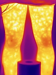
Shear stress caused by massage promotes the release of vasoactive substances, e.g. histamine and bradykinin, increasing the permeability of blood vessels and resulting in local and general tissue vasodilation.49 Although data on histamine release were not collected, the infrared images show the characteristic blotchy redness of the skin typically observed after massage (Figure 3). Furthermore, a recent study by Needs et al. found that the use of antihistamine medication resulted in a nonsignificant increase in popliteal blood flow following percussion therapy, whereas local percussion therapy without antihistamine resulted in a more pronounced popliteal blood flow.50
_displays_characteristi.jpeg)
This mechanism could explain the lack of increase in SmO2 observed with WBV therapy. In WBV therapy, the vibration is applied to the sole of the foot and transmitted vertically to the muscles, rather than being applied directly to the muscle or tendon as with local PT. This likely results in less shear stress and, consequently, a reduced release of vasoactive substances. The release of vasoactive substances is likely the primary mechanism behind the enhanced blood flow and SmO2 values observed with Theragun™ application. This conclusion is based on the lack of external pressure applied during treatment and the findings by Needs et al., which demonstrated an effect of antihistamine medication.
Limitations
In the present study, only women were recruited, and local perfusion and skin temperature changes after standardized TheragunTM application were determined. Potential differences in outcomes for a male population must be considered due to differences in body composition and the absence of hormonal fluctuations associated with the menstrual cycle and contraception use.51–53 Research has shown sex differences in vascular control mechanisms, such as cutaneous vasomotion and sudomotion.54–57 Females typically have a higher proportion of subcutaneous white adipose tissue, particularly around the hips and thighs, which could influence perfusion results.58 Additionally, contraceptive methods used by women may alter the balance and release of hormones, affecting body temperature and potentially impacting skin temperature measurements compared to those in a male population.59 Data on the menstrual cycle of the female participants were collected, but the small sample size precludes any conclusions. Furthermore, this aspect was not part of the research question in this study, so no further interpretations regarding menstrual cycle data were conducted.
A possible side effect of repeated mechanical stress, such as shear forces applied to muscle tissue, is erythrocyte damage.60 Consequently, repetitive mechanical stress from local TheragunTM application could potentially evoke erythrocyte hemolysis. This study did not include a control group, and subjective data regarding “thermal sensation” and “thermal comfort” were not obtained. Although no adverse events were reported, participants of local TheragunTM treatment remain unknown.
This study demonstrated that the application of Theragun™ influenced physiological parameters at multiple tissue levels, including skin temperature (Tskin), cutaneous blood flow (flux and speed), and muscle oxygenation (SmO₂). While these findings provide initial insights into the physiological effects of percussion therapy, further research is needed to evaluate its practical applications in rehabilitation and sports contexts fully. For example, future studies should investigate whether the increased perfusion and oxygenation following the use of Theragun™ lead to measurable benefits for recovery or performance. Studies with extended follow-up periods would help clarify how long the observed physiological changes persist and whether they contribute to enhanced functional outcomes. Comparative research is also warranted to explore the differential effects of side-alternating versus vertical vibration, particularly concerning shear stress and its role in promoting vasodilation. Additionally, identifying the most effective frequency and duration of application concerning specific target tissues and desired outcomes (e.g., recovery vs. warm-up) would contribute to establishing an evidence-based dose-response model for percussion therapy.
CONCLUSION
The results of this study demonstrate that a 4-minute localized Theragun™ application to the vastus medialis significantly enhances physiological responses in the cutaneous, subcutaneous, and muscle tissues in females. The treatment not only increases local skin temperature but, more interestingly, also improves deep tissue blood flow, the speed of red blood cell movement, and muscle oxygenation. These findings suggest that Theragun™ has a profound effect on deeper tissue layers. They provide valuable insights into the physiological mechanisms of PT, helping athletes and healthcare professionals make more evidence-based decisions regarding the local application of Theragun™. From a practical perspective, the results support the use of Theragun™ treatment for optimizing athlete’s performance and recovery through enhanced blood flow and oxygenation. Based on the observed time course of effects, applying Theragun™ approximately 10 minutes before activity may maximize physiological benefits.
ACKNOWLEDGMENTS
The authors would like to thank the Thim van der Laan Foundation for financial support and Carlina Deflorin for their practical assistance in the data assessment. The datasets generated and analyzed during the current study are not publicly available but are available from the corresponding author who was an organizer of the study. The experiments comply with the current laws of the country where they were performed.
Disclosure
The authors declare no competing interests.