INTRODUCTION
The overhead throwing motion is a relatively unnatural human movement that requires training and practice. Baseball pitchers and football quarterbacks rely on an ideal overhead throw for success in their sport. Biomechanical analysis can provide information on the most efficient and effective throwing motion kinematics; however, most research has been published on baseball pitching mechanics.
Few researchers have investigated the throwing motion in quarterbacks. Biomechanists have identified similarities between baseball pitching and football quarterback throwing mechanics. Fleiseig et al.1 and Rash et al.2 provided the foundations of what is known about quarterback kinematics, while Kelly et al.3 provided the foundation of electromyography (EMG) analysis through four phases of the quarterback throwing motion. Gaining a better understanding of the kinematics of quarterbacks passing a football may provide sports medicine professionals with useful information for preventing, treating, and rehabilitating football-related throwing injuries.
A 2018 study of professional football quarterbacks reported that rotator cuff tendonitis was the most common shoulder injury related to throwing, followed by biceps tendonitis.4 The authors noted that an understanding of football passing is essential in managing upper extremity injuries in quarterbacks. In addition, knowledge of the throwing dynamics may improve performance.
Phases of Football Quarterback Throwing
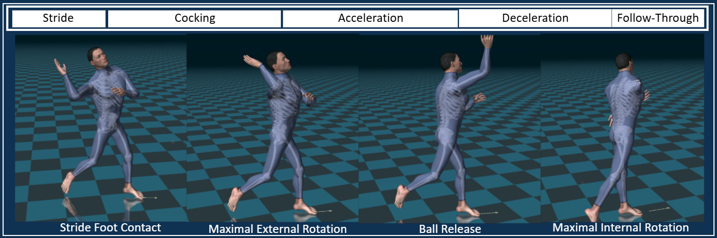
Football quarterbacks generate force in their throwing arm through a combination of movements broken down into phases. Previous authors have utilized EMG and three-dimentional (3D) cameras to quantify range of motion (ROM), velocity, and torque at specific intervals within the phases of the quarterback throwing motion.1–3,5 The consecutive phases of the football pass are generally 1. Stride, 2. Cocking, 3. Acceleration, 4. Ball Release, 5. Deceleration, and 6. Follow-through. Phases can be identified using key points of interest (POI): stride foot contact, maximal shoulder external rotation, ball release, and maximal shoulder internal rotation. These POI represent the kinematic sequence of the quarterback football pass.
Figure 1 illustrates the phases and POI during a football pass. The stride phase begins when the lead leg is lifted and ends when the front foot contacts the ground. The cocking phase begins when the front leg contacts the ground and ends when the throwing shoulder reaches maximal external rotation. The arm acceleration phase begins at maximal external rotation and ends at ball release. The arm deceleration phase begins at ball release and ends at shoulder maximal internal rotation. The follow-through phase begins at maximal internal rotation and ends when reaching a balanced position to continue playing.
_and_points_of_interest_(bottom)_during_a_quarterback_football_pass.png)
The body executes movement of adjacent joints in various planes of motion through consecutive phases to transfer forces from the legs to the arm. This sequence of movement is known as “kinematic sequencing.” This kinematic analysis provides spatial variables (joint range of motion in planes of movement) and temporal variables (time and velocity) that are reported in the sequence of individual segments throughout the body. In football quarterbacks, the stride knee, the trunk and pelvis, and the throwing elbow and shoulder contribute to the football pass sequence throughout the five phases.
Kinematic Analysis with Inertial Motion
Football quarterback throwing patterns typically involve a succession of drop-back steps, high-speed trunk rotation, and throwing arm mechanics that may require more space than a typical lab can offer. Traditional camera-based kinematic analysis and optical motion capture systems are widely used in laboratory settings; however, these systems have limitations, including the cost and maintenance of fixed cameras, and the need for an indoor lab setting to function optimally.6 These space constraints are inherently limited, which may restrict the path and range of motion that can be analyzed.
The limitations of traditional optical motion capture systems necessitate exploring alternative technologies that can capture realistic, high-speed movements in natural environments.6 Advances in technology have led to the development of other motion capture devices that do not require fixed cameras or lab settings. Inertial measurement units (IMUs) are small sensors placed on body parts to capture movement data wirelessly. IMUs use gyroscopic, magnetometer, and accelerometer data at high speeds in three planes to attain a three-dimensional motion model. Advantages of IMUs include their portability and ease of use in various settings, which supports data collection both inside the lab and outside on the athletic field. IMUs offer flexibility in sensor placement, allowing the system to target individual body parts and capture a wide range of movement data without being constrained by the fixed positions required by camera-based systems. The disadvantages of IMUs include data artifacts caused by poor IMU stabilization or skin movement, electromagnetic interference, and signal drift. Inertial motion units are ideal to capture the kinematic sequence of quarterbacks as they do not require a fixed setting, facilitating their use on the field compared to camera-based systems in a laboratory. An understanding of kinematic sequencing in the extremities and trunk during football passing can provide valuable insights for optimizing performance and inform the creation of specialized training and rehabilitation protocols for quarterbacks. The purpose of this pilot descriptive study was to identify the kinematic sequencing of a football quarterback’s pass using wireless inertial motion technology.
MATERIALS AND METHODS
Participants
Data were analyzed from eight healthy, right-handed, pain-free quarterbacks with at least two years of experience, four of whom were from a high school team and the other four from a collegiate team. The kinematic assessment was part of a voluntary, routine pre-season screening performed at a sports medicine facility providing medical coverage to the schools. Deidentified data was used for analysis by Franciscan Missionaries of Our Lady University as IRB exempt.
Procedure
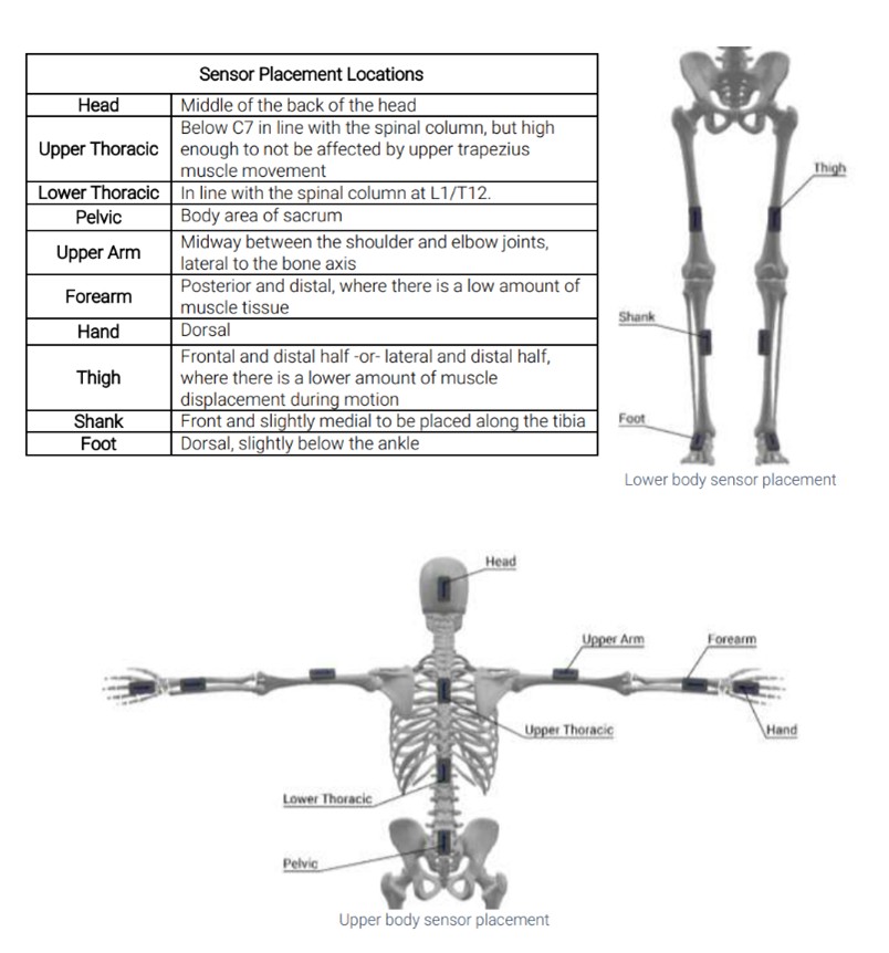
Sixteen Noraxon Ultium IMUs (Noraxon, Scottsdale, AZ) were fitted to each quarterback at the lower extremities, trunk, and upper extremities (Figure 2). Each quarterback performed three drop-back passes at three increasing distances (10, 20, and 30 yards), resulting in nine passes per participant. Passes were performed in order and not randomized. Quarterbacks wore athletic shorts and shirts without a helmet.

The kinematic data from each pass were analyzed using Noraxon MyoMotion 3.18 software. A kinematic sequence algorithm was designed to identify the timing and sequencing of joint angles during the throwing motion at four key POI of the throwing motion: stride foot contact, maximal shoulder external rotation (MaxER), ball release, and maximal shoulder internal rotation (MaxIR). The four POI were automatically detected by the software using the segmental acceleration signals. The mean angles for shoulder abduction, shoulder external rotation, elbow flexion, knee flexion, trunk inclination, lumbar extension, trunk rotation, pelvis rotation, and hip-shoulder separation were calculated for each participant across all nine throws. Hip-shoulder separation was calculated as the difference in transverse plane angles of the pelvis and shoulder girdle.
RESULTS
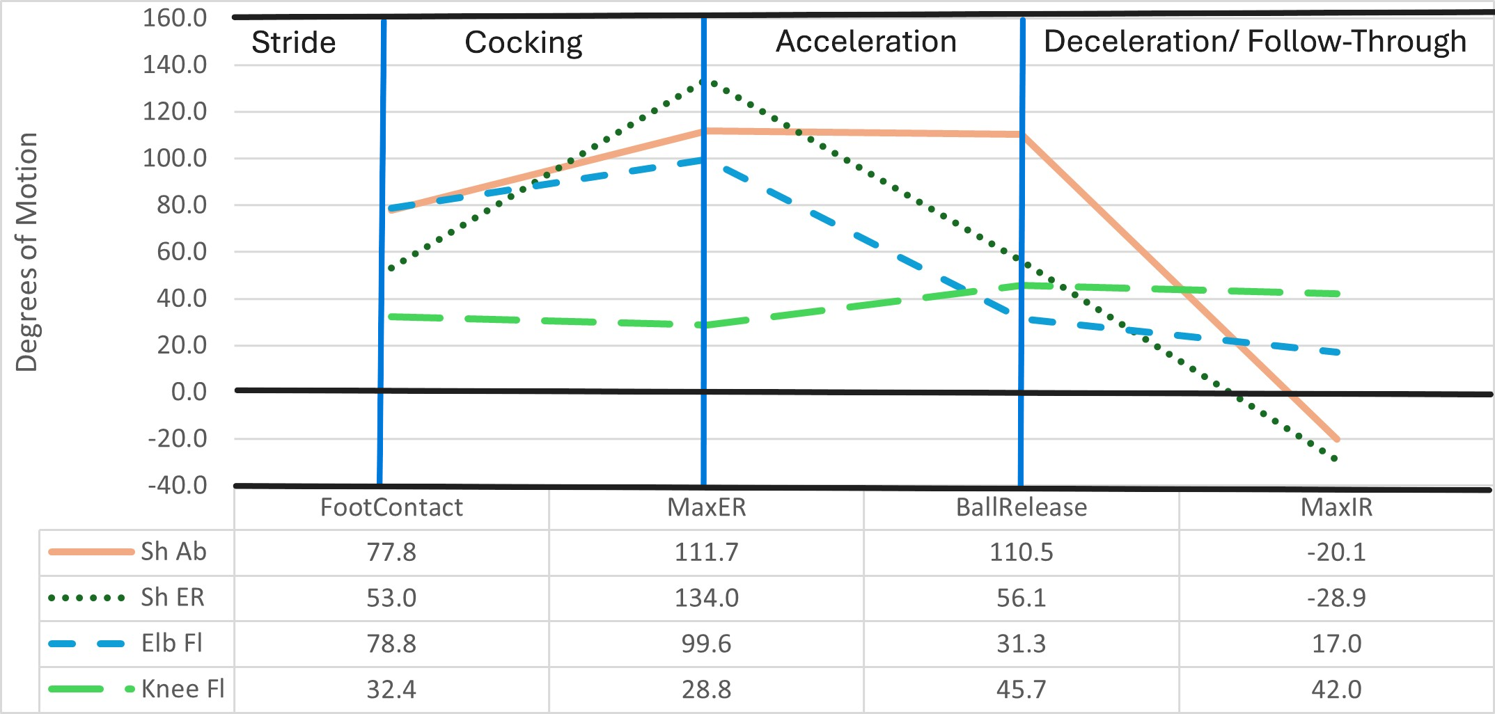
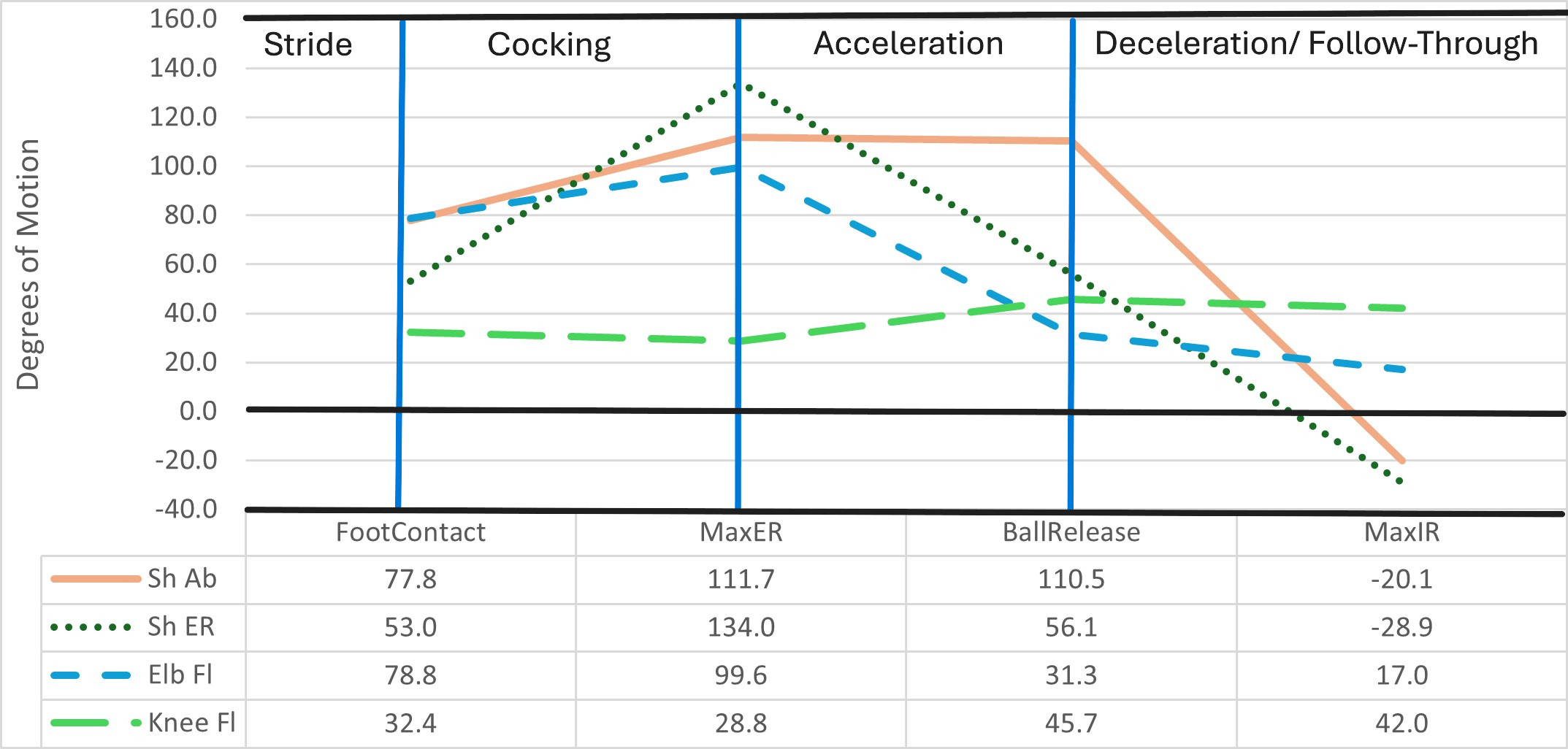
The extremity kinematic sequencing within the football pass was analyzed during the four phases of throwing (Figure 3). Just prior to the acceleration phase, shoulder abduction and external rotation peaked at an average of 112° and 134°, respectively. Shoulder external rotation was 56° at ball release. Shoulder abduction was maintained near 110° through the acceleration phase as the shoulder internally rotated, and the elbow extended from 99° to 31°. Stride knee flexion remained relatively consistent, averaging from 29° to 46°, peaking at ball release. Each variable decreased during the deceleration and follow-through phases.
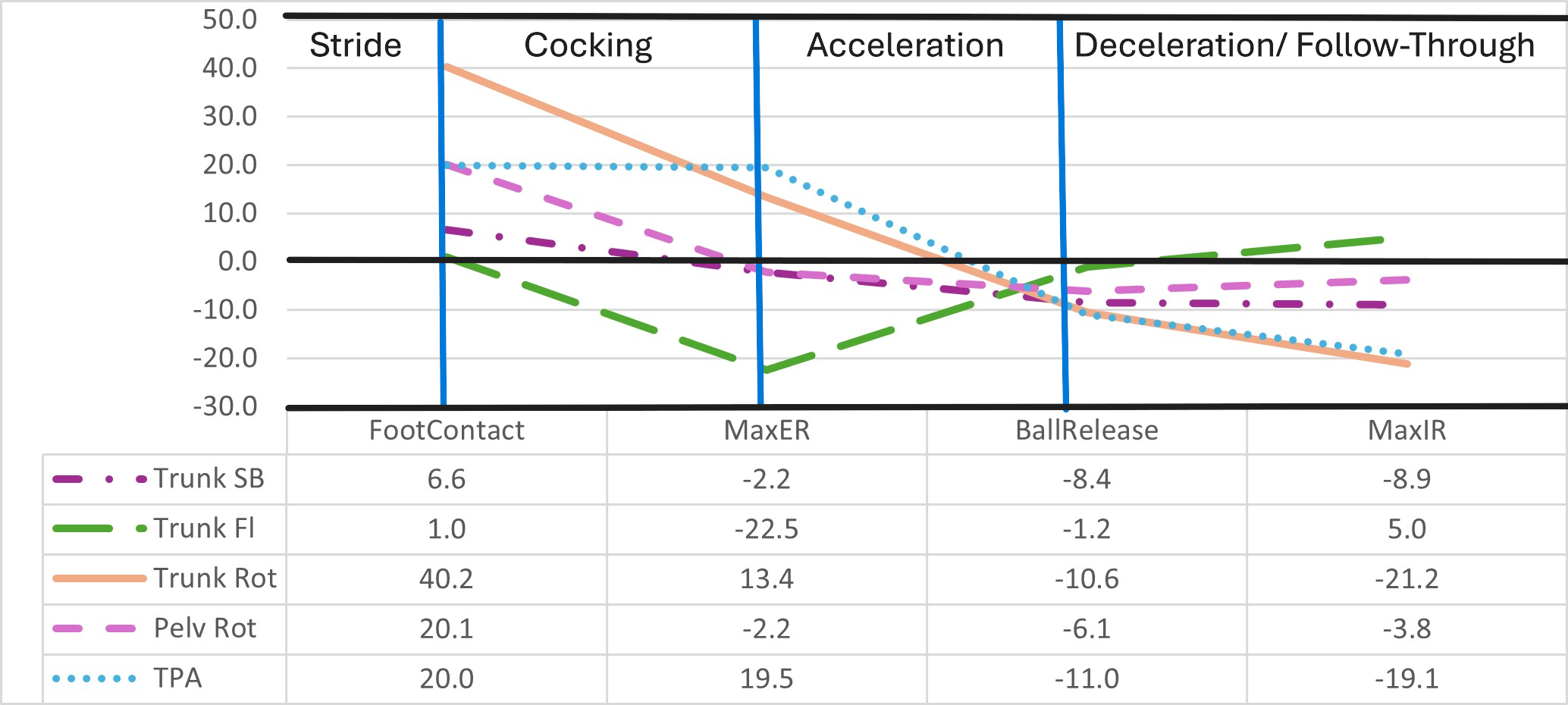
The trunk remained relatively upright throughout the passing motion, generally leaning away from the throwing arm by 2° to 9° (Figure 4). Quarterbacks experienced an average of 23° of lumbar extension at maximal shoulder external rotation. Most trunk motion occurred in the transverse plane, totaling approximately 60°. Trunk-initiated rotation occurred in the right-handed quarterbacks with an average of 40° to the right at stride foot contact, which reversed during the acceleration phase to a maximum of 21° to the left.
The pelvis followed a similar sequence, although the rotation of the pelvis toward the target began earlier in the cocking phase and generally faced the target for the remainder of the throw, with 2 to 6° of left rotation during the right-handed pass. This sequence was also observed in hip-shoulder separation (the “Trunk-Pelvis Angle”), which remained about 20°, initially favoring right trunk rotation in the cocking phase but quickly reversing to 20° favoring left trunk rotation in the follow-through. Minimal hip-shoulder separation (11°) occurred at ball release. Interestingly, all trunk measures converged to near neutral just prior to ball release with trunk extension, side-bending, and rotation toward the non-throwing side.

DISCUSSION
The purpose of this pilot descriptive study was to identify the kinematic sequencing of football quarterbacks using wireless inertial motion technology. Through the kinetic sequence from lower to upper extremity, stride knee flexion remained stable, while trunk motion generally moved away from the throwing side, converging to a point near upright neutral in all three planes just prior to ball release. Elbow flexion, shoulder external rotation and abduction peaked at the maximal external rotation POI when acceleration began after the cocking phase; all ranges decreased until the end of the follow-through.
Kinematic Sequencing
The kinematic sequencing of football passing demonstrates a clear sequence of joint motions to generate the throwing force. Lower body kinematics, particularly knee flexion, averaged 32.4° to 42° throughout the throwing motion, suggesting that quarterbacks maintain a stable lower body posture to generate force. The trunk and pelvis variables converged near neutral alignment at ball release, suggesting that quarterbacks square their hips to optimize force transfer from the legs through the pelvis into the throwing arm. The trunk remained relatively upright throughout the throwing motion, generally side-bending away from the throwing arm by 2° to 9° beginning at maximal external rotation. Quarterbacks extended their trunk an average of 23° during the cocking phase which persisted until maximal shoulder external rotation.
Trunk-initiated rotation occurred with an average of 40° to the right (toward the throwing side) at foot contact, which reversed during the acceleration phase to a maximum of 21° to the left, demonstrating how the trunk plays a crucial role in the transfer of power from the lower extremities to the upper extremities through the throwing motion. Similarly, the pelvis followed sequence with the rotation of the pelvis toward the target beginning earlier in the cocking phase and generally facing the target for the remainder of the throw, with 2 to 6° of left rotation. This sequence was observed with hip-shoulder separation, which remained about 20° initially favoring right trunk rotation in the cocking phase but quickly reversed to 20° favoring left trunk rotation in the follow-through. It was noted that minimal hip-shoulder separation (11°) occurred at ball release, which suggested that greater degrees of hip-separation occurred before ball release to aid in generating power, as hip-shoulder separation is an important determinant in trunk rotational velocity during throwing,7 thereby generating greater throwing force and velocity.
Another finding of this study was the kinematic relationship between shoulder external rotation and elbow flexion. Both variables peaked simultaneously at MaxER and declined rapidly as the shoulder moved into internal rotation during ball release. This simultaneous peak and decline suggest that the shoulder and elbow movements are closely synchronized, potentially to maximize throwing efficiency and power. Additionally, shoulder abduction increased at MaxER and remained stable through ball release before declining at MaxIR, highlighting the importance of shoulder stability during critical phases of the throw.
Comparison to Baseball Pitching
Several similarities and differences emerge when comparing these kinematic sequencing results to baseball pitching. In both sports, shoulder ER and elbow flexion peak at similar points during the throwing motion, suggesting a universal need for synchronization between these two joints to optimize throwing velocity. However, the degree of shoulder rotation and trunk involvement may differ due to the different demands of football versus baseball. For instance, while both sports involve hip-shoulder separation, football quarterbacks may rely more on trunk flexion and rotation given the weight and size of the football compared to a baseball.
Rash et al.2 highlighted a notable difference regarding the degree of elbow extension at ball release. Baseball pitchers achieve higher elbow extension angles (150-160°) than football quarterbacks in this study (121°). This difference is attributed to their different throwing requirements: pitchers throw from an elevated mound, necessitating a lower release point, while quarterbacks throw over linemen, requiring a higher release point. Different shapes, sizes and weights of the ball used (football vs. baseball) may contribute to kinematic differences as well.
Comparing the current results to those reported by Diffendaffer et al.,8 baseball players reach maximum lead knee flexion earlier in their pitching motion. Baseball pitchers achieve their maximum flexion of around 45° at foot contact and extend at ball release. The data from the current study show a more stable lower body posture with a peak average knee flexion found at ball release.
Inertial Motion Measurement
While IMU technology shows potential for monitoring, rehabilitation, and injury prevention in throwing athletes, caution should be exercised when comparing IMU data to the gold standard camera-based marker systems. Neither camera-based systems nor wearable IMU sensors provide absolute certainty of all kinematic measurements. Both systems measure physical quantities at the skin overlay and estimate quantities for underlying rigid bodies, such as bones. Soft tissue artifacts (STA) remain a significant source of error in motion analysis studies, especially in ballistic motions like pitching.9 STAs manifest as the inertial reaction of the sensor against elastic skin and sensor rocking as muscles move and deform during extreme athletic gestures.
Camp et al.10 aimed to validate the use of wearable IMUs against the gold standard of marker-based motion capture system during a baseball pitching motion. The authors suggested that IMUs are not valid for measuring arm slot (position of the arm relative to the ground) and shoulder rotation when compared to marker-based motion capture. Despite the lack of validity in some areas, the IMU system demonstrated consistency and reliability for shoulder rotation and was relatively reliable for arm slot. Therefore, IMUs may be useful for internal consistency within thrower comparisons and tracking athlete performance over time.
Implications for Training and Rehabilitation
Understanding the biomechanical distinctions between different sports and skills is essential for developing sport-specific training and conditioning programs. The high level of shoulder external rotation observed in football throws suggests that exercises focusing on shoulder flexibility and stability should be emphasized. Sequencing the throwing shoulder with the opposite hip will facilitate hip-shoulder separation; therefore, shoulder strengthening should be considered with hip muscle activation through the kinetic chain. Additionally, monitoring hip-shoulder separation could provide insight into an athlete’s power generation, and tracking changes in trunk rotation could inform both performance enhancements and injury prevention strategies.
Limitations and Future Research
This study had several limitations that should be addressed in future research. The small sample size, which consisted of only right-handed high school and college quarterbacks, may limit the generalizability of the results, particularly when directly comparing football with baseball throwing. This study included various throwing distances but did not isolate the throwing mechanics for each distance. Future research should separate data based on throwing distance to investigate how joint angles and angular velocities change depending on the length of the throw.
Limitations inherent to IMU technology, such as potential skin artifact and reliance on baseline calibration may have affected the accuracy of the data. Further research is needed to refine the use of IMUs in sports biomechanics and address these limitations as technology continues to improve. The lack of IMU data from baseball pitching also allows future research to be conducted studying baseball throwing to gain more accurate cross-sport comparisons.
Further, there may be ‘real-world’ application concerns, as football requires spontaneous movements due to external stimuli and situational reaction. These decisions occur within a live game, which cannot be replicated in a controlled lab setting or with planned throws. While baseball research often relies on optical motion capture systems, this study’s use of IMUs introduces a new approach to analyzing throwing biomechanics. Although direct comparisons between IMU and optical data may be challenging, this study highlights the potential for IMUs to provide real-time, in-game data that could revolutionize the analysis of throwing mechanics.
CONCLUSION
Football quarterback throwing mechanics and kinematic sequencing warrants further study. This pilot study provided valuable insights into the kinematic sequencing of the overhead throw performed by football quarterbacks, highlighting the unique biomechanics required for their position. These results may lead to insights for performance optimization and assist in the development of training and rehabilitation protocols tailored to quarterbacks. IMU technology in this context not only enhances our understanding of throwing mechanics but also holds potential for evaluating injury risk throughout a season by establishing a baseline of individual quarterback movements in a small sample. Future research should expand upon these findings by addressing the small sample size and variable throwing distances. Continued exploration of quarterback biomechanics may impact athlete care, performance enhancement, and injury prevention strategies.