Loading [Contrib]/a11y/accessibility-menu.js
1.
Manske RC, Wolfe C, Page P, Voight M. Diagnostic Musculoskeletal Ultrasound in the Evaluation of the Metacarpal Phalangeal and Proximal Phalangeal Extensor Tendons. IJSPT. 2025;20(9):1420-1431. doi:10.26603/​001c.143602

Abstract

Injuries to the hand and fingers are among the most common injuries in orthopedics and general practice, as they are the part of the human body that interacts most frequently with our environment. Diagnostic musculoskeletal (MSK) ultrasound offers a portable, real-time, and cost-effective alternative that is very useful for obtaining information about small structures like the finger extensor tendons. MSK ultrasound is adept at detecting changes in both tendon tissue composition and integrity. This manuscript will review the utility of MSK ultrasound in evaluating finger extensor tendons and include a review of its anatomy, common injury patterns, sonographic techniques, and clinical implications for professional rehabilitation. By integrating MSK ultrasound into clinical practice, providers can improve the accuracy of diagnosis, enhance diagnostic confidence, monitor healing progression, and guide rehabilitation strategies to optimal patient outcomes.

Introduction

Given their location on the most mobile, intricate portion of the human body, the susceptibility of the finger to injury is relatively high. Injuries to the fingers occur from repetitive injury from work or sports activities, or acute trauma, with trauma accounting for 28% of musculoskeletal injuries.1

Anatomy of the Extensor Tendon

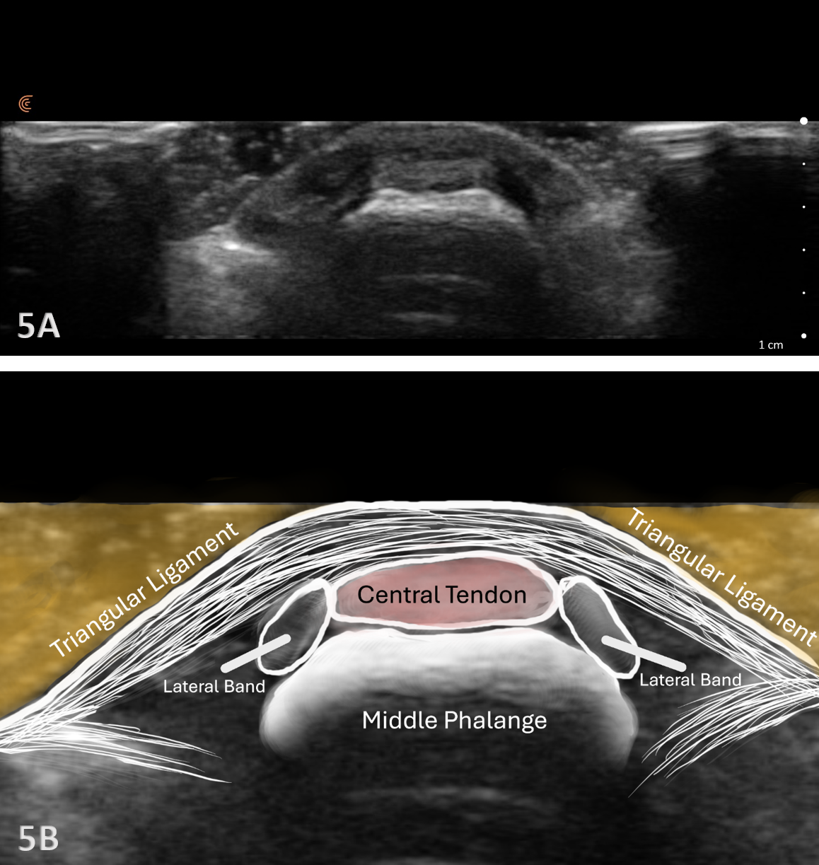
The extensor mechanism of the fingers involves 5 different zones. This article will mainly discuss the area around the metacarpals or zones III and IV. The extensor tendon runs along the dorsal aspect of the phalanges and inserts distally at the base of the middle phalanx (MP) to extend the proximal interphalangeal joint (PIP). The tendon requires other vital components to help stabilize the tendon and obtain active extension at the distal interphalangeal joint (DIP). These structures will be discussed in a later article.

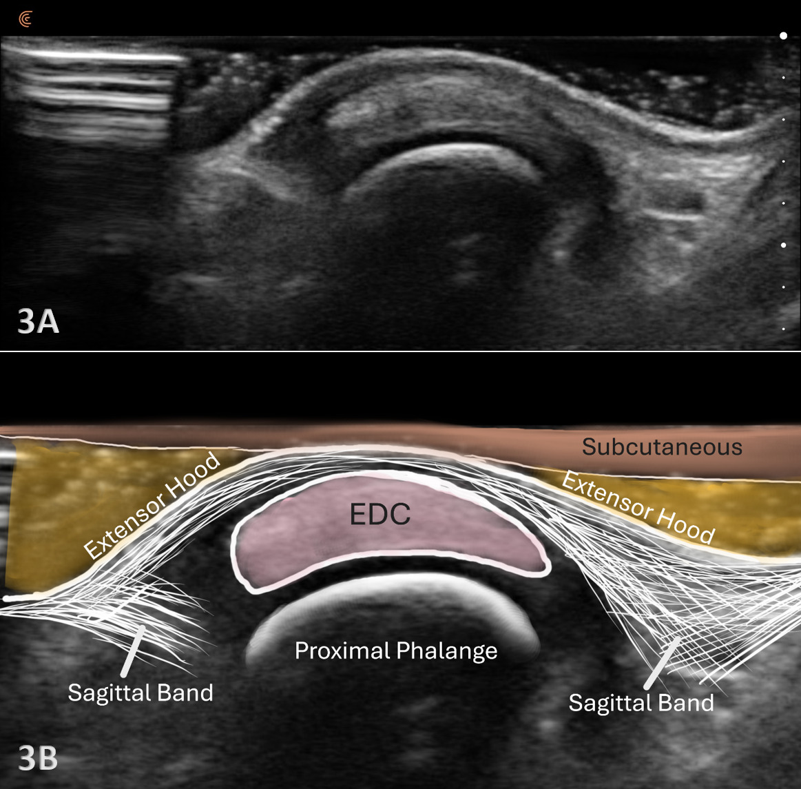
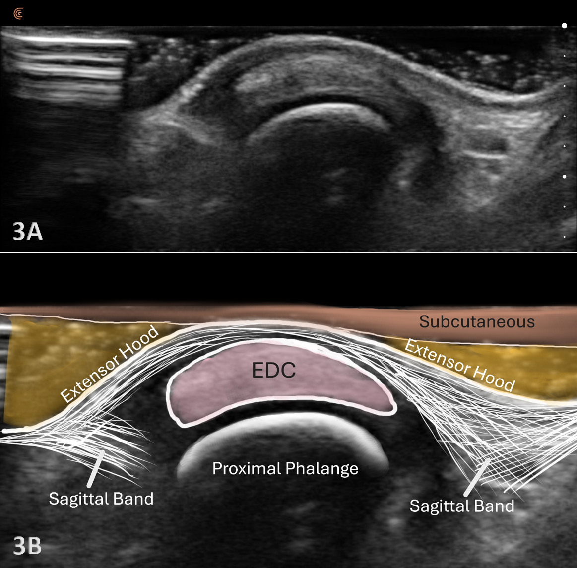
At the level of the metacarpophalangeal (MCP) joint, the extensor digitorum tendon is joined by the sagittal bands, which are part of the extensor hood. The sagittal bands on the extensor surface are further attached volarly to the palmar plate of the MCP joint. The sagittal bands are critical for stabilizing the tendon, limiting proximal excursion of the tendon, and assisting with extension at the PIP joint.

Because the superficial skin on the dorsal surface of the hand is thin, and the tendons are located superficially, they are susceptible to laceration and or avulsions from trauma. A sagittal band rupture known as “Boxer’s knuckle” can also be seen due to direct trauma or repetitive microtrauma to the MCP joint.1 With Boxer’s knuckle, the most affected finger is the 3rd, with the tendon seen dislocating ulnarly. With lacerations, obviously, an incision will be seen. However, with Boxer’s knuckle and an avulsion fracture, the injury may be closed. Pain, swelling, and diminished function of the suspected tendon will be present as the major signs and symptoms.

The Role of MSK Ultrasound in MCP/DIP Extensor Tendon

Advantages

  • Real-Time Imaging: Allows dynamic evaluation of extensor tendon integrity while the wrist and hand can be moved through the available range of motion.

  • High-Resolution Visualization: Provides detailed images of soft tissue structures, including the extensor tendon, sagittal bands, and their attachment to the PIP joint.

  • Accessibility and Cost-Effectiveness: MSK ultrasound is portable, widely available, and less expensive than magnetic resonance imaging (MRI).

Limitations

  • Operator Dependency: Requires skill and experience for accurate interpretation of findings. The ability to sonograph is to a large extent influenced by the operator and the availability and technical considerations of state-of-the-art equipment.

  • Depth Limitations: Because of the superficial location of the extensor tendons, visualization is very effective, and depth is not a limiting factor for their examination.

  • Artifacts and Shadows: Bone and calcifications may create image artifacts, requiring adjustments in probe positioning and frequency.

Sonographic Technique for Evaluating the Extensor Tendons
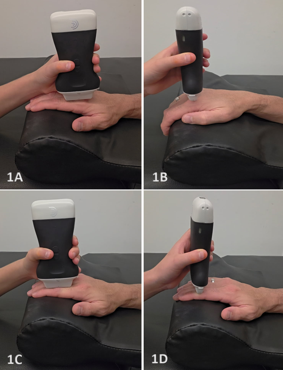
Equipment Setup

  • Transducer Type: Because of the superficial depth of the extensor tendons, a high-frequency, hockey stick transducer is commonly used. A standard linear array transducer may be used, but may not be optimal due to its size.

  • Patient Position: Patient is supine or seated with the hand resting on a pillow or supported on a table. Adjust the arm and hand position to allow optimal access to the dorsal surface of the hand.

  • Dynamic Assessment: Stress maneuvers, such as wrist and phalangeal passive or active flexion and extension, can be applied during MSK ultrasound to reveal tendon or disruptions, instability, or changes in echogenicity that may indicate partial tears, tendon ruptures, avulsions, or instability.

Examination Protocol Normal Sonographic Appearance

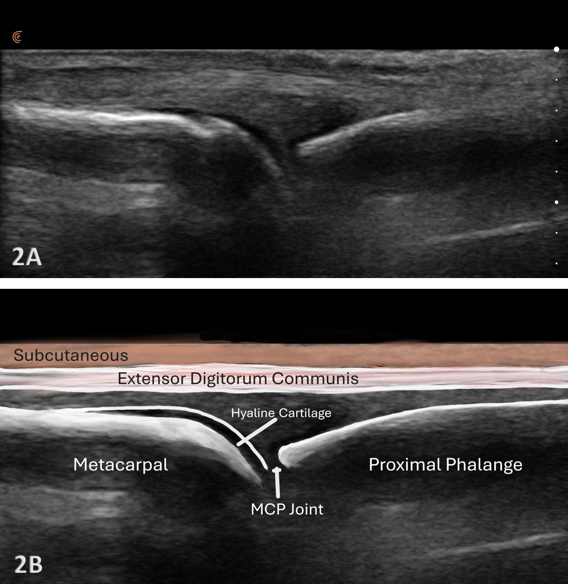
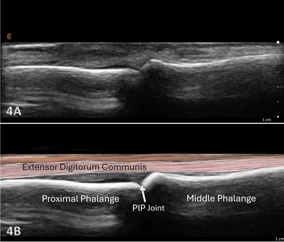
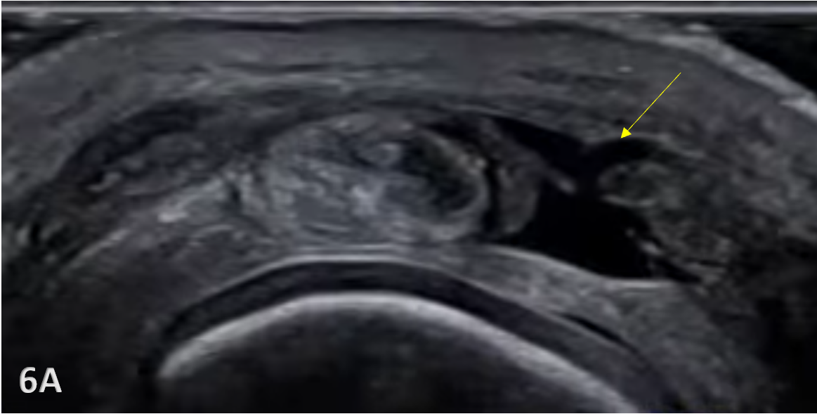
The extensor tendon can be examined in both long axis (LAX) and short axis (SAX) views. The tendons can each be seen at the MCP and PIP joints. The hyperechoic outline of the metacarpal and proximal phalanx will be seen at the MCP joint, while the proximal phalanx and middle phalanx will be visualized at the PIP joint. The extensor tendon will be an echogenic fibrillar structure in appearance. In the LAX view, the tendons will become smaller as they run more distally, making them slightly more difficult to visualize. In the SAX view, the tendon should be oval and fibular and should sit in the central position over the metacarpal. The soundhead may need to be toggled at the distal tendons’ insertion to avoid anisotropy. Dynamic visualization can be performed with resisted or active movement of the wrist or fingers.

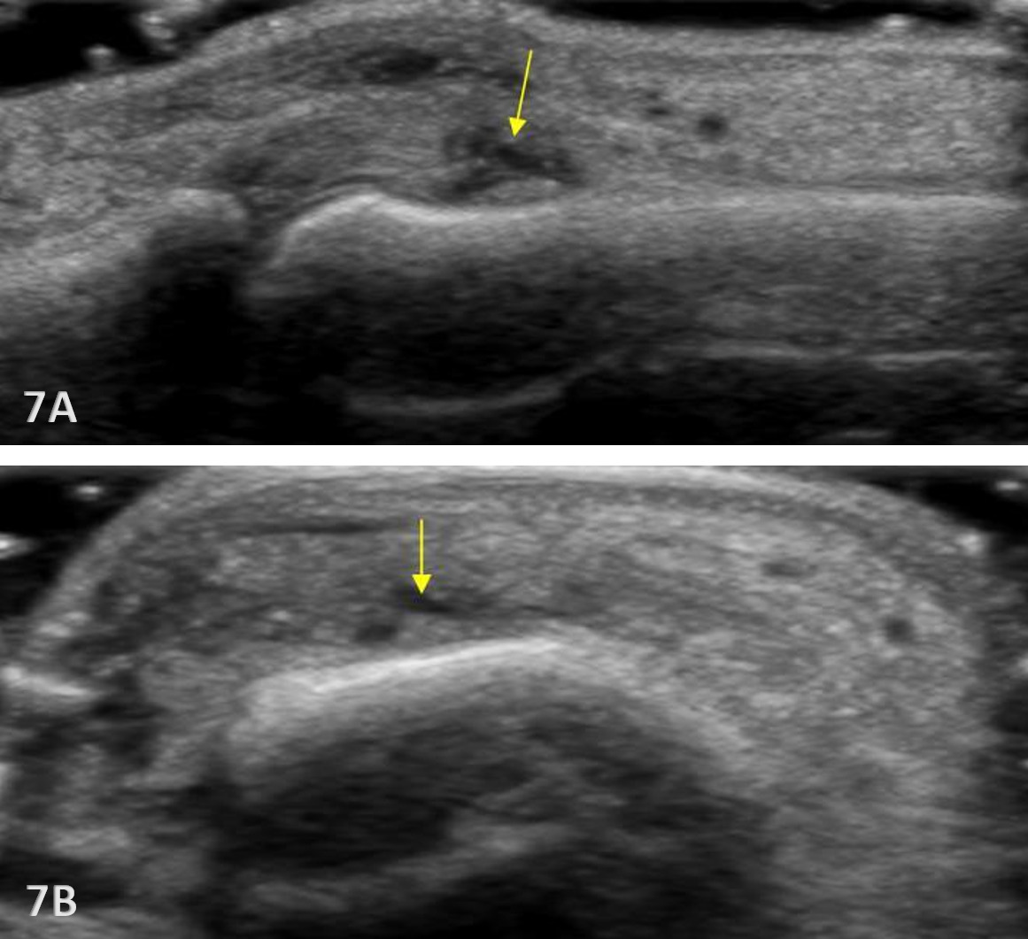
Pathologic Findings in Extensor Tendon Injury

  • Hypoechoic thickening due to swelling and edema.

  • Disruption of fibrillar pattern in partial tears and ruptures.

  • Cortical irregularity and interruption in bony avulsions of insertion.

  • Associated edema and effusion.

Clinical Implications for Rehabilitation Providers

MSK ultrasound provides real-time feedback for rehabilitation professionals, facilitating early diagnosis and intervention. Key applications include:

  • Early Detection of Injury / Accurate Injury Grading: MSK ultrasound can help guide treatment planning by quantifying tendinopathy, tendon ruptures, and bony avulsion of the insertion.

  • Dynamic Functional Testing: Rehabilitation professionals can use MSK ultrasound during physical therapy sessions to monitor recovery and assess tendon function dynamically. Serial MSK ultrasound imaging aids in assessing tissue remodeling, thickness and readiness for rehabilitation progression.

  • Guided Interventions: Ultrasound imaging assists in precision-guided injections, such as corticosteroids for inflammation.

  • Patient Education: Real-time imaging serves as a visual aid to explain the nature of the injury and set realistic expectations for recovery.

Limitations and Challenges

Despite its advantages, MSK ultrasound cannot entirely replace MRI for all finger injury cases. Additionally, the expertise required for optimal imaging technique limits its immediate adoption across all rehabilitation settings.

Conclusion

Diagnostic MSK ultrasound is a valuable tool for the evaluation of the extensor tendon injuries of the phalanges, offering fast, accurate, and cost-efficient imaging for rehabilitation professionals. Its ability to provide real-time, dynamic assessments makes it particularly suited for rehabilitation providers who can integrate the MSK ultrasound findings into clinical decision-making, optimizing treatment strategies and improving patient outcomes. However, practitioners must be adequately trained to maximize it’s diagnostic potential. By integrating MSK ultrasound into practice, rehabilitation providers can enhance patient care, improve outcomes, and reduce the burden of false diagnoses or delayed treatment.

References

1.
Quílez Caballero E, Bueno Horcajadas ÁL, Cebada Chaparro E, De Iruarrizaga Gana M, López-Vidaur Franco I, Martel Villagrán J. Ultrasound (US) of the fingers: anatomy and pathology. Quant Imaging Med Surg. 2024;14(11):8012-8027. doi:10.21037/​qims-24-591
Google Scholar


Evaluation of the Metacarpal Phalangeal and Proximal Phalangeal Extensor Tendons

A collage of hands holding a device AI-generated content may be incorrect.

Figure 1A: Transducer Placement over the MCP Extensor Tendon in LAX

For the LAX view, shown in Figure 1A, rotate the transducer 90° to align with the tendon, then follow the EDC from the distal metacarpal shaft across the MCP joint to the dorsal base of the proximal phalanx. Toggling the transducer will help to improve the image and reduce anisotropy. A gel mound or stand-off pad can improve near-field contact and visualization.

Figure 1B: Transducer Placement over the MCP Extensor Tendon in SAX

For the SAX view, shown in Figure 1B, place the transducer transversely over the MCP joint of interest. Toggling the transducer will help to identify the EDC tendon centered by the sagittal bands. A gel mound or stand-off pad can improve near-field contact and visualization.

Figure 1C: Transducer Placement over the PIP Extensor Tendon in LAX

For the LAX view over the PIP joint, shown in Figure 1C, place the transducer longitudinally along the tendon over the proximal interphalangeal (PIP) joint. A gel mound or stand-off pad can improve near-field contact and visualization.

Figure 1D: Transducer Placement over the PIP Extensor Tendon in SAX

For the SAX view over the PIP joint, shown in Figure 1D, place the transducer transversely over the MCP joint of interest. Toggling the transducer will help to identify the EDC tendon centered by the sagittal bands. A gel mound or stand-off pad can improve near-field contact and visualization.

Patient Positioning

Patient seated with the shoulder in neutral and the elbow flexed to approximately 90°. Position the forearm in pronation with the dorsum of the hand resting on the table. Allow the fingers to relax over a towel roll to place the MCP and PIP joints in mild flexion, which reduces anisotropy and opens the dorsal recess.

NORMAL VIEW OF THE EXTENSOR TENDON AT THE MCP AND PIP JOINTS

A close-up of a medical scan AI-generated content may be incorrect.

Figures 2A and 2B: LAX View of Extensor Tendon at the MCP Joint

Refer to Figure 1A for patient and transducer placement. For the LAX view of the EDC, identify the continuous hyperechoic cortical line of the metacarpal head and proximal phalanx, the thin dorsal capsule/recess superficial to the cortex, and the fibrillar EDC as it broadens into the extensor hood/central slip. With gentle active MCP flexion–extension or resisted extension, confirm smooth gliding and centralization without bowstringing, snapping, or subluxation, and survey for focal hypoechoic defects or discontinuity.

A close-up of a ultrasound AI-generated content may be incorrect.

Figures 3A and 3B: SAX View of Extensor Tendon at the MCP Joint

Refer to Figure 1B for patient and transducer placement. Maintain a perpendicular beam, SAX is highly anisotropic, using subtle heel-toe toggling, tiny rotational adjustments, liberal gel, and light pressure to avoid effacing the dorsal recess. In this plane, the EDC appears as a compact, oval bundle of tightly packed hyperechoic fibrillar echoes superficial to the bright cortical line of the metacarpal head. The radial and ulnar sagittal bands are seen as echogenic restraints flanking the tendon, keeping it midline across the MCP during motion. Just superficial to the cortex, identify the thin capsular line of the dorsal recess, which normally contains minimal or no anechoic fluid. With slow MCP flexion–extension or a gentle fist, confirm centralized tracking without snapping or radial/ulnar subluxation.

A close-up of a medical scan AI-generated content may be incorrect.

Figures 4A and 4B: LAX View of Extensor Tendon at the PIP Joint

Refer to Figure 1C for patient and transducer placement. For a LAX view the transducer is placed over the dorsal PIP. The central slip appears as a continuous, fibrillar echogenic band inserting on the dorsal base of the middle phalanx, superficial to the bright cortical line and thin dorsal capsule. With active PIP extension, flexion and gentle resisted extension, the slip should tauten and relax smoothly without gapping, step-off, or focal hypoechoic defects.

A close-up of a ultrasound AI-generated content may be incorrect.

Figures 5A and 5B: SAX View of Extensor Tendon at the PIP Joint

Refer to Figure 1D for patient and transducer placement. For a SAX view over the PIP joint, the central slip lies midline as an echogenic structure superficial to the smooth, continuous cortical contours of the proximal phalanx head and middle phalanx base. The lateral bands may not be individually resolved in every patient; when visible, they appear as thin echogenic strands that shift dorsolateral with motion. The dorsal recess is typically minimal, and the joint cortex should remain smooth and uninterrupted. Subtle heel–toe adjustments can help minimize anisotropy if these structures are indistinct.

Pathology: Sagittal Band Injury (MCP) – “Boxer’s Knuckle”

A close up of a grey and white image AI-generated content may be incorrect.

Figure 6A: At the MCP joint, a sagittal band injury called a “boxer’s knuckle” typically shows on SAX view as disruption or thinning of the radial or ulnar sagittal band with peritendinous hypoechoic fluid and decentering of the EDC from the metacarpal head. Dynamically, with MCP flexion, the EDC subluxes—often toward the ulnar side in the long and ring fingers. In the LAX view, expect focal peritendinous hypoechogenicity with a possible small avulsion fragment; evaluate the continuity of the tendon.

Pathology: Central Slip Injury (PIP) – Boutonnière Pattern

Ultrasound of a fetus AI-generated content may be incorrect.

Figures 7A and 7B: At the PIP joint, a central slip injury consistent with a Boutonnière pattern is shown above. It appears in LAX, Figure 7A, as a hypoechoic gap, thinning, or a step-off at the central slip insertion on the dorsal base of the middle phalanx, with the discontinuity often widening on attempted extension. In SAX shown in Figure 7B, there will be a loss of the symmetric midline tendon profile and reactive fluid within the dorsal recess. Dynamically, the central slip fails to tighten with extension; in later cases, there may be attenuation of the dorsal hood and altered positioning of the lateral bands.