
Introduction
Shoulder and elbow pain are common issues during pitching in baseball. Lyman et al.1 reported a 26% incidence of elbow pain, with 68% of the cases occurring on the medial side. In a systematic review, Bullock et al.2 reported that elbow valgus torque (EVT) was positively associated with the occurrence of elbow injuries. Regarding factors that increase EVT, Khalil et al.3 reported a relationship between increased shoulder external rotation range of motion (glenohumeral external rotation gain) and glenohumeral internal rotation deficit. In addition, Sakata et al.4 reported that the thoracic kyphosis angle is a risk factor for elbow joint disorders; therefore, the spinal angle, especially the thoracic angle, may be related to EVT.
Recent advancements in technology have enabled the measurement of maximum EVT (MEVT) using inertial measurement units (IMUs) alongside smartphone applications.5,6 This innovation has allowed the convenient measurement of MEVT during repetitive pitching on the field, instead of under controlled laboratory conditions.
Hattori et al.7,8 reported an increase in the medial elbow joint width after over 60 pitches and a decrease in ulnar collateral ligament (UCL) stiffness after 100 pitches. Okoroha et al.9 reported that increased EVT was associated with increasing number of innings in a game format. This suggests that EVT increases with increasing number of pitches, leading to a greater load on medial elbow tissues, especially the UCL. Although a relationship between the condition of the medial elbow joint and physical characteristics after repeated pitching has been reported,10 no studies have investigated this relationship while incorporating mechanical factors such as EVT. Therefore, in addition to the results of previous studies,10 the relationship between changes in the elbow joint and repeated pitching can be clarified by examining multiple factors, including spinal curvature and MEVT, which are associated with elbow joint disorders.
The present study aimed to investigate the relationship between the rate of change in medial elbow conditions and the factors related to physical characteristics, including the maximum elbow valgus torque (MEVT) after 100 repetitive pitches. The authors hypothesized that the greater the measured MEVT, the greater the medial elbow gap distance and the lower the stiffness of the UCL and forearm flexor pronator muscles (FPMs) would be after repeated pitches.
Methods
Study participants
Twenty-six male high school students were recruited and enrolled between January 2023 and February 2024. The inclusion criteria were 1) membership on a baseball club and participation in at least five practices or games per week and 2) experience as a pitcher or presently playing as a pitcher. Participant characteristics are presented in Table 1. The exclusion criteria were 1) history of surgery on the pitching arm and 2) pitching in a game or practice within the last 24 h.
This study was approved by the Research Ethics Review Committee of Bunkyo Gakuin University Health and Medical Sciences (approval number 2022-08) and performed in accordance with the Declaration of Helsinki. In addition, the purpose and content of the study were explained to the participants, and their consent was obtained before measurements were taken.
Pitching task
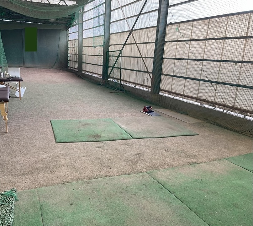
The pitching conditions were based on a study by Hattori et al.7,8,10 The throwing distance was 18.44 m. Participants made a full pitch from the pitcher’s mound toward the catcher (Figure 1). The participants started pitching from a set position and performed five sets of 20 pitches each, totaling 100 pitches. The pitch interval was 30 s, and the rest between sets was 5 min. Ball velocity was measured using a pitching analysis device (PITCHING 2.0; Rapsodo LLC, Kanagawa, Japan). The device was positioned 4.75 m from the front edge of the home plate, as recommended in the user manual.
Measurement of MEVT during pitching
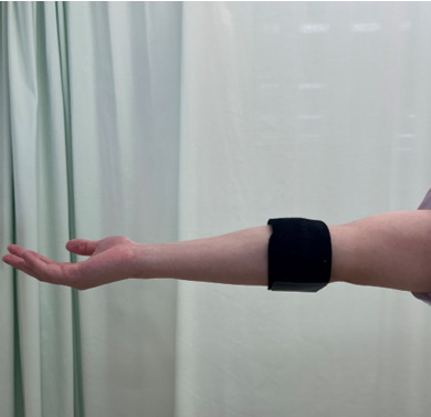
MEVT, the main outcome, was measured using an IMU (Pulse Throw, Driveline Baseball, WA, USA) device. The device was placed 5 cm distal to the medial epicondyle of the humerus (Figure 2).11 The average value of the MEVT in each participant when 100 balls were thrown was calculated. In addition, according to the method described by Aguinaldo et al.12 the measured MEVT was divided by height (m) and body mass (kg) to exclude the effect of body size, and the result was termed normalized MEVT (nMEVT).

Ultrasound image measurement
Ultrasound images were captured using an ultrasound 18-5 Hz linear array transducer (L64; Hitachi Aloka Medical, Tokyo, Japan), with the B-mode setting and real-time sonoelastography technique. The ultrasound transducer was installed as previously described10,13 and in accordance with the guidelines of the American Society of Ultrasound in Medicine to ensure image consistency. The test position was confirmed using a goniometer, with the participant in the supine position, the shoulder joint 90° abducted, the elbow joint 90° flexed, and the forearm in the mid-position (test position). The medial elbow gap distance and strain rate of the UCL and FPMs were measured, as previously described.10 The average value of the three measurements was used for data analysis.
All ultrasound measurements were performed by a single physical therapist (H.H.) with over 12 years of experience in ultrasound imaging.
Other measurements
Passive range of motion (PROM) was measured for shoulder flexion, shoulder external and internal rotation at 90° abduction, shoulder internal rotation at 90° flexion, and elbow flexion and extension.10 PROM was measured by passively moving the joint to the point of first end feel, where initial resistance was detected without overpressure. Measurements were taken using a goniometer in 5° increments, and all PROM assessments were performed in the supine position. When measuring the internal and external rotation angles of the shoulder joint in 90° abduction, a towel was placed under the distal humerus and adjusted to the same height as the scapular plane. For internal rotation measurements, the coracoid process was stabilized by applying posterior pressure.14,15 Elbow joint PROM was determined by measuring the flexion angle and extension PROM, with the forearm in a supinated position.
Hip joint PROM was evaluated following the method described by Saito et al.16 Hip joint internal rotation and external rotation PROM were measured in the supine position, with the hip flexed to 90°, the knee flexed to 90°, and the lower leg positioned at midline (0°) as the starting point, verified with a goniometer. The straight leg raise angle was measured by raising the lower leg with the knee joint fully extended, and the angle between the long axis and the thigh from the side of the trunk was measured. The straight leg raise angle was measured by passively moving the joint to the point of first end feel, where initial resistance was detected without overpressure. Muscle strength was measured using a handheld dynamometer (microFET2; Hoggan Industries Inc., West Jordan, Utah, United States). Shoulder abduction strength was measured in the standing position, with the shoulder at 90° abduction in the scapular plane. Shoulder external and internal rotation strength were both measured in the supine position, with the shoulder at 90° abduction and the elbow at 90° flexion. Participants were verbally instructed to exert maximal muscle force for 2 s.17 The results obtained (N) were normalized by dividing them by body mass (kg), yielding a weight ratio (N/kg). Each movement direction was measured three times, and the average value was used as the representative value.
Spinal curvature angles were measured using a spinal shape measuring device (Spinal Mouse; Index Ltd., Tokyo, Japan). The reliability of this device is good within and between examiners.18,19 Measurements of spinal curvature angles were performed in two conditions: resting standing and maximum shoulder flexion positions. The sensor was placed on the spinous processes from C7 to S3 and moved from the head to the caudal side. The thoracic kyphosis angle (Th1-12) and lumbar lordosis angle (L1-5) were measured at each measurement position. A change in an angle was determined by subtracting the result of the resting standing position from that of the maximum shoulder flexion position. An increase in the thoracic kyphosis angle was considered positive, whereas an increase in the lumbar lordosis angle was considered negative.
Statistical analysis
A priori power analysis was conducted to determine the sample size needed to achieve statistical significance with a power of 80% (1 – β). This analysis was performed using G*Power 3.1.9.4 (http://www.gpower.hhu.de/). To compare the condition of medial elbow tissues before and after pitching, the sample size was calculated using the following settings: t-test; effect size, 0.5; α, 0.05; power (1 – β), 0.8. A total of 26 participants were required for this study. The sample size for multiple regression analysis was calculated with the following settings: f test; effect size, 0.35, α, 0.05; power (1 – β), 0.8. As a result, a total of 31 participants were required for this study.
Data are reported as mean ± standard deviation. For statistical processing, the normality of each measurement item was confirmed using the Shapiro–Wilk test, and a paired t-test was used to compare the medial elbow joint width and UCL and FPMs stiffness before and immediately after pitching. In addition, Cohen’s d was calculated as an effect size (ES) for items with significant differences.
Factors related to the level of change in medial elbow tissue due to repeated pitching (post-pitching/pre-pitching) were examined using stepwise multiple regression analysis. The rate of change in the medial tissue of the elbow joint due to repeated pitching was used as the dependent variable. Meanwhile, the independent variables were participant characteristics such as obesity level, height, weight, years of baseball experience, medical history of the medial elbow joint, measurements of the medial elbow joint before pitching, immediately after pitching, PROM, muscle strength, average pitch speed, MEVT, nMEVT, and spinal curvature angle. The significance level was set as ˂5%.
Results
Table 1 presents the descriptive information regarding the 26 male high school baseball players included in the study.
The average maximum ball speed was 121.6 ± 7.4 km/h. The mean value of MEVT was 40.2 ±8.7 Nm, and the mean value of nMEVT was 0.33 ± 0.06 Nm/(m*kg).
Table 2 shows the ultrasound measurement results before and after pitching. Medial elbow joint width (mm) significantly increased after pitching compared the before pitching value (3.93 ± 0.76 before pitching; 4.77 ± 0.86 after pitching; p = 0.001, ES: 1.04). UCL stiffness (arbitrary units) significantly decreased immediately after pitching compared with the stiffness before pitching (before pitching 5.06 ± 2.04; after pitching 3.42 ± 1.23; p = 0.001, ES: 0.97). The stiffness of FPMs significantly decreased immediately after pitching compared with the stiffness before pitching (before pitching 0.49 ± 0.21; after pitching 0.4 ± 0.15; p = 0.001, ES: 0.49).
Table 3 shows the post-throwing change rate of the medial elbow joint and the results of multiple regression analysis. The pre-medial elbow joint width was extracted as a factor independently related to the change rate of medial elbow joint width (β = -0.46, p=0.02). The UCL stiffness change rate was extracted as a dependent variable; multiple regression analysis showed that the nMEVT (β = 0.82, p < 0.001) and shoulder abduction muscle strength (β = -0.4, p = 0.008) were independently related factors. The FPMs stiffness change rate was extracted as a dependent variable; multiple regression analysis showed that the level of thoracic kyphosis angle variation (flexion – rest position) (β = -0.53, p = 0.004) and thoracic kyphosis angle in shoulder flexion (β = 0.36, p = 0.04) were independently related factors.
Discussion
This study clarified the relationship between the condition of the medial elbow following 100 repetitive pitches and other objective measures, including the MEVT measured using an IMU. The results showed that after 100 pitches, the medial elbow gap distance increased significantly and the stiffness of the UCL and FPMs decreased.
The analysis identified nMEVT and shoulder abductor muscle strength as predictors of change in the UCL. The UCL and FPMs provide stability that resists EVT. EVT applies an extensional load to the UCL, and excessive EVT can cause damage.20 Regarding the relationship between the condition of the UCL and EVT, Hurd et al.21 reported that the elbow valgus torque was significantly greater in patients with magnetic resonance imaging-based abnormalities compared to those without. Although the author did not diagnose UCL pathology in the same cohort as the current study, prior work has linked greater elbow valgus moment to indicators of medial elbow compromise. In the current study the data suggests that the MEVT measured using an IMU reflects the condition and changes in the medial elbow tissue of baseball players. Furthermore, in the present study, the findings were normalized by height and weight, to minimize the effects of bone structure and muscle mass between subjects. Therefore, the authors believe that only the rate of change in UCL stiffness, a tissue within the elbow joint not affected by muscle mass, is relevant.
Regarding the finding that shoulder abductor strength was extracted as a significant factor, the shoulder abductor muscles function to maintain shoulder joint stability and act as prime movers for humeral elevation during pitching. When shoulder abductor strength is reduced, shoulder stability may not be sufficiently maintained during repetitive pitching, increasing susceptibility to fatigue and leading to changes in arm position. Aso et al.22 reported that the shoulder joint horizontal adduction angle during maximum external rotation decreased in the latter inning compared to the first inning. Such fatigue-related positional changes may increase the load on the elbow joint, particularly on the UCL. Therefore, this may explain the observed association with changes in UCL stiffness.
The increase in the medial elbow gap distance after repeated pitching was similar to that which has been described in previous studies.7,8 In multiple regression analysis using the post-throwing change rate of the medial elbow gap distance as the dependent variable, the medial elbow gap distance before pitching was extracted. A possible explanation may be that the rate of change in medial elbow gap distance due to pitching may be greater in athletes with a shorter medial elbow gap distance before pitching. However, if the medial fissure distance of the elbow joint is larger, ligament loosening may occur. Therefore, it is unclear whether this increase in the rate of change in the medial elbow gap distance is a negative factor.
Regarding the FPMs, Hodgins et al.23 reported that among patients with FPM strain, 19.4% underwent UCL reconstruction within one year. This highlights the importance of preventing damage to the FPMs. In the current study, a correlation was identified between FPMs and changes in the thoracic angle, as well as the thoracic kyphosis angle in the shoulder flexion position. Specifically, the current findings indicate that the amount of change in thoracic kyphosis angle variation (flexion – rest position) and the maximum thoracic kyphosis angle in shoulder flexion were important variables associated with the change in FPMs stiffness. This relationship is clinically significant because the thoracic spine contributes to scapulothoracic posterior tilt and upward rotation, which facilitate glenohumeral external rotation during pitching. When thoracic mobility or posture is limited, kinetic demand can shift distally, increasing valgus loading at the elbow and dependence on the flexor–pronator mass to maintain medial stability. Indeed, previous studies have highlighted the importance of thoracic spine mobility in overhead athletes. For instance, Gonno et al.24 reported that athletes with medial elbow disorders had a smaller thoracic spine extension angle during standing trunk extension than did athletes without such disorders. This thoracic kyphosis angle is related to the scapular and humeral angles during arm elevation.25,26 Miyashita et al.27 reported that maximum external rotation during pitching involves not only the external rotation angle of the glenohumeral joint but also scapular retroversion and thoracic spine extension. A decrease in thoracic kyphosis angle (extension motion) may reduce glenohumeral external rotation, thereby decreasing the valgus torque applied to the elbow joint.
Regarding the measurement accuracy of the IMU used to measure nMEVT, Camp et al.5 reported that IMU-derived measurements were lower than those obtained from motion capture (MC), a finding that should be considered when interpreting the accuracy of IMU measurements in the present study. Nevertheless, the measurement accuracy of IMUs during repetitive fastball pitching range from 96.9%28 to 100%,29 and IMUs are more practical to use than MC systems under repetitive pitching conditions such as those in the present study. Therefore, although IMU measurements are not identical to those obtained with MC, their use is considered appropriate when evaluating elbow valgus torque in a repetitive pitching protocol. However, the authors believe that dividing the value by weight and height and using the nMEVT as an index,12 instead of the actual IMU value, can prove useful in the management of the UCL condition.
A limitation of this study is that the muscle activity of the FPMs, which protects the UCL from nMEVT, was not measured. Regarding the activity of the forearm muscles during pitching, the activity of the flexor carpi ulnaris muscle during the early cocking phase has been reported to be low in athletes with UCL injuries.30 In the future, the authors believe that it will be necessary to examine the relationship between nMEVT and FPMs muscular activity and to consider this relationship together with UCL stiffness. Another limitation of this study is that the participants were limited to male high school baseball players. Compared with professional players, high school athletes generally demonstrate lower ball velocity.12 In addition, pitching mechanics differ depending on the competitive level.31,32 Therefore, it remains unclear whether the present findings can be generalized to other age groups. Future studies including athletes of different age groups are warranted.
Finally, the sample size did not reach the initially targeted number of participants. Therefore, a post hoc power analysis was conducted for the multiple regression analysis. Based on the model with the lowest coefficient of determination (R² = 0.213), one predictor, a sample size of 26, and α = 0.05, the effect size was calculated as f² = 0.27. The achieved statistical power (1 – β) was 0.72. Because this value is below the generally recommended threshold of 0.80, the possibility of insufficient power should be considered, and the interpretation of the present findings should be made with caution.
Conclusion
The results of this study indicate that repetitive pitching of 100 throws induced changes in the medial structures of the elbow joint, specifically, the UCL. Future studies examining how these changes relate to injuries are warranted. In addition, a relationship only between nMEVT measured using an IMU and the rate of change in UCL stiffness after repetitive pitching was observed. These findings support monitoring ultrasound-based medial elbow changes after pitching and warrant prospective evaluation of whether their magnitude and the IMU-derived metric relate to later symptoms or injury.