INTRODUCTION
Shoulder pain is one of the most common musculoskeletal complaints among tennis players, both recreational and elite level.1,2 The lower body is the most common region for injuries in tennis, followed closely by the upper extremity with the elbow and shoulder being the most frequently affected areas.2–5 Like other overhead athletes, the repetitive, explosive, and forceful overhead motions involved in tennis make the shoulder susceptible to chronic overuse injuries such as internal impingement, Superior Labrum-Anterior to Posterior (SLAP) tears, and tendinopathies of both the shoulder and elbow are common.3,4
Scapular function is an important factor in optimizing the function of the shoulder by virtue of its effect on the glenohumeral and acromioclavicular joints.6–8 During overhead motions, correct alignment of the scapula provides optimal muscle activation of the scapular stabilizers and rotator cuff, as well as efficient energy transfer from the hips and core into the upper extremity.9 However, weakened or improper activation of the scapular stabilizing muscles may alter scapular function, contribute to glenohumeral instability, and promote inefficient movement patterns such as scapular malpositioning.6,10,11 Narrative reviews of the current literature have suggested that scapular dyskinesis is identified when the scapula demonstrates atypical or dysfunctional patterns with recognizable features.7 These key features include an increased posterior protrusion of the inferior angle of the scapula and a protracted and medially rotated scapula. The shoulder may appear lower on the involved side when compared to the uninvolved side, and the coracoid process may tilt inferiorly and tender when palpated.8,12,13 However, as with many shoulder injuries, research has suggested that the frequency, nature, and mechanism for scapular dyskinesis in tennis players is dependent on the competitive level (e.g., junior, elite, professional) and both chronological and training age.1,14
According to Kibler et al.7 scapular dyskinesis is associated with almost every pathological injury in the shoulder and arm in throwing athletes and is a common contributor to shoulder pain in the overhead athlete.1,14,15 Scapular dyskinesis may contribute to an unstable platform and altered scapular positioning, reducing glenohumeral joint stability during shoulder and upper extremity activities and increasing risk of soft tissue pathology.9,16,17 Furthermore, scapular dyskinesis may be related to a narrowing of the subacromial space, although this is inconsistent when compared across individuals who are symptomatic or asymptomatic for shoulder pain.17,18 Scapular dyskinesis is not itself an injury and considerable debate exists regarding whether it is a concurrent symptom or a cause of shoulder-related pathological conditions or pain.8,18 For example, a meta-analysis that included a total of 419 athletes showed that 35% of the athletes with scapular dyskinesis also had shoulder pain, whereas 25% of the athletes with pain did not have scapular dyskinesis, and overall, scapular dyskinesis presented a 43% increased risk of shoulder pain.14 More recently, Salamh et al.15 showed that 81% of athletes symptomatic with shoulder pain and 42% of asymptomatic athletes had scapular dyskinesis in a systematic review of 19 studies with 700+ athletes. In elite, professional tennis players specifically, scapular dyskinesis was found in 57.7% and 45.9% of dominant and non-dominant shoulders, respectively, with 13% of these individuals also exhibiting long bicep head tendonitis and synovitis, and glenohumeral internal rotation deficiency.1 This inconsistency in presentation of pain and other co-morbidities supports the need for individualized evaluation and treatment approaches to patients who present with probable scapular dyskinesis.
When evaluating an individual for the presence or absence of scapular dyskinesis, medical or allied health professionals (e.g., physician, athletic trainer, physical therapist) first observe the static resting position of the scapula, followed by observation and assessment of dynamic motion of the scapula using the scapular dyskinesis test (SDT).19 The SDT is a clinical assessment to evaluate scapular movement patterns during arm elevation and involves observing an individual’s scapular movement while they perform bilateral shoulder flexion and abduction with weights.7 The SDT is considered a reliable clinical tool to establish the presence or absence of scapular dyskinesis and its use by clinicians has been well documented.19,20 Corrective maneuvers of scapular motion or positioning using the scapular assistance test or scapular retraction test is advised to alter symptoms and look for correlation of scapular movement with pain provocation and assess the total picture of dysfunction.7 Kibler et al.7 recommends concluding the evaluation process with a comprehensive checklist of possible causative factors including evaluation of core stability and strength, manual muscle testing of periscapular muscles, testing of flexibility of periscapular muscles, assessment of glenohumeral motion and joint structures, clavicle and A-C joint assessment, and a neurologic evaluation of the long thoracic and accessory nerves. Scapular dyskinesis and shoulder pain may present because of numerous factors including but not limited to acute injury, overuse, repetitive improper form on dynamic movements, underlying postural and asymmetry or misalignment.8,9,21 Therefore, addressing the inefficiencies in a therapeutic setting may help improve muscle activation of the scapula stabilizers and rotator cuff complex, improve scapula positioning, and stability of the glenohumeral joint. If the cause of the inefficiency is not addressed (e.g., biomechanical inefficiencies or limitations during functional activities like groundstrokes or serve actions) the problem may recur.
Exercise-based therapy has been shown to be effective in restoring normal or pain-free scapular kinematics.22–25 For example, a six-week stretching, stability, and strengthening program in 36 elite female tennis players diagnosed with scapular dyskinesis significantly improved range of motion and strength of the shoulder internal and external rotators, although the direct benefit on the extent of scapular dyskinesis was not measured.24 A meta-analysis conducted by Khodaverdizadeh et al.26 also supported the use of scapular-based therapeutic exercise programs as tools to improve risk factors or symptoms associated with scapular dyskinesis. However, the therapeutic exercise program showed minimal improvement in scapular angular alignment.
The shoulder and upper extremity represent the distal portion of the kinetic chain, with each segment of the body contributing to the total amount of energy needed to execute a tennis serve.9,27 Effective overhead motions, such as throwing or performing a serve, rely on all parts of the kinetic chain (including, in order, the legs, hips, trunk, shoulder, elbow, and the wrist), to move in a coordinated and efficient sequence.28,29 For optimal tennis serve execution, the kinetic chain needs to be coordinated with normal scapular function and intact stabilizers of the scapula and glenohumeral joint for a tennis player to execute efficient energy transfer from the lower extremity and core through the upper extremity to the racquet.27,30 Therefore, it is essential for a tennis player to maintain optimal scapular mechanics during the serve and overhead stroke in tennis. These movements, however, are repeated frequently throughout a tennis match and require substantial velocity to be executed correctly.9,30 The earlier onset of fatigue associated with scapular dyskinesis has been associated with decreased serve performance.31
A systematic literature review by Lambrich and Muehlbauer32 on the analysis of serve and groundstroke techniques revealed that multiple approaches have been used to ascertain the biomechanical demands in adults (18-62 years of age) across a wide range of competitive levels (e.g., recreational, college, elite). These approaches have included force-plates, pressure-detection devices within shoes, motion capture systems, video recordings, electromyography, and inertial measurement unit sensors to determine outcomes such as ground reaction forces (horizontal and vertical), muscle activation, plantar pressure application, racket and limb velocity, angular displacement through multiple joints, joint rotation and other joint kinematics.32 These measurement tools help researchers and practitioners (e.g., strength and conditioning coaches, tennis coaches) understand the kinetic, kinematic, and electromyographic patterns used to execute strokes with different techniques (e.g., spin versus slice serve), and across different populations to help identify adjustments that may help improve technique and consequently performance.
The Novel Stroke Efficiency Rating (SER) is a relatively new evidenced-based tool that uses a point-based system to assess efficiency and biomechanical motion of the three major tennis strokes (forehand, backhand, and serve). The SER includes an Injury Risk Assessment (IRA) subsection that relates the biomechanical patterns to injury risk because of possible increased stress on various joints and other movement inefficiencies.33 It is not designed to predict injury in tennis players, but to serve as a tool to identify movement patterns that may predispose a tennis athlete to an injury and to provide a functional approach for exercise specialists to help relate rehabilitative or corrective exercise programming to tennis performance and promote complimentary conditioning by coaches.33 The purpose of this case report is to describe the differential diagnosis for a tennis athlete with scapular dyskinesis and shoulder pain, integrating physical therapy interventions with a SER/IRA to identify biomechanical faults during the serve.
CASE DESCRIPTION
A 23-year-old male right-handed recreational tennis player and tennis coach with right shoulder pain was referred to physical therapy by his tennis coach without any formal diagnosis or prior physical examination performed by a physician. The subject had no significant prior medical or surgical history documented in the healthcare organization’s electronic medical records and he denied any relevant history. The subject reported he had been playing tennis, on average, two to three times a week since the age of eight consisting of junior development programs and United States Tennis Association (USTA) tournaments. As he got older, he competed at both the high school and college club tennis levels. The subject reported that he continued to play tennis recreationally and competed in USTA league tennis two to three times a week. He also coached tennis players of all ages and levels, though his primary focus was teaching younger children to play.
The subject reported his right shoulder pain began when he was 14 or 15 years of age. His shoulder pain had been intermittent throughout the years, consistently aggravated by serving and persisting after playing tennis. He explained he attempted to manage the shoulder pain on his own by taking over-the-counter anti-inflammatory medication and icing his shoulder after play. However, these self-management techniques provided only temporary relief. The subject sought physical therapy because his shoulder pain had become more intense and more frequent since starting to coach regularly. Additionally, the increase in intensity and frequency of pain resulted not only in diminished ability to coach tennis but also affected his activities of daily living (ADLs). His short-term goals included returning to his activities of daily living and work-related duties as a tennis coach with 50% limitation, reducing his level of pain to 4/10 with decreased frequency during tennis, and playing with minimal restriction. His long-term goals were to resume both work and ADLs without limitations or pain and to maintain a consistent, pain-free status daily.
EVALUATION AND DIAGNOSIS
The subject was evaluated through direct access to an outpatient sports medicine orthopedic physical therapy office without a referral or prescription. In the state of New York, physical therapists with three years of experience are permitted to treat patients without a referral.34 Additionally, a licensed physical therapist can only treat a patient for ten or less visits or 30 days, whichever comes first, without a prescription or referral from a physician. If the patient wants to continue physical therapy after the first 10 visits or 30 days, the patient is required to be evaluated by a physician and provide the physical therapist with a valid prescription. Patient and Institutional Review Board approval was sought prior to the inclusion of any information regarding diagnosis or intervention in the current case report.
Initial Examination: Subjective Measures and Medical History
The subject complained of pain along the anterior proximal aspect of his right arm that consistently occurred during the serve and after playing tennis. The subject completed a Numerical Pain Rating Scale (NPRS)35 and rated his worst pain an 8/10 during and after playing tennis, describing it as sharp and aching. At rest, he rated the pain a 1-2/10 on the NPRS and described the pain as achy. He stated he always had some level of pain daily with intensity varying dependent on his activity level. In addition to his pain, he reported the arm felt weak. He denied any acute injury, falls, numbness and tingling. The subject completed the Upper Extremity Functional Score (UEFS)36 and the Disabilities of the Arm, Shoulder, and Hand (DASH)37 to assess baseline functional limitations and track progress throughout the rehabilitation process. The UEFS is a patient reported outcome measure used to evaluate the level of disability in individuals with musculoskeletal upper extremity injuries or disfunction. The questionnaire has 20 items consisting of questions on daily activities requiring the use of the upper extremity ranging from household work, lifting groceries, dressing, and throwing a ball. The patient rates each activity from 0 to 4, where 0 is extremely difficult or unable to perform the activity and 4 is no difficulty. Therefore, the lower the score, the higher the level of disability.36 The UEFS has demonstrated good test-retest reliability and validity (relative to pain change scores) in adults with an upper-extremity dysfunction beginning physical therapy treatment (both 20-item and the refined 15-item version).38 The DASH self-report questionnaire is a 30-item questionnaire evaluating the patient’s ability to perform specific upper extremity activities. Additionally, it contains two optional 4-item modules about work and sports/performing arts. The work module is used for patients whose disability affects their ability to work, and the sports/performing arts is used for athletes and musicians.37 A higher score indicates a greater level of disability and severity, whereas a lower score indicates a lower level of disability. The DASH questionnaire has demonstrated excellent test-retest reliability and acceptable construct validity compared to the Medical Outcomes Study Short Form in 240 industrial workers and patients in a physical therapy practice presenting with an upper extremity musculoskeletal dysfunction.39–41 Content validity of the DASH and Quick DASH questionnaires for musculoskeletal concerns throughout the whole body has been supported in feedback from 172 registered allied healthcare users.42 The DASH questionnaire has also been used as the comparison outcome tool for evaluating reliability and validity of other similar questionnaires in athletic populations.43 On the initial visit, the subject scored a 68/80 on the UEFS and a 26.67 on the disability/symptom score and a 75 on both the work and sports/performing arts module of the DASH.
Initial Evaluation: Objective Measures
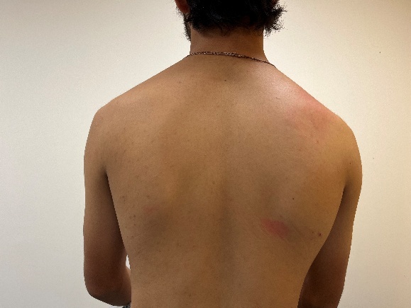
Observational postural assessment from a lateral view revealed a significant decrease in thoracic kyphosis and lumbar lordosis, with an increase in cervical lordosis and forward head posture. Anterior postural observation revealed rounded shoulders with medial rotation of bilateral humeri and the right shoulder girdle depressed with an anterior tilt compared to the left shoulder. Posterior postural observation revealed bilateral scapular protraction, downward rotation, and medial border prominence (Figure 1). The right scapula was more depressed and downwardly rotated compared to the left. Scapular motion during active range of motion (AROM) into abduction lacked coordination and smoothness. The scapula failed to upwardly rotate completely at end range abduction resulting in a slight elevation of the shoulder girdle with the overhead motion. During lowering of the arm, scapular downward rotation was also not smooth and appeared uncoordinated. Medial border prominence of the scapula was observed, along with a lack of smoothness during the last 20° of lowering the arm to the original starting position. The subject had full active cervical range of motion in all planes which did not provoke any symptoms. Muscle tightness and decreased flexibility were noted of the right pectoralis minor, right upper trapezius, right latissimus dorsi and right subscapularis.

AROM of left glenohumeral motions were full and did not provoke any symptoms. Minimal left scapula border prominence was noted also along the last 20° of AROM when the subject lowered the left arm to the original starting position. Gross assessment and observation of AROM of the right shoulder was assessed in a seated position. Because the subject had a loss of right shoulder AROM accompanied with pain during the screen, it was deemed necessary to obtain specific and quantifiable data using goniometric assessment. AROM and passive range of motion (PROM) of the right shoulder were performed with the subject in the supine position. Pre-intervention AROM, PROM, and strength values for both upper extremities are shown in Table 1.
Tenderness to palpation was noted along the right bicipital tendon, the right subscapularis, and the right upper trapezius. Additionally, the subject had tenderness to palpation along supraspinatus, infraspinatus, and teres minor musculotendinous junctions. Assessment of the right glenohumeral joint revealed hypomobility of the humeral head inferiorly and posteriorly. The scapula was hypermobile along the thoracic cage exhibiting increased medial and lateral glide along the frontal plane. Special tests including the Hawkins Kennedy, Neer’s test, and Yergason’s test, were positive on the right. The drop arm and lift off tests were both negative.
Initial physical therapy diagnosis and plan of care
Based on the subjective history and the initial physical examination, the primary physical therapy working diagnosis was secondary shoulder impingement with tendinopathy of the long head of the bicep and the rotator cuff, attributed to scapular dyskinesis. This clinical diagnosis was supported based on reports of pain along his anterior and superior shoulder with overhead motion, tenderness along the rotator cuff tendons and bicep tendons, provocation of symptoms with Hawkins Kennedy, Neer’s test, and Yergason’s test. Other contributing findings for this working diagnosis were poor seated posture with forward head and rounded shoulders, altered static scapular positioning of protraction, anterior tilt, and downward rotation, along with poor scapulohumeral rhythm in higher angles of flexion and abduction and on return from those positions. With these observed patterns, scapular dyskinesis was present and aligned with features historically described as a Type II pattern. This pattern is characterized by prominence of the entire medial scapular border at rest with abnormal pattern of motion during overhead movement and represents abnormal rotation around a vertical axis.6 Although literature often describes the use of the binary approach of using ‘present or absent’ ratings when assessing scapular dyskinesis,29,44 a Type II scapular dyskinesis presentation was used in this case report to provide a more specific description and understanding of the pathological context relevant to the findings.45 Based on these findings and development of a physical therapy diagnosis, conservative treatment was recommended. Since this was a direct access case, it was recommended to the subject to schedule an appointment with a sports medicine physician for a medical evaluation. It was also planned that the primary physical therapist would evaluate the subject’s service motion using the SER/IRA during the intervention to assess his serving mechanics and identify biomechanical faults that may be contributing to his anterior shoulder pain. The subject was educated and informed that he would be monitored throughout the physical therapy treatment process using regular assessments of pain, strength, range of motion, and performance. The rehabilitation program was structured in progressive phases based on the subject’s symptom presentation, functional goals, and tissue healing timelines.
INTERVENTION
Based on the findings of the initial comprehensive examination, subject goals, consideration of insurance visit coverage and restrictions, and anticipated carryover into post rehabilitation setting, a physical therapy plan of care was developed. Exercises were selected based on evidence supporting their safety and effectiveness for glenohumeral and scapulothoracic musculature, scapular dyskinesis, and improving shoulder function. Based on the subject’s capabilities on evaluation, baseline impairments found on evaluation, and performance related goals, the plan of care focused on (1) improvement of glenohumeral joint mobility, (2) strengthening of the rotator cuff and peri-scapular muscles, (3) restoring dynamic shoulder stability, and (4) improving neuromuscular control of the shoulder complex and upper extremity in all planes of motion for the shoulder and upper extremity to support pain-free return to ADLs and tennis performance.
Phase 1 (Weeks 1–3)
The subject reported he had an appointment with a sports medicine physician one week after the initial physical therapy evaluation and was diagnosed by the physician with a rotator cuff strain and scapular dyskinesis. The subject was not referred for any imaging and was not provided with any medical intervention such as injections or prescription medications. He was instructed to continue with physical therapy. Therefore, the goals of the program in the first three weeks were to (1) decrease pain in his shoulder when performing ADLs and when playing tennis, (2) correct and improve postural awareness, (3) improve glenohumeral joint mobility, (4) improve soft tissue mobility and flexibility, (5) decrease tenderness, (6) achieve full AROM and PROM, (7) activate lower trapezius and serratus anterior, and (8) address neuromuscular coordination. Phase 1 included the introduction of manual therapy techniques, flexibility and strengthening exercises, and closed-chain neuromuscular re-education strategies. A full description of the exercises within each category, including volume and dosage, is provided in Table 2. Dosages for all therapeutic interventions were selected to be appropriate for the initial phase of rehabilitation.
In this phase of the rehabilitation program, overhead activity was limited to avoid narrowing of the subacromial space and to allow for time to address underlying impairments of strength, stability, and joint mobility while restoring neuromuscular control and movement quality. Because of the subject’s early stage of recovery and presence of pain and dysfunction, the physical therapist determined it inappropriate to utilize the SER/IRA to assess the serve for biomechanical faults in this phase.
Patient Education: In addition to the physical therapy sessions, the subject was provided with guidance on at-home practices. The subject was cleared to continue coaching and playing tennis, however serving was restricted to avoid exacerbation of symptoms. Therefore, the subject was educated to use self-pain management strategies including the use of over-the-counter medication as recommended by his physician and icing his shoulder after playing tennis or at any time during the day when he was in pain or discomfort.
The subject was provided with a written home exercise program that included descriptions, pictures, durations, and repetitions for each prescribed exercise. He was instructed to perform the stretches daily, preferably when his body was warm and specifically after playing tennis, and to complete the strengthening exercises two to three times per week. If he attended physical therapy twice a week, he was instructed to perform strengthening exercises one additional time at home. As he progressed through this phase, he was continually provided with updated written exercise programs that mirrored the activities performed in the clinic. Given that the subject could only attend physical therapy one to two times per week, emphasis was placed on the importance of consistent exercise performance at home and/or the gym.
Phase 2 (Weeks 4-6)
After the sixth session in physical therapy, the subject started to report a steady decrease in the intensity of pain he was experiencing. He rated his pain a 5/10 on the NPRS when performing ADLs and when working and hitting groundstrokes. He stated that the pain after playing tennis had also decreased and was minimal. Pain at rest and when performing low level ADLs and activities not requiring his arm to go overhead were rated a 0/10 on the NPRS. Therefore, the goals for the second phase of rehabilitation were to (1) improve scapular muscle strength and endurance, (2) improve scapular control in increased degrees of glenohumeral flexion, scaption, and abduction between 100 and 120 degrees, and (3) improve scapulothoracic motion in straight planes. Emphasis was placed on improving postural awareness by maintaining a scapular retracted position 90% of the time during performance of the therapeutic exercises. Phase 2 continued with manual therapy, flexibility, strengthening, and neuromuscular reeducation treatment strategies. Phase 1 strengthening exercises were continued with progression of load as previously described and supplemented with TheraBand resisted standing exercises. Phase 1 neuromuscular reeducation activities were also continued in Phase 2, with the addition of the Body blade and small weighted ball activities. A full description of the exercises within each category, including volume and dosage, is provided in Table 3.
Serve Evaluation: In the latter portion of Phase 2, the SER/IRA was used by the subject’s physical therapist, the primary author, as an assessment tool to rate the efficiency of the subject’s service mechanics. There is a total of 102 points, divided between the serve (48 points), forehand (30 points), and backhand (24 points). The IRA can be divided between serve (24 points), forehand (12 points), and backhand (6 points).33 The SER/IRA has demonstrated good inter-rater reliability for the overall score and for the individual analysis of the serve.33 The SER/IRA was used for the serve only for this case report since this was the stroke the subject had associated with ongoing pain presentation and the stroke which mimics the ADLs that most frequently elicited pain. This assessment was included in Phase 2 as opposed to Phase 1 to allow the physical therapist to observe sufficient progression to recommend undertaking the service actions needed for SER/IRA implementation.
As the subject successfully progressed through Phase 2 and demonstrated improvement in joint mobility, scapula and glenohumeral neuromuscular control, there was a gradual reintroduction of overhead activity, including the service motion. Performing the serve motion in a controlled environment during the latter portion of Phase 2 of rehabilitation was considered more appropriate to allow for a more accurate assessment of biomechanical faults. This timing of the assessment allowed for the physical therapist to make more accurate and informed recommendations to the subjects coach regarding possible modifications of the service motion based on the SER/IRA findings, the physical therapy initial evaluation, and the subject’s progression through the first two phases of the physical therapy rehabilitation program.
The SER/IRA was implemented using the standardized procedures recommended in prior research.33 The subject’s serve was recorded by the primary physical therapist using a video camera from a handheld mobile device and was uploaded into a password-protected file. The subject, a right-handed player, served two flat serves, and two topspin/slice serves on the deuce side (right side of the court) with the camera positioned 30° behind to the left on the opposite side of the court. Once the videos were completed, the primary author of this case report evaluated the video recordings and scored the SER for the subject’s service motion. The serve portion of the SER/IRA has a total of 48 points, with 24 points reflecting the SER score and 24 points reflecting the injury risk score. The subject scored 21/24 for stroke efficiency and 19/24 on the IRA, resulting in a total score of 40/48. Three biomechanical faults were identified with the serve analysis and SER/IRA scoring:
-
Significant drop in elbow position during the preparation phase demonstrating suboptimal alignment. Being in this position puts excessive force on both the shoulder and elbow.7,33 During the preparation phase of the serve, proper biomechanics of the shoulder and elbow should be in alignment without any valgus positioning of the elbow. When moving from the preparation phase to the acceleration phase of the service motion, the elbow should be at the height of the shoulder at 90° of abduction.9,33
-
During the cocking phase, the shoulder was positioned sub optimally at approximately 60° of abduction, demonstrating notable shoulder lag. Delayed shoulder abduction to 90 degrees during this portion of the serve can create unnecessary torque on the anterior aspect of the shoulder increasing a player’s risk for shoulder impingement.33
-
Fair energy transfer from the lower to the upper body due to full extension of the knees before ball contact. This indicated that lower body force generation was not well-sequenced or coordinated with upper limb mechanics.33 This uncoordinated transfer of energy demonstrated by the subject made the service motion less efficient, therefore putting more force on the anterior aspect of the shoulder to produce the force required to serve.
After the video session and scoring of the SER/IRA by the physical therapist, the results were discussed with both the subject and his coach. The coach expressed full agreement with the findings and highlighted the need for targeted adjustments in the athlete’s take-back mechanics of his right arm from the start of the serve to the release stage of the ball toss. The athlete was instructed during the preparation phase of the service motion to move the racquet in a smooth continuous arc from the front of the body to a position behind the body moving the arm into an abducted and externally rotated position more naturally. This adjustment allows for improved positioning during the preparation phase and cocking phase of the elbow and shoulder reducing the shear forces on the anterior aspect of the shoulder resulting in a more effective and efficient execution.
Phase 3 (Weeks 6-10)
In phase 3, the subject attended four sessions. When not coming to physical therapy, the subject was compliant with his home exercises. He stated he was mainly feeding tennis balls to students during instruction and was minimally playing and serving. He stated the pain intensity and frequency had both decreased significantly and was starting to feel better when performing ADLs. He rated his pain a 3/10 at worst on the NPRS. All treatments were tolerated well, with his chief complaint feeling some soreness from the exercises. Therefore, the goals for the third phase of rehabilitation were (1) the elimination of pain with daily activities, (2) full participation in daily activities, (3) significant reduction of scapular dyskinesis patterns demonstrating improved scapular retraction, (4) decreased anterior tilt and a more smooth coordinated upward rotation and posterior tilt during active shoulder elevation to 150° without scapular winging or compensatory trunk movement, and (5) normal strength of the scapular stabilizers as quantified by a 5/5 on manual muscle testing. Phase 3 continued with manual therapy, flexibility, strengthening, and neuromuscular reeducation treatment strategies. Some interventions from Phase 2 were carried over into Phase 3, while additional exercises and interventions were introduced to progress the subject in his rehabilitation program. A full description of the exercises within each category, including volume and dosage, is provided in Table 4.
Phase 4 (Weeks 11-14)
In this last phase of rehabilitation, the subject attended physical therapy for five sessions. Like the previous phase, the subject continued to be compliant with the prescribed home exercise program outside of scheduled sessions, which was continually modified as he progressed through the physical therapy program. The subject reported notable improvement in shoulder symptoms, with only minimal pain experienced during the service motion and post-tennis activity. He rated his pain a 2/10 at these moments on the NPRS. He frequently had no pain throughout the day when performing ADLs and functional activities. This phase of rehabilitation emphasized the subject’s return to tennis, including a gradual reintroduction to serving, along with education on the importance of maintaining an exercise routine to reduce the risk of future injury following discharge. Therefore, the goals for the fourth phase of rehabilitation were (1) the return to full participation of all activities, including tennis, without any pain or feeling of weakness, (2) no flexibility limitations of periscapular muscles, (3) 5/5 strength for rotator cuff and scapular muscles, and (4) complete elimination of scapular dyskinesis-related signs and symptoms. Phase 4 continued with manual therapy, flexibility, strengthening, and neuromuscular reeducation treatment strategies, with supplementary interventions added to advance the patient’s rehabilitation. Tennis specific functional activities using the cable column were introduced and advanced through this phase. A full description of all exercises including volume and dosages can be found in Table 5.
OUTCOMES
This subject had twenty physical therapy sessions over a four-month period. By the final treatment session, the subject’s pain was rated 0/10 on the NPRS with all activities. The subject reported being able to serve with maximum effort with a minimal pain rating of a 1/10. He reported no pain consistently when hitting forehand and backhand strokes and after playing. He was able to play two to three times a week without any symptoms. On discharge, the subject completed the UEFS and the DASH to assess any functional limitations. He scored 79/80 on the UEFS, an increase of 11 points from his initial evaluation UEFS score, demonstrating a clinically significant improvement in functional status. For a score to be clinically significant, a score needs to improve by a minimum of 9 points.46 He also scored a 5.8 on the disability/symptom score, a decrease of 20.87 points, and an 18.75 on both the work and sports/performing arts module, a decrease of 56.25 points, both significantly surpassing the MCID threshold for this outcome measure.47
At the time of discharge from physical therapy, the subject had full AROM for flexion, abduction, external and internal rotation. Right shoulder manual muscle tests for flexion and abduction were graded a 4/5, where external rotation was graded a 5/5. Strength of the right lower and middle trapezius as well as the serratus anterior were graded a 4/5. Left shoulder strength of all muscles were graded 5/5. Strength of the left middle and lower trapezius and left serratus anterior were graded a 4/5. These grades, assessed on the day of discharge, demonstrated improvement from the initial evaluation measurements and fell within optimal and clinically accepted parameters, therefore demonstrating notable improvement in previously identified impairments.
Serve biomechanical faults were identified and discussed with the subject and the subject’s coach during the second phase of rehabilitation. Towards the end of the second phase and through the remainder of rehabilitation, the subject worked with the coach to improve these mechanics. Emphasis was placed on improving lower limb drive, sequencing and coordinating his tennis service motion to improve kinetic chain function, maintaining a continuous racquet head arc of motion path during the preparation phase, and improving shoulder positioning to a height of 90 degrees of abduction to optimize lower body force generation and minimize shoulder and elbow injury risk.33 The subject stated that he had increased awareness of his biomechanical faults when serving and was actively working to improve them with his coach and independently during practice.
Given these outcomes of the subject’s progress and functional status, he was discharged from the formal physical therapy program. The subject felt confident continuing independently with his current home exercise program of shoulder strength, shoulder stabilization, scapula stabilization, eccentric loading of the posterior rotator cuff, and upper extremity endurance activities.
Six months after discharge from the physical therapy program, the lead author/treating physical therapist contacted the subject. The subject reported over the phone that his shoulder still felt good and had no pain during serve actions or ADLs. He reported consistently going to the gym to perform the home exercises; however, he stated he does not perform all the exercises that were performed when in physical therapy. He can coach and play tennis throughout the week and states “overheads and serves feel natural and has no strain” at all on the shoulder.
DISCUSSION
The purpose of this case report was to describe a physical therapy intervention of a tennis coach and player with right shoulder pain and scapular dyskinesis and the integration of the SER/IRA to identify biomechanical faults during the tennis serve. Clinical decisions throughout the phases of this rehabilitation program were informed and supported by evidence-based research and best practices. An understanding of the biomechanical activation patterns throughout the kinetic chain during a serve action, such as optimal energy transfer from trunk to the distal portion of the upper extremity, balance of glenohumeral mobility with stability, eccentric-concentric stretch and shortening cycle, and timing, coordination, and sequencing of muscle contraction, further supports the exercise choices made in each phase.9,19 Optimal results for individuals with scapular dyskinesis are most likely demonstrated when focus is primarily placed on decreasing areas of tightness and loss of flexibility and strengthening weak muscles.7,9,24,25,48 For example, the most restricted areas of tightness with this diagnosis are often the inferior and posterior glenohumeral capsule, the pectoralis minor, and internal rotators of the rotator cuff.6,9,21,25 These impairments were addressed in this case report with manual therapy techniques and flexibility therapeutic exercises prescribed through the phases.
Strengthening of weak muscles should progress from single plane motions to multiplanar motions with focus especially on the serratus anterior and lower trapezius and restoration of scapular force couples.7,21 Avoiding movements that recruit muscles that perpetuate compensatory patterns is important. The addition of weight bearing and closed kinetic chain exercises for the upper extremity such as serratus slides, ball rolls in varying glenohumeral joint angles, and scapular clocks, early in the rehabilitation program can be important in order to promote scapular control and co-contraction of glenohumeral stabilizers.7,21,24 Progression of movement into higher planes of motion with increased resistance as strength, kinesthetic awareness, and neuromotor control improve can help facilitate a return to functional and sport specific activities.7,24 Furthermore, multiplanar motions should be progressed to include hip and trunk motions.21 Functional and sport-specific activities are added towards the end stages of rehabilitation when multiplanar motions are performed with full motion, adequate strength, and improved neuromuscular control.7,24 Functional activities using the cable column were added in this case report to optimize kinetic chain function and minimize the load placed on the scapulothoracic joint and the glenohumeral joint by making the kinetic chain more efficient. Impairments were identified during this case study that were potentially contributing factors to the subject’s scapular dyskinesis and shoulder pain. Identifying and recognizing these impairments allowed the treating physical therapist to better understand the relationship between dyskinesis, symptoms, and the subject’s movement dysfunction and allowed for treatment to be directed appropriately. Upon discharge, the change in outcome measures reflected clinically meaningful improvement. These improvements are consistent with the rehabilitation program focusing on glenohumeral mobility and stability, improving scapular control, rotator cuff and scapular stabilizer muscle strength, and incorporating tennis functional activities to improve efficiency of the kinetic chain. In addition to these quantifiable gains, the subject reported a 0/10 on the NPRS scale with all activities and was able to serve consistently when playing tennis with minimal pain. This change also exceeded the MCID consistent with prior research.35 He also reported no pain when hitting forehand and backhand strokes. More importantly, he was able to play tennis two to three times a week without any exacerbation of symptoms and resume all work-related duties as a tennis coach. These outcomes indicate a true restoration in movement and function and were clinically meaningful.
It is important for health professionals to appreciate that an individual’s posture cannot be changed without addressing adjacent and supporting structures.49 Poor thoracic and cervical posture are often associated with protracted scapula and internal rotation of the humerus.24,49 This positioning further up in the human body kinetic chain is likely related to poor control and range of motion of the hips, pelvis, and lumbar spine.49 Although assessment of the hips, core, and lumbar spine was not included in this case study, an assessment of the hips, pelvis, and lumbar spine may be a beneficial addition for future initial evaluations especially in individuals who engage regularly in sports like tennis. Any impairments discovered in the evaluation process should be included in the initial treatment plan and progressed through the phases to allow for quicker recovery and return to play. Furthermore, inclusion of trunk activities and kinetic chain activities are beneficial for muscle activation and timing.48 Restoration of scapular muscle force couples and scapulohumeral rhythm requires core strength and facilitation of kinetic chain activation. It is important to establish a stable base along the hips and core initially in the rehabilitation program prior to proceeding with recommended guidelines for scapular rehabilitation.20,24 Training of the kinetic chain was included in the tennis functional training portion of Phase 4.
One limitation of this study was that a post rehabilitation SER/IRA serve analysis video was not obtained. Although the outcomes for this individual case report were favorable based on clinically significant changes in pain level, outcome scores, and the subject’s ability to return to all functional activities without limitation, the lack of a follow up video prevents a comparison of the subject’s service motion. Not performing a post rehabilitation SER/IRA restricts the ability to quantifiably assess any change in efficiency and biomechanics of the subject’s serve.
Another factor limiting and challenging the addition of advanced interventions in the current case study may have been the systemic constraints of a fast-paced outpatient environment. Such outpatient, referral-based settings rely on reimbursements from private insurance companies leading to a higher patient volume resulting in time pressures and constraints on the therapist when working with multiple patients simultaneously. Time pressures, administrative demands such as documentation in Electronic Medical Record systems, billing procedures, insurance verification, and insurance visit limitations compromise personalized care, thorough and efficient treatment sessions, and can potentially reduce the ability to deliver high quality evidenced based care. It is important for other allied healthcare professionals such as exercise physiologists, athletic and personal trainers, and strength and conditioning coaches to understand these constraints and clinical barriers when intercepting and assuming care of these patients after discharge from traditional physical therapy settings.50
This case report used both movement analysis with a targeted rehabilitation plan to address biomechanical faults and muscle imbalances to support safe and healthy return to play. This approach was unique in that it integrated evidence based physical therapy evaluation and treatment strategies for scapular dyskinesis with an SER/IRA scale to provide a comprehensive and performance focused approach for rehabilitation and optimization of the serve for a tennis athlete. While the results were favorable, this approach may not be generalizable to other tennis players with a similar injury.
CONCLUSIONS
This case report showcases the successful rehabilitation of a tennis coach and player using targeted and evidence-based interventions and treatment strategies guided by a biomechanically informed approach. The subject demonstrated improvement in range of motion and strength, alongside clinically meaningful improvements in functional outcome scores of the DASH and UEFS. He was discharged pain free and was able to return to unrestricted tennis participation and coaching.
This case highlights the effectiveness of integrating individualized stroke analysis with evidenced based rehabilitation approach for the subject in this case study with scapular dyskinesis experiencing shoulder pain. The use of the SER/IRA provided insights into the subject’s movement quality, guided treatment progression, augmented his rehabilitation, and promoted collaboration between the physical therapist and coach in identifying factors influencing shoulder pain. Collaborative use of these assessment tools may help promote long term reduction or elimination of pain, improve likelihood of quicker return-to-play potential, and optimize performance for tennis players presenting with shoulder pain.
Conflicts of Interest
The authors report no conflicts of interest.