


Introduction
Hamstring injuries may be among the most common muscle injuries of the thigh in young, active individuals.1 Out of the three possible hamstrings that can be injured, the biceps femoris long head is the most commonly affected. A recent study assessing injuries in English football has shown that the hamstrings account for almost 40% of all muscle strains and 16% of all injuries.2 These injuries typically occur when the muscle goes through an eccentric contraction as part of dynamic movement like sprinting or kicking.3 It is in the late portion of the swing phase of sprinting that appears to be the most vulnerable position for the biceps femoris, as the hamstrings produce a violent eccentric contraction to decelerate the forward-moving tibia.4 The ability to contract so aggressively may be because the biceps femoris, like the semimembranosus muscle, has larger pennation angles, suggesting that it may have more force-generating fibers within each muscle fascicle.5–7 Furthermore, this pennation angle does not vary across the various portions of the biceps femoris, indicating that the entire muscle has similar contractile properties.8–10
Anatomy of the Lateral Proximal Hamstring Tendon
The proximal hamstring complex consists of three main muscles—the biceps femoris, semitendinosus, and semimembranosus, which all originate from the ischial tuberosity of the pelvis. The biceps femoris have both short and long heads. The short head originates from the distal and lateral part of the thigh along the linea aspera of the femur. The long head is approximately 6-9 cm long. These muscles share a common proximal tendon that anchors them to the bone before diverging into their respective muscle bellies. The biceps femoris long head and semitendinosus arise more medially and posteriorly to their cojoined attachment to the ischial tuberosity. The proximal attachment of the hamstrings is more complex than classically described and may help describe why different people have varying symptoms at varying locations.11
The hamstring tendons and muscles are surrounded by connective tissue, including the fascia lata and the ischiogluteal bursa, which reduce friction during hip extension and knee flexion. This proximal portion of the biceps femoris attachment and configuration allows the hamstrings to play a key role in generating powerful hip extension, controlling knee flexion, and stabilizing the pelvis during gait and athletic activity.
The Role of MSK Ultrasound in Tendon and Muscle Evaluation
Advantages
-
Real-Time Imaging: MSKUS allows dynamic evaluation of the lateral proximal hamstring tendon while the knee can be manipulated through the available range of motion.
-
High-Resolution Visualization: MSKUS provides detailed images of the lateral proximal hamstring tendon and its proximal enthesis at the ischial tuberosity.
-
Accurate: MSKUS has been proven to be both a valid and reliable method for examining hamstring architectural properties at rest.9,12–14
-
Accessibility and Cost-Effectiveness: MSKUS is portable, widely available, and less expensive than magnetic resonance imaging (MRI).
Limitations
-
Operator Dependency: MSKUS requires skill and experience for accurate interpretation of findings. The ability to sonograph tendons and their respective muscles is largely influenced by the operator and by the availability and technical considerations of state-of-the-art equipment.
-
Depth Limitations: Visualization is usually not a problem; however, in some individuals, a curvilinear probe may be indicated due to depth.
-
Artifacts and Shadows: Bone and calcifications may create image artifacts, requiring adjustments in probe positioning and frequency.
Sonographic Technique for Evaluating the Lateral Hamstring
Equipment Setup
-
Probe Type: Because of the depth of the proximal hamstring tendon and muscle, a standard high-frequency, linear array probe can usually be utilized. However, in some individuals who have more adipose tissue or larger muscle mass, a lower-frequency curvilinear probe may be required to achieve depth.15–17
-
Patient Position: The patient is in the prone position with the feet over the edge of the table.
-
Dynamic Assessment: A passive or active movement of knee flexion and extension can be applied during ultrasound assessment to evaluate for lateral hamstring contractile and excursion properties.
Examination Protocol
Normal Sonographic Appearance
The starting point for examining the proximal hamstring tendon and muscle is at the ischial tuberosity. The ischial tuberosity can almost always be palpated, providing the examiner with a perfect starting point for their scan. The proximal hamstring can be scanned in both the long axis (LAX) and the short axis (SAX). In the LAX view, depending on the probe width and size, one should start proximally to visualize the hyperechoic reflection of the bony cortex of the ischial tuberosity, with its large acoustic shadow underneath. In LAX, the proximal hamstring tendon fibers should be easily seen coming off the attachment of the ischial tuberosity with a clear hyperechoic fibrillar structure running distally from the insertion site. The probe can be turned 90 degrees for viewing in the SAX. If the probe is moved slightly distally in the SAX view, the biceps femoris will appear as a triangular shape. As the examiner moves distally along the biceps muscle belly, the size will decrease until it appears to disappear. In both views, some toggling may be required to reduce anisotropy.
Pathologic Findings in Lateral Proximal Hamstring Tendon and Muscle Injury
-
Disruption of fibrillar pattern in partial tears and ruptures. Proximally, it is important to determine whether the injury is a free-tendon injury or a purely myotendinous injury.18
-
Associated swelling and effusion or ischial tuberosity bursitis
-
Calcifications or retraction of the tendon near the enthesis sites.
Clinical Implications for Rehabilitation Providers
MSKUS provides real-time feedback for rehabilitation professionals, facilitating early diagnosis and intervention. Key applications include:
-
Early Detection of Injury / Accurate Injury Grading: MSKUS can quickly differentiate between a tendinopathy versus a strain, or more severe tendon rupture or muscle tear, to help guide treatment planning.
-
Dynamic Functional Testing: Rehabilitation professionals can use MSKUS during physical therapy sessions to monitor recovery and assess tendon and muscle function dynamically. Serial MSKUS imaging aids in assessing muscle healing and remodeling, helping determine readiness for rehabilitation progression.
-
Guided Interventions: MSKUS imaging assists in precision-guided interventions such as dry needling or injections.
-
Patient Education: Real-time MSKUS imaging serves as a visual aid to explain the nature of the injury and set realistic expectations for recovery.
Limitations and Challenges
Despite its advantages, MSKUS cannot entirely replace MRI for complex cases, especially when examining the origin at the ischial tuberosity.19 Additionally, the expertise required for optimal imaging technique limits its immediate adoption across all rehabilitation settings.
Conclusion
In summary, diagnostic musculoskeletal ultrasound offers rehabilitation professionals a powerful, clinically relevant extension of the physical examination when evaluating the lateral proximal hamstring, particularly the biceps femoris long head. Its ability to provide high-resolution, real-time, and dynamic visualization of tendon and muscle architecture enhances diagnostic precision, supports early clinical decision-making, and allows serial monitoring of tissue healing and load tolerance. When integrated with a thorough understanding of proximal hamstring anatomy, injury mechanisms, and functional biomechanics, MSKUS becomes more than an imaging modality—it becomes a performance-informed clinical tool. While operator skill and certain anatomic limitations must be acknowledged, the thoughtful incorporation of MSKUS into sports and orthopedic practice has the potential to elevate assessment accuracy, refine rehabilitation progression, and ultimately improve return-to-play outcomes for athletes with lateral proximal hamstring injury.
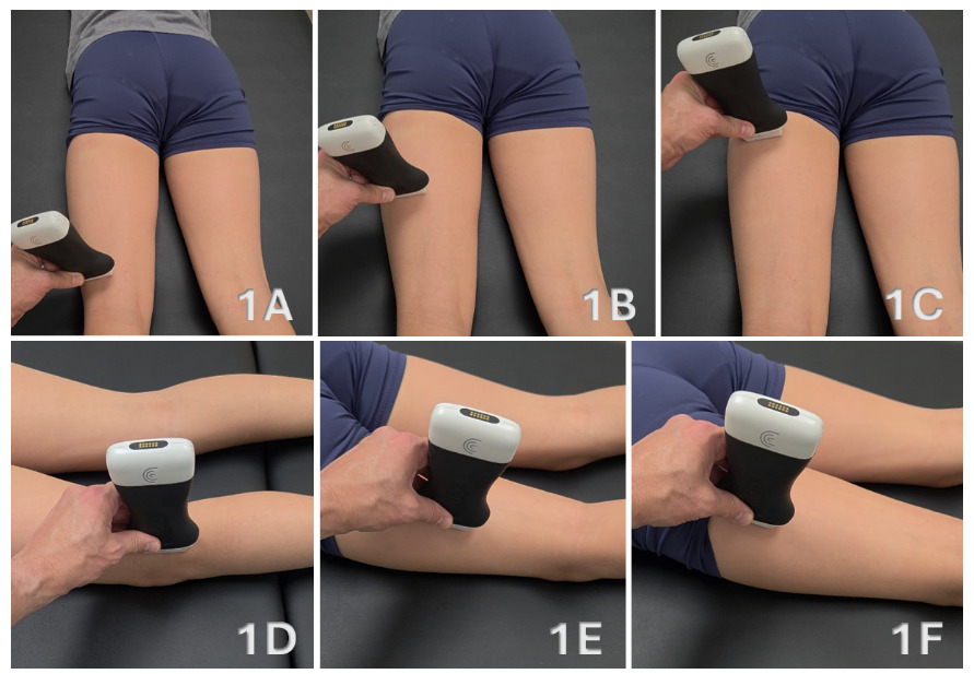
Patient Positioning
The patient is positioned prone with the feet extending slightly beyond the edge of the examination table to allow passive tension across the hamstring musculature. The knee may be placed in slight flexion to reduce passive tension if needed. Imaging is typically performed at rest; however, dynamic knee flexion may be utilized to enhance visualization of muscle contraction and fascial movement. Slight internal rotation of the hip may improve access to the lateral hamstring compartment.
Figures 1A-1C: Transducer Placement for Biceps Femoris in SAX
For SAX imaging, a high-frequency linear transducer is placed transversely at the posterior lateral knee, just proximal to the fibular head. This location serves as a reliable starting point for identifying the distal biceps femoris tendon. The transducer is then translated proximally along the posterior lateral thigh while maintaining a transverse orientation. Medial-to-lateral sweeping and proximal progression should be performed to evaluate muscle morphology, fascial planes, and compartmental organization.
Figures 1D - 1F: Transducer Placement for Biceps Femoris in LAX
For LAX imaging, the transducer is rotated 90 degrees with the marker oriented proximally and aligned parallel to the muscle fibers. Subtle heel-toe angulation is required to maintain perpendicular insonation and minimize anisotropy. LAX imaging is used to confirm fiber continuity, evaluate the myotendinous junction, and assess the distal tendon insertion.

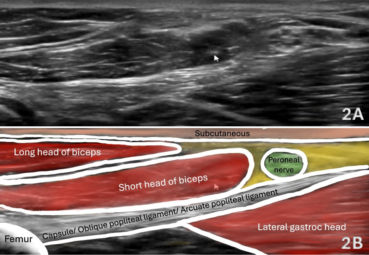
Figures 2A - 2B: Distal Biceps Femoris in SAX
Evaluation is initiated distally at the lateral posterior knee, where the distal biceps femoris tendon can be identified as it prepares to insert onto the fibular head. See Figure 1A above for the transducer placement. This location serves as a reliable anatomical landmark for orientation and facilitates identification of adjacent structures, including the common peroneal nerve. At this level, the long head of the biceps femoris tendon appears as a superficial hyperechoic structure while the short head of the biceps femoris is visualized as a deeper muscle belly. Both structures merge to form the tendinous attachment at the fibular head providing a bright osseous landmark deep to the tendon. The common peroneal nerve is identified posterolateral to the fibular head and should be consistently recognized as a key neurovascular structure.
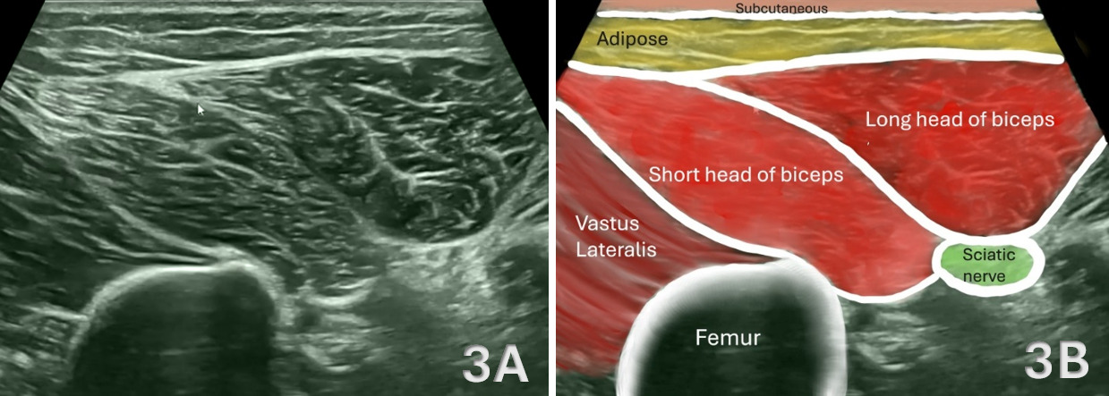
Figures 3A - 3B: Mid-Thigh Biceps Femoris in SAX (T Junction)
From the distal starting point, the transducer is translated proximally along the posterior lateral thigh while maintaining a transverse orientation. See Figure 1B above for the transducer placement. At the mid-thigh level, the biceps femoris demonstrates its characteristic dual-head morphology. The long head is positioned superficially and posteriorly, while the short head lies deeper and more anterior. These structures are separated by distinct fascial planes, which appear as hyperechoic linear interfaces and serve as critical landmarks for identifying the T-junction which is a region where the two heads converge and a common site of injury. The sciatic nerve is visualized deep to the musculature and provides an important anatomical reference during scanning.
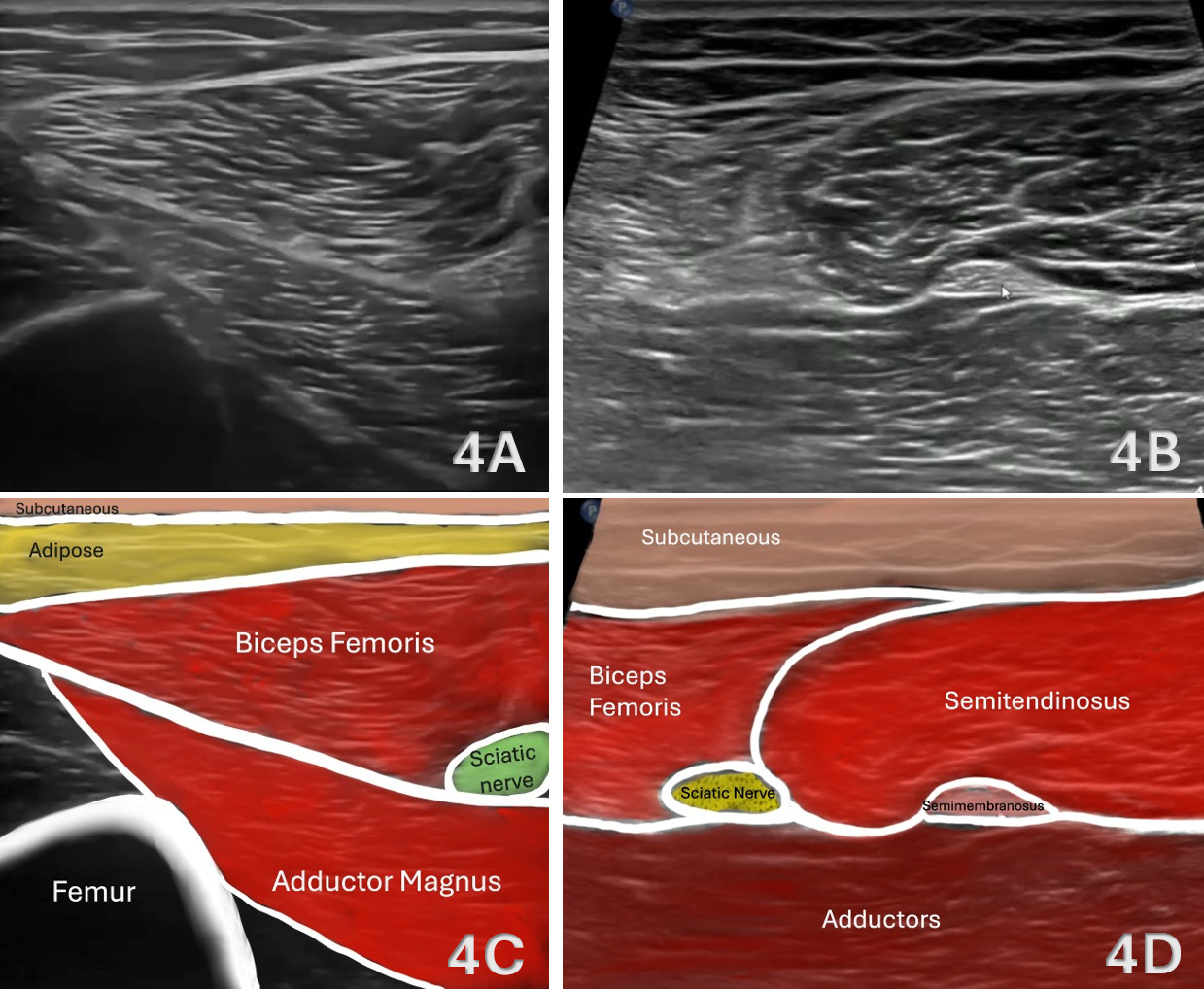
Figures 4A - 4D: Proximal Mid-Thigh Biceps Femoris in SAX
The transducer is advanced proximally beyond the level of the T-junction, as shown in Figure 1B above, where the biceps femoris demonstrates progressive transition in morphology. At this level, the short head becomes less prominent, while the long head of the biceps femoris increases in relative cross-sectional area and assumes a more dominant, superficial position within the posterior lateral thigh. The previously distinct fascial interface between the two heads becomes less conspicuous as the muscle transitions toward its proximal tendon contribution. This view allows for improved visualization of the sciatic nerve, which remains deep to the musculature and serves as a consistent anatomical landmark. Subtle transducer angulation is required to maintain perpendicular insonation and minimize anisotropy, particularly as the fibers begin to orient more obliquely toward their proximal attachment.
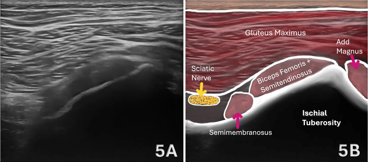
Figures 5A - 5B: Proximal Biceps Femoris in SAX
In SAX imaging at the level of the ischial tuberosity, the ischium serves as a hyperechoic cortical landmark with posterior acoustic shadowing. See Figure 1C above for the transducer placement. The biceps femoris long head contributes to the lateral portion of the conjoint tendon, which it shares with the semitendinosus and appears as a superficial hyperechoic structure spanning the ischial tuberosity. Deep and slightly anterior, the semimembranosus tendon is identified as a distinct, thicker structure. The sciatic nerve lies lateral and deep to the conjoint tendon and should be consistently identified as a key landmark. As the transducer is advanced proximally, the biceps femoris contribution becomes more apparent, highlighting its relationship with the medial hamstrings at their origin. Subtle transducer angulation is required to maintain perpendicular insonation due to the oblique fiber orientation. A normal proximal hamstring origin demonstrates homogeneous echogenicity and preserved fibrillar organization without evidence of tendon disruption or peritendinous fluid.
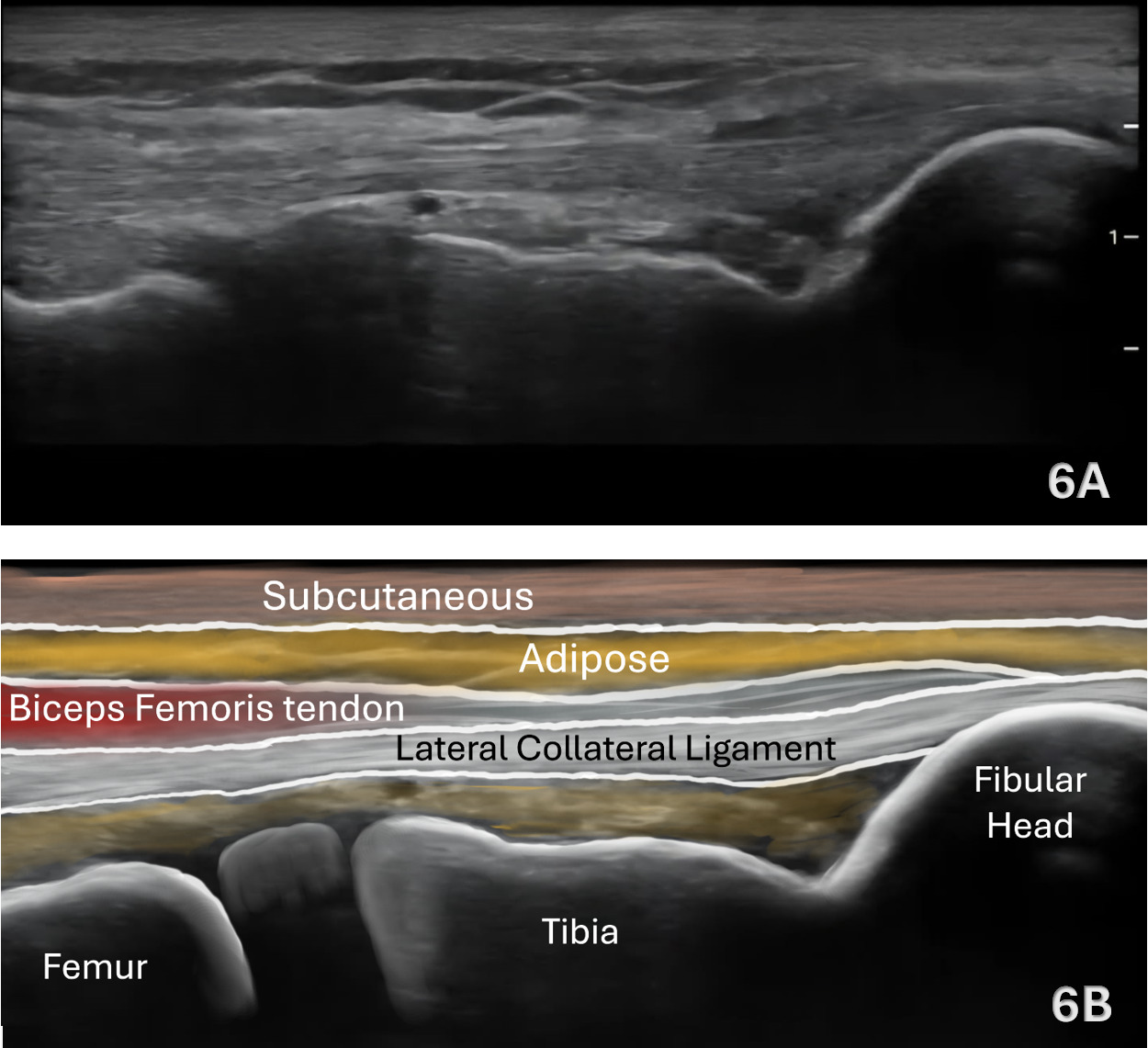
Figures 6A - 6B: Distal Biceps Femoris in LAX
LAX imaging is performed to assess fiber continuity, myotendinous junctions, and tendon integrity. See Figure 1D above for the transducer placement. Distally, the biceps femoris tendon demonstrates a linear fibrillar architecture as it courses toward its insertion on the fibular head. The fibula serves as a deep hyperechoic landmark, and subtle heel-toe angulation is required to maintain perpendicular insonation and minimize anisotropy. This view is particularly useful for confirming tendon integrity and identifying subtle changes in fibrillar organization.
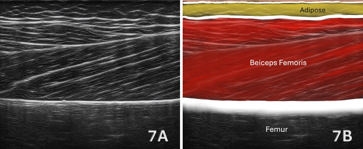
Figures 7A - 7B: Mid-Thigh Biceps Femoris in LAX (T-Junction)
At the mid-thigh, LAX imaging highlights the convergence of the long and short heads of the biceps femoris. See Figure 1E above for the transducer placement. The fascial interface between these structures may appear as parallel hyperechoic lines and represents the T-junction. Dynamic scanning techniques, including gentle transducer compression or active knee flexion, may be utilized to enhance visualization of compartmental movement and fascial interfaces. This region is of particular clinical importance due to its susceptibility to injury and should be carefully evaluated for disruption of normal architecture.
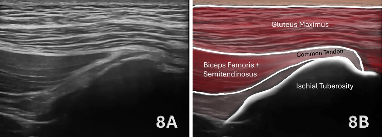
Figures 8A - 8B: Proximal Biceps Femoris Insertion in LAX
In LAX imaging, the proximal biceps femoris is assessed at its attachment to the ischial tuberosity as part of the conjoint tendon shared with the medial hamstrings. See Figure 1F above for the transducer placement. In this orientation, the ischial tuberosity serves as a deep hyperechoic osseous landmark, with the overlying tendon demonstrating a converging fibrillar pattern as it approaches its origin. The biceps femoris long head contributes to the lateral aspect of the conjoint tendon, while the semitendinosus contributes more medially, and the semimembranosus remains deeper and slightly anterior. Due to the oblique curvature of the tendon around the ischium, optimal visualization requires slight lateral probe positioning with heel-toe angulation to maintain perpendicular insonation and minimize anisotropy. The tendon may appear triangular or fan-shaped in this view, reflecting its broad attachment. The sciatic nerve is visualized deep and lateral to the tendon and should be identified as a key anatomical landmark. This view is critical for assessing proximal hamstring integrity, allowing for evaluation of tendon thickness, fiber continuity, and detection of proximal strains or partial avulsion injuries.

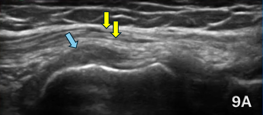
FIGURE 9A: BURSITIS
Ultrasound imaging demonstrates adventitious bursitis deep to the distal biceps femoris tendon adjacent to the lateral femoral condyle. The long head and short head of the biceps femoris are identified and outlined in yellow, while the bursitis is indicated by the blue arrow as a hypoechoic fluid collection deep to the tendon complex.
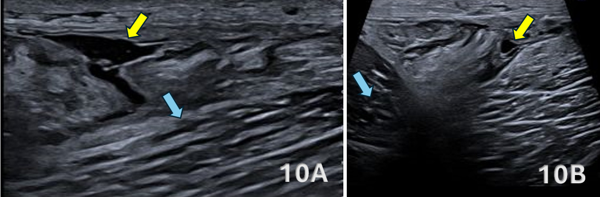
FIGURES 10A-10B: BICEPS FEMORIS TEAR
Ultrasound examination of the posterior thigh demonstrates a full-thickness, partial-width rupture of the long head of the biceps femoris at the myotendinous junction. Figure 10A shows a LAX view of the long head of the biceps femoris and Figure 10B shows a SAX view. The lesion highlighted by the yellow arrow demonstrates disruption of the normal fibrillar architecture with an associated hypoechoic hematoma/edema collection. The short head of the biceps femoris, highlighted by the blue arrow, remains intact with preserved echogenicity and fibrillar pattern.