INTRODUCTION
Anterior cruciate ligament (ACL) rupture is a common and debilitating injury in both competitive and recreational athletes. The annual incidence of these injuries is estimated at 68.6 per 100,000 person-years in the US.1 ACL reconstruction (ACLR) is the standard of care following ACL rupture, but there is a high rate of reinjury upon return-to-sport (RTS).2 The overall risk of re-injury to the ipsilateral graft ranges from 3-25%,3–8 and risk of contralateral ACL injury ranges from 2-24%.3–8 These epidemiological data are worrisome and warrant research to determine the risk factors for reinjury following RTS post ACLR.
There has been some exploration of risk factors that contribute to second ACL injury, defined here as ipsilateral graft failure or contralateral ACL rupture following RTS, including young age,4 return to high level of activity,4 musculoskeletal factors,9 lower psychological readiness,10 and not meeting specific discharge11 or RTS9 criteria. One systematic review demonstrated that both young age (<25y/o) and return to a high level of activity are factors associated with an increased risk of second ACL injury.4 Musculoskeletal factors including asymmetrical quadriceps strength9 and a decreased hamstring to quadriceps strength ratio11 have been found to have an association with second ACL injury. Recent authors have demonstrated that lower psychological readiness in younger patients (<20y/o) is associated with increased risk of second ACL injury.10 And, as expected, not meeting specific discharge11 or RTS9 criteria also lead to increased risk. Despite the success with the identification of risk factors, second ACL injury remains a concern upon RTS.
One potential area of research that may be helpful is the examination of sex differences in potential risk factors for second ACL injury. This area of research has contributed to the development of successful sex-specific injury prevention programs for primary ACL injury.12–17 This was prompted by the significant amount of epidemiological data demonstrating sex differences in primary ACL injury rates18–20 with female athletes at greater risk than male athletes when competing in similar sports.18 Previously studied sex differences that likely contribute to this disparity include anatomic, neuromuscular and biomechanical factors.19–26 Current data is not conclusive regarding sex differences in second ACL injury rates, but there are some preliminary data suggesting that they may be present. While two previous systemic reviews showed no differences between men and women incurring ipsilateral graft injury,27,28 a more recent meta-analysis found women to have a higher overall rate of secondary ACL injury and increased contralateral ACL injury rate while men had a higher rate of ipsilateral graft injuries.8 Given these epidemiological data, it is appropriate to examine if similar sex differences are present in biomechanical, neuromuscular, and musculoskeletal characteristics at the time of RTS following ACLR.
The purpose of this study was to determine if there are sex-specific differences in potential risk factors for second ACL injury at the time of clearance for RTS. The hypothesis that females would demonstrate more dangerous (less knee flexion, more valgus, and greater ground reaction forces) landing biomechanics; lower hip and knee strength; better static postural stability; and worse dynamic postural stability scores was tested. The findings of significant differences in neuromuscular, biomechanical, or musculoskeletal variables between sexes in the post ACL reconstruction population could be used to inform current guidelines for physical therapy or timelines for RTS in order to prevent second ACL injury.
MATERIALS AND METHODS
Participants
Male and female athletes were recruited for participation at the time of RTS following an initial ACLR surgery. Participants were recruited from Duke University. All participants must have been cleared by their provider to return-to-sport prior to participation. All participants provided written informed consent prior to enrollment and testing. Inclusion criteria included a minimum age of 12, participation in a sport at any level prior to ACL injury, and intention to RTS following ACLR. Exclusion criteria included any history of any major lower extremity injury, greater than one ACL injury or surgery, significant back injury or surgery, or any conditions affecting balance.
Instrumentation
Ground reaction forces (GRF) during static and dynamic postural stability testing were collected at 1000 Hz with an AMTI force plate (Advanced Mechanical Technologies, Inc., Watertown, MA). Knee isokinetic strength was measured using an isokinetic dynamometer (Biodex Medical systems, Inc., Shirley, NY). Hip and knee isometric strength were measured using a handheld dynamometer (Lafayette Instrument Company, Lafayette, IN). Knee flexion and knee valgus angles were quantified using a camera based motion analysis system with an integrated force plate system (Vicon Motion Systems, Centennial, CO).
Data Collection
Single-leg static postural stability was tested under eyes open (EO) and eyes closed (EC) conditions. Participants assumed a single-leg stance on the force plate with their hands on their hips and were asked to remain as still as possible for each ten second trial. They focused on a marker approximately ten feet in front of them at eye height for the EO condition and assumed the same stance with their eyes closed for EC condition. Participants completed one practice trial for each condition before three ten second trials were collected for data analysis. Trials were discarded and recollected if the participant’s non-stance limb touched the stance limb or the ground around the force plate, or if there was any movement of the foot of the stance limb relative to the force plate. This protocol has been previously described and found to be reliable.29–33 Following data reduction, primary variables included standard deviation of the ground reaction forces across the ten seconds of data collection in the anterior-posterior (AP), medial-lateral (ML), and vertical (V) directions32 along with a combined value for both EO and EC.
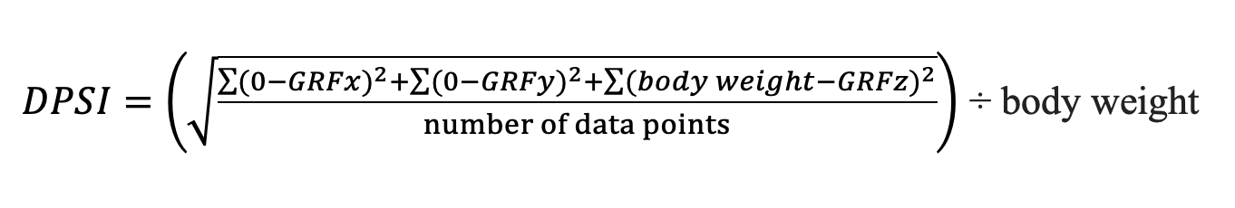
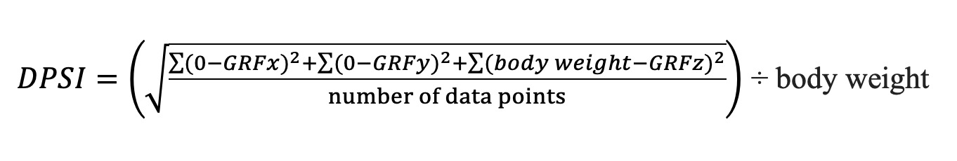
Dynamic postural stability was calculated during a single-leg landing. Participants were instructed to jump forward off two feet over a 30.5 cm hurdle and land on foot on a force plate located at a distance of 40% of their height. Individuals were asked to stick the landing and hold the stance for at least five seconds. Participants completed a minimum of one practice trial on each foot, and then three trials landing on each foot were collected for data analysis. Trials were discarded and recollected if the participant’s non-stance limb touched the stance limb or the ground around the force plate, or if there was any movement of the foot of the stance limb relative to the force plate after sticking the landing. This method was performed as previously described.32,34,35 The variables analyzed following data reduction were an index of resultant ground reaction force based on each of the three force directions (AP, ML, and V) along with the calculated dynamic postural stability index (DPSI) shown in Figure 1.
Knee flexion and extension strength were assessed with an isokinetic dynamometer concentrically at 60° per second. Testing was performed with the participant in a seated position with torso straps and a thigh strap (for the tested lower extremity) in place in order to reduce accessory motion and isolate knee flexion and extension performance. Range of motion limits were set for knee flexion and extension with maximum flexion measured at 90 degrees and maximum extension measured at zero degrees. On each limb, participants performed three practice trials of knee flexion and extension at 50% of their maximum strength, followed by three practice trials at maximum strength. Following one minute of rest, participants proceeded with five consecutive repetitions of flexion and extension at maximum strength. Reliability of the knee protocol has been previously established and determined to be excellent.29 Variables calculated included knee flexion average peak torque normalized to body weight and knee extension average peak torque normalized to body weight.
A handheld dynamometer was used to assess isometric knee flexion and extension strength along with hip abduction strength. For knee flexion, participants were tested in the prone position on an exam table with their knee in 30-45° of flexion. The participant then flexed the knee to full flexion strength while the examiner resisted the participant’s flexion using a handheld dynamometer placed on the distal one-third of the calf. For knee extension, participants sat on the edge of the exam table with their legs hanging off in 30-45° of flexion. With the dynamometer secured using a gait belt on the distal one-third of the tibia, participants extended the knee to maximum strength. For hip abduction, the participants were tested in the side-lying position on the examination table and the dynamometer was placed on the distal one-third of the lateral side of the upper leg while the knee was kept fully extended as the participant abducted the hip to full strength. Each trial with the handheld dynamometer was repeated three times on each limb, with the average of the three trials used for data analysis. Handheld dynamometry has been previously described and validated for reliability.36–40
Kinematic and kinetic analysis of a single-leg stop-jump (SLSJ) task was performed bilaterally. Retroreflective markers were placed on specific anatomic landmarks of the lower body Plug-in Gait marker set (bilateral anterior-superior iliac spine, posterior-superior iliac spine, iliac crest, lateral thigh, lateral femoral epicondyle, lateral mid-tibia, posterior superior calcaneus, lateral malleolus, and first metatarsal).41 All tasks were completed in the participant’s own athletic footwear or footwear was provided by the lab if necessary. The SLSJ task was performed with participants standing 40% of their height from the force plate. Participants were instructed to jump forward off of two legs onto a force platform landing on the instructed limb on a single force plate, and immediately perform a maximum vertical jump. The task was completed three times for each leg. Trials were repeated if the participant did not land completely on the platform, paused between landing and vertical jump, or did not perform a vertical jump. This method has been previously described in the literature.42
Data Reduction
Following data collection, a custom MATLAB (MathWorks, v7.0.4, Natick, MA) script file was used to filter and process the static postural stability data. A low-pass Butterworth filter was used to filter the data with a cutoff frequency of 20 Hz. Three successful trials were averaged for each lower extremity. All ground reaction forces from successful trials were normalized to body weight. The standard deviation of the ground reaction forces was calculated in three directions (AP, ML, V). A combined value for both EO and EC was calculated by taking the square root of the sum of squares of the three primary variables.
The DPSI was calculated to quantify dynamic postural stability in each participant. Three successful trials of the dynamic postural stability task were averaged for data analysis. A stability index was created for each anatomical direction in addition to a composite score of all three directions. The first three seconds following initial contact was used for calculation. Initial contact was defined as the point where the vertical ground reaction force exceeded 5% of the participants’ body weight. Following data collection, a custom MATLAB script filtered and processed the data. A low-pass Butterworth filter was used to filter the data with a cutoff frequency of 20 Hz.
Lower extremity kinematic and kinetic data calculated using Nexus Software (version 1.8.5: Vicon Motion Systems) according to the Plug-In Gait (version 1.9: Vicon Motion Systems) biomechanical model. This is the Vicon version of the conventional gait model and is based on the Newington-Helen Hayes gait model.41 Raw marker trajectory data were filtered using a Woltring filter routine. Ground reaction force data were not filtered to avoid producing errors in peak GRFs, joint moments, and joint force calculations. Using an anatomic reference system, the Plug-In Gait model uses relative Euler rotation angles and inverse dynamics to calculate joint kinematic and kinetic measurements. A custom MATLAB script (version R2014a; The MathWorks Inc, Natick, MA) was used to identify joint angles and forces at initial contact as well as maximum values during landing. The values of maximum knee flexion angle, knee flexion angle at initial contact, maximum knee valgus angle, and maximum vertical force were averaged across three successful trials and used for statistical analysis.
Data Analysis
The means, medians, and standard deviations were calculated for all variables for each sex. The data for each variable was assessed for normality utilizing the Shapiro-Wilk test. Comparisons between sexes were made utilizing independent sample t-tests or their non-parametric equivalent (Mann-Whitney U test) depending on the results of the Shapiro-Wilk test. Significance was set at p < 0.05 a priori. We used SPSS (version 24; IBM Corp, Armonk, NY) for all statistical analysis.
RESULTS
A total of ten male and eight female athletes who had undergone primary unilateral ACLR were enrolled. Demographic information for the entire group and for males and females is presented in Table 1. At the time of testing males averaged 10.4 months post-op and females 10.0.
The means, medians, and standard deviations for all variables are presented based on sex in Table 2(ACLR Leg) and Table 3(Healthy Leg). Significant differences are noted by grey shading. In the ACLR leg, males had significantly greater knee strength for isokinetic flexion (IKKF) and extension (IKKE) as well as isometric flexion (IMKF) and extension (IMKE) compared to females. In the SLSJ task, females had a significantly greater valgus angle at initial contact (KVAIC) (valgus = + direction). Male mean initial knee contact angle was 4.44 degrees varus and female 0.59 degrees valgus. Males had a greater maximum vertical force (MVF) after normalization to body weight compared to females. All other comparisons of the ACLR leg were not insignificant.
In the healthy leg, males had significantly greater knee strength for isokinetic flexion and extension, as well as isometric flexion and extension compared to females. In the SLSJ task, females had a significantly greater valgus angle at initial contact compared to males. Male mean initial knee contact angle was 2.1 degrees varus and female 2.4 degrees valgus. All other male to female comparisons for the healthy leg were not significant.
DISCUSSION
Second ACL injury continues to be a problem in the ACL reconstruction population. While there has been some exploration of risk factors that contribute to second ACL injury, further exploration into sex differences as a potential risk factor could provide valuable information to support sex-specific injury rehabilitation programs for the prevention of second ACL injury. The hypothesis that there would be sex-specific differences in potential risk factors for second ACL injury was explored by testing postural stability, strength, and stop-jump biomechanics at the time of clearance for RTS. There were significant differences between male and females in isometric knee strength, isokinetic knee strength, and knee valgus angle at initial contact in a stop-jump task in both the ACLR and healthy leg. There was also a significant difference in the ACLR leg for maximum vertical ground reaction force in a stop-jump task. These results illuminate sex-specific differences in potential risk factors that may contribute to ACL injury in the male and female ACLR populations. These differences may provide specific evidence to support how physical therapy programs for this population may need to be adjusted based on the sex of the individual.
As expected, females were found to have lower knee strength in both isokinetic and isometric flexion and extension in both the ACLR and healthy leg. The lower knee strength in females at RTS is consistent with the lower knee strength in female athletes prior to ACL injury.43,44 Although not studied in this investigation, lower knee strength of both the extensor and flexor muscle groups may contribute to increased difficulty in successful RTS or potential for second ACL injury.
Females were found to have a greater valgus knee initial contact angle compared to males in both the ACLR and healthy leg. The increased knee valgus angle in females is consistent with male to female differences prior to ACL injury.45–47 Even though a significant difference was found between the males and females, this is not likely clinically relevant. The means for both groups were close to a neutral (safe) position in both the ACLR and healthy legs for both sexes. The largest mean valgus knee initial contact angle of 2.4° found in the female healthy leg group is still near neutral. The minimally increased valgus angle is unlikely to contribute to the increased risk of contralateral side second ACL injury in females.8 Of note, a consistent shift of approximately 2° in the varus direction was found in both the male and female groups when comparing the ACLR leg (2° increased varus) to the healthy leg.
There were no significant differences observed in static postural stability testing between sexes in either the ACLR leg or the healthy leg. This result did not support the hypothesis which was based on previous research showing females to have better static postural stability scores.33,48 Previous research included a similar population of active military professionals33 and college level athletes,48 but these participants were not at the time of RTS from ACLR. There were also no significant differences observed in dynamic postural stability testing between males and females in either the ACLR leg or the healthy leg. This did not support the hypothesis that males would have better dynamic postural stability based on gender differences observed in strength, components of joint stability, and landing biomechanics.48–50 Previous research comparing dynamic postural stability between sexes in healthy populations has been equivocal33,48,51–53 which is consistent with these findings of no significant differences at the time of RTS.
After adjusting for body weight, males showed a statistically significant greater maximum vertical force when landing in the stop-jump task. This finding was only statistically significant in the ACLR leg and may be a potential factor increasing the risk of second ACL injury in males. This result is interesting considering that a recent meta-analysis found males to have a higher rate of second ACL injury of the ipsilateral graft even though women were found to have a higher overall rate of second ACL injury.8 The increased vertical force observed in males in the ACLR leg could increase stresses experienced at the knee and be a potential risk factor for ipsilateral graft injury potentially contributing to the differences in injury rates observed.
There are a few limitations to the study. First, there was a small sample size of 18 individuals. This included comparisons between the groups of eight females and ten males. While significant differences were found, a larger sample size could increase the power of the study and strength of the results. Second, postural stability trials during which participants failed to complete the task (for example stepping off of the force plate) were not counted. The number of failed trials per participant may provide additional insight into that participant’s static balance performance. Third, this study did not take into account the specific rehabilitation that each participant went through prior to clearance for RTS. Further research could focus on the effect of physical therapy rehabilitation programs after ACLR on biomechanical and neuromuscular risk factors for second ACL injury.
CONCLUSIONS
The results of the current study indicate a significantly lower knee strength in both the ACLR and healthy leg in females as well as a significantly greater maximum vertical force in the ACLR leg in males when landing in a stop-jump task. These findings of sex differences in potential risk factors between males and females could contribute to the current preliminary data8,27,28 regarding differences in second ACL injury rates based on sex. Given these findings, it may be important to individualize rehabilitation programs based on the sex of the individual. Examples of modifications or changes to physical therapy programs could include an increase in the volume, frequency, or length of training of the specific difference observed. Given the decreased hamstring and quadricep strength found in females, a greater emphasis on hamstring and quadricep strengthening exercises for females may be warranted. Given the data showing males landing with a proportionally greater ground reaction force, rehabilitation programs emphasizing landing techniques to decrease the maximum vertical force in males may also be warranted.
Conflict of Interest Statement
The authors report no conflicts of interest.