INTRODUCTION
Avulsion of the distal biceps brachii tendon represents about 3% of bicep tendon injuries.1 The typical mechanism of these injuries occurs in a flexed elbow and supinated forearm position,1,2 which may be associated with acute tensile overload.3 Patients frequently report a history of an audible pop and acute pain at the antecubital fossa after an eccentric contraction of the biceps caused by unexpected extension force applied to a flexed elbow with the forearm in a supinated position.3,4 The majority of patients with distal bicep tendon ruptures are males in the fourth to fifth decade of life, and 52-86% occur in their dominant extremity.2,5,6 Common risk factors include increased body mass index (BMI), use of anabolic steroids, smoking, weightlifting, and bodybuilding.1,4–7 Distal biceps rupture (DBR) can result in functionally significant loss of supination and elbow flexion strength, as well as decreased resistance to fatigue.1–8 Immediate surgical repair is the recommended course of treatment, but delaying surgery for a few weeks after diagnosis has been found to be equally beneficial compared to patients who have early surgical intervention within a week of injury.9 In other words, immediate repair is advocated in ideal circumstances but a short delay may not necessarily lead to worse outcomes.
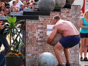
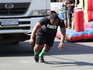
Strongman is a competitive sport where athletes perform a variety of tasks with very high loads to test physical strength and stamina. The first World’s Strongest Man competition took place in 1977. Most strongman competitions consist of six to eight events. Contestants are awarded points for each event based on their position in standings.10 Strongman competitions typically have four key components – the overhead or push press, the deadlift, grip strength, and anaerobic capacity.11 Strongman has some nuances in competition compared to other weightlifting sports. Contrary to powerlifting and weightlifting where only a loaded bar is used, strongman events often have implements like logs (Figure 1 ), atlas stones (Figure 2), axles, sandbags, or devices that allow high loads to be carried over distances, like the farmer’s walk (Figure 3) or the yoke walk (Figure 4). Tire flips (Figure 5) and pushing/pulling high loads like sleds, vehicles or trucks (Figure 6) or sustaining isometric contractions with high loads such as the “Hercules Hold” (Figure 7) are also often a part of strongman competitions. Strongman competitors face a high relative risk of bicep injury due to their body mass coupled with lifting high loads.
Among strongman competitors, in a retrospective review12 of 213 strongman athletes, 82% reported injuries. Most (24%) common were in the low back, followed by bicep and knee (11% each), with most being strains of muscle (38%) and tendon (23%). 68% of the injuries were acute.12 There were significantly more competition injuries for those under 30 years of age compared to those over the age of 30.12 Training with implements almost doubled the injuries compared to traditional weight training methods using barbells, dumbbells, or universal weight training machines.12 Ninety-one percent of those injured sustained injuries when lifting loads 90% of 1RM or greater. The incidence of bicep injury in the strongman athlete was higher than for weightlifting,13 powerlifting,13,14 and bodybuilding.15 Events like the tire flip and stone lifts suggest that bicep weakness or fatigue may limit the transfer of force produced from the larger muscle groups about the shoulder and torso and increase bicep injury risk.12 Deadlifting has also been implicated as a mechanism of bicep rupture. A study evaluating mechanisms of distal bicep found that all ruptures occurred in the supinated arm in “mixed grip” lifters when the elbow was in extension (“mixed grip” is when one hand is supinated and the other is pronated). As such, researchers proposed that eccentric loading on an extended and supinated elbow may be an alternative mechanism of injury.4
Due to the nature of the sport, the extreme loads lifted, as well as the pre-morbid strength levels of these athletes, adjusting rehabilitation protocols to accommodate the physical capabilities of the athlete is warranted. Alternatively, there may be implications for healing rate and strength of repair due to pre-morbid status. A published post-operative protocol limited loading to 5-10 pounds for the first several weeks and limited biceps isotonics till 12 weeks,8 but protocols vary among surgeons. The reattachment site is at the greatest risk for failure during the first one to two weeks after surgery.8 Normal tension of the bicep with the elbow at 90° against gravity is about 50 Newtons.8,15 Mazzocca et al.16 evaluated four different distal bicep repair techniques and cyclical load to failure varied from 232 Newtons to 440 Newtons. Kettler and others17 evaluated the linear load to failure strength of thirteen different methods for distal biceps tendon repair and found that the EndoButton had a significantly higher failure load than other techniques (259 +/- 28 N), with a mean failure rate of 180 N among all methods. The transosseous suture technique, used for the subject in this case, showed a mean failure of 210 +/- 29 N. In Kettler et al.,17 no tendon failure was seen in any transosseous or suture anchor repair when using an Ethibond No. 2 suture. Of note, the subject in this case was fixated with FiberWire, rather than Ethibond. A previous study by Miller et al.18 comparing orthopedic sutures found that FiberWire had higher ultimate load to failure and resisted the greatest number of cycles to failure compared to Ethibond and other suture types. The clinical utility of cadaveric study information is questionable however because the Mazzocca et al16 study was in “much older” elbows with low bone density and it included cyclic loading at 3600 cycles with 50 Newtons. In contrast, the Kettler et al.17 study was linear load to failure, but also in cadaveric elbows with an age range of 79 +/- 13 years. Older, cadaveric elbows with low bone density are arguably not a proper comparison with the patient in this case.
From a rehabilitation perspective, it has been suggested that unrestricted or early range of motion may begin earlier since repair strength is greater than the force of an unweighted forearm in a splint or brace.19–21 Restrictions typically include lifting no more than five pounds and no supination against resistance. At six weeks, gradual strengthening of the upper extremity and aerobic conditioning may begin.8 Strength training commences usually about two to three months post-operative.22 Return to heavy lifting is allowed at three to six months after surgery. The reader is referred to rehabilitation plans that have been outlined previously.23–25
Given that each patients’ demands are unique, should this same loading restriction in the initial phases be used for a strongman as it would be for a recreational athlete? In Srinivasan et al.,8 return to heavy lifting is suggested at three to six months after surgery. What defines “heavy?” A heavy load for one patient may be maximal for another, and a general warm-up for yet another. Therefore, the purpose of this case report is twofold. The primary objective of this case report is to share the progressive loading strategy used in the rehabilitation of a strongman athlete following a DBR repair. An additional objective is to highlight the need for individualized protocols and progressions with respect to patient goals and sport demands, as well as the need for shared decision making (SDM) between the medical doctor, patient, and rehabilitation provider.24
CASE DESCRIPTION
The subject (age = 39 years old, height= 187 cm, weight= 125kg) is a right-hand dominant male who ruptured his right distal bicep tendon doing a tire flip. The subject underwent successful surgical repair (DBR) approximately 14 days later. Post-operatively, he was placed in a splint at 90° flexion with the forearm supinated. Per the physician protocol for this subject, the first two weeks required the elbow brace to be locked at 90° when not performing rehabilitation activities. Exercises included elbow extension to 45° with the forearm supinated, passive elbow flexion to tolerance, passive forearm pronation and supination with the elbow flexed to 90°, and maintenance of range of motion of the shoulder, wrist, and hand. From two to six weeks post-operative, the elbow brace was to remain locked at 90° when not doing rehabilitation exercises. Exercises during this time frame included elbow active extension and passive flexion. Extension was allowed to be increased by 15° per week. While braced, the protocol advised light progressive resistance exercise for the musculature of the shoulder and grip strength exercises, but no active or resisted biceps work. From weeks six to eight, he was to wean from the brace and begin active elbow flexion without resistance. If needed, more aggressive treatments to get full extension could be utilized. From weeks 8-12, resisted bicep isotonic exercises could be initiated, and at twelve weeks post-operative, the protocol indicated that sport-specific activities could commence.
The subject’s first physical therapy visit was approximately three weeks post-operatively. The subject presented to physical therapy with his brace at 90° flexion. The wound was healed, wrist and hand motion were symmetrical and pain free. Left elbow range of motion was 0-150°, while the right was 11-142° passively (note, lacking 11 degrees from full extension).There were no other objective measures performed on this date because a lengthy discussion commenced about his displeasure with what he felt was a generic protocol that didn’t suit his needs. He felt that he should not be doing the same protocol as the typical patient would. The subject was very frustrated with his medical provider and the lack of guidance he received, and he was dismayed by the fact that the protocol read “updated in 2015,” about six years before his injury. Further, he felt like advances had to have been made since then. He struggled with compliance as he believed there had to be more current knowledge and subsequently adjustments or updates to treatment protocols. As the discussion progressed, he revealed that he was doing some active flexion out of his brace in the early phases and had not been very compliant with his brace. The subject was educated on the need for compliance with brace use and avoiding active flexion range of motion to protect his repair and healing until told otherwise. The potential adverse effects of non-compliance were highlighted by discussing graft failure. The physical therapist also talked about long-term planning and goals and a timeline was discussed for getting back into his desired level of high loading activities necessary for training for strongman events. The physical therapist also made it clear that in order to continue working together, there had to be some mutual respect and compromise on progression of activities.
On the second visit three days later, gentle isometrics of the bicep at 90° flexion using two-fingers of resistance mid-forearm and multi-angle tricep isometrics were initiated. Even though the protocol at the time called for no active bicep work, gentle two-finger isometrics with a short lever arm at mid forearm was used due to the patient’s pre-morbid status and to retard muscle atrophy.
Blood flow restriction (BFR) training (Delfi Medical, Vancouver, BC) with supine tricep extension to 30° of extension utilizing a resistance band was performed to help accommodate the subject’s desire to “somehow get some arm work in.” He was pre-occupied with the level of atrophy compared to his uninvolved arm already. Given the subject’s typical workout routine and level of effort he was accustomed to, BFR to the triceps was a reasonable compromise to simultaneously protect the repair and potentially provide some psychological benefit to the patient by enhancing low-load training. BFR is a training modality that utilizes low loads to promote hypertrophy and strength gains in muscle when higher loads are not appropriate.26 Sessions closed with neuromuscular electrical stimulation (NMES) to the bicep with the arm resting at his side in approximately 90° flexion, followed by ischemic preconditioning (IPC) to the involved arm inferior to the deltoid tuberosity. Contrary to BFR being performed with exercise at a percentage of arterial occlusion pressure for three to five sets of an exercise, IPC is performed with full occlusion at rest for three to five minutes followed by reperfusion for three to five minutes, and this cycle is repeated three times. IPC has been shown to increase muscle perfusion,27,28 oxygen uptake and force in strength-trained athletes,26,27 increase microvascular blood flow,28 provide an ergogenic benefit,29 increase muscle performance when performed prior to resistance training,30 and help with recovery.31 IPC was used in this case after the session to potentially help with recovery and the muscle physiology benefits listed above, but also to maximize individualized patient care time. While the benefit of IPC for this subject is debatable, he was grateful for the progressive approach to his rehabilitation that went beyond the general protocol. For his home program, he was also encouraged to perform high-load isolated biceps isotonics to his uninvolved side to potentially realize the benefit of cross-education. Cross-education is the use of unilateral resistance training to increase the strength of the contralateral non-trained side.32 Sato et al.32 found that progressive eccentric or concentric elbow flexor activity performed twice a week for five weeks showed strong cross-education effects on involved side maximum voluntary isometric contraction (MVIC) and one-repetition maximum (1RM).
The subject was seen only once a week due to his schedule and the distance he travelled for his appointments. Sessions involved soft tissue and scar mobilization, passive range of motion, elbow joint mobilizations, isometrics, and exercises and modalities described previously. At week six, despite the initial protocol limiting resisted bicep activities till eight weeks, he was cleared by his physician to begin resisted exercise at week six with a five-pound restriction and was “released to his PT.” The physical therapist was unable to reach the provider for confirmation. The subject was again frustrated by the minimal guidance received by his medical provider, stating that he was only told not to “go too heavy too fast.” For this subject in particular, “too heavy” for him would far exceed a maximal attempt for a typical patient. For rehabilitation professionals in a number of settings, there is a delicate balance between tailoring protocols to individual histories and physical qualities prior to injury or surgery and respecting the healing process. Arbitrary guidelines are provided (such as the statement above) with no sound progression or template for patients or their rehabilitation providers.
At this physical therapy visit, the subject’s range of motion was 2-141° actively, and 0-145° passively. Bicep strength was measured with a hand-held dynamometer (HHD) in sitting with elbow flexed to 90°. The subject was instructed to push to comfort without pain or pulling sensation over the repair site. Testing consisted of three, five-second flexion tests against a rigid dynamometer placed in his hand. His uninvolved side averaged 48 pounds while the involved side averaged 26 pounds. The rationale for performing HHD testing at 90° because the bicep is more vulnerable the closer the lifting load is to extension based on previously discussed mechanisms of injury. The test was not intended to be a maximum force assessment but rather a test of force to tolerance without pain. Interestingly, the uninvolved side values seemed rather low given his pre-morbid status. These lower-than-expected values may be due to the position of the elbow during testing or a decline in strength due to limited resistance training of the uninvolved side since the surgery. At this time frame, sled pushes were added with the elbow was locked in extension and the movement was driven by the legs. Additionally, isometric mid-thigh pull (IMTP) (Figure 8) was added with the involved side in pronation due to previous studies showing the supinated grip position has been implicated in DBR.4 The IMTP is a useful exercise in rehabilitation because it has been correlated to athletic capabilities of strength, maximal sprint speed, countermovement jump, and change of direction tests.33–35
Bench press as well as barbell military press were initiated at six weeks post-op due to the subject’s previous experience and desired goals as well as the limited involvement of the bicep in these activities. Saeterbakken and colleagues36 previously found that that flat bench press resulted in 48.3%-68.7% less bicep activity than incline bench position and that a narrow grip (biacromial distance) elicited lower bicep loads than a wide grip (50% more than biacromial distance). For the military press, Saeterbakken and Fimland37 found similar EMG of the biceps during seated barbell and dumbbell shoulder press, and about 16% greater bicep activation in standing barbell versus dumbbell shoulder presses. Loads used were either 30% of previous one-repetition maximum (1RM), 30% bodyweight, or comfort, whichever came first. The American College of Sports Medicine (ACSM) has previously stated38 that for muscular endurance training, the loads should be about 50% of 1RM and this was supported by Schoenfeld et al39 in a later review. These guidelines are in the healthy general population. 30% was used because it is about half of the load used in the healthy population as suggested by the ACSM.38 The rationale here was to establish a load that was pain free and that the subject felt comfortable/confident with while facilitating proper technique. For this subject, previous best on the bench press was close to 400 pounds. He worked up to 185 pounds on the first day after initial sets of five repetitions each at 95, 135, and 165 pounds. Previous military press 1RM was 270 pounds, and the subject worked up to 95 pounds in the first training session. Given the subject’s extensive training experience, he was given the freedom to load within a subjectively comfortable range on these specific lifts with the guidelines of limiting to no more than 50% of previous 1RM. This approach allowed the subject to have input into his progression limited by his subjective analysis of limb confidence, comfort, and pain or pulling sensation at the distal bicep during the lifts (had to be absent). It was theorized that using pain, a subjective increase in “pulling sensation” at the location of the repair, or breakdown in exercise technique would be an adequate clinical basis for judgment of load tolerance. In this case, the subject’s pre-morbid status along with surrounding healthy tissue stress shielding the bicep as well as these being multi-joint, total body lifts made this a plausible guideline. Additionally, almost all the exercises performed were not bicep exercises in isolation or where the bicep is the prime mover, as would be the case in bicep curls or pull-ups. In the exercises performed, the biceps acted as stabilizers or synergists.
When isolated bicep isotonics commenced at week six, BFR was used due to the ability to improve strength and hypertrophy with low loads. The use of BFR enabled the physical therapist to load the bicep in isolation but mitigating risk of injury by using heavier loads without BFR. An initial load of five pounds was used due to it being the physician recommendation. The subject performed the suggested repetition scheme of 30/15/15/15 with 45 seconds rest between sets and the cuff remaining inflated. If the subject did not achieve failure or close to it on the final set, weight was increased one to two pounds for subsequent sessions.
At week eight, seated rows with a pronated grip were added, along with hammer curls using a rope with the forearm pronated at the start and ending the concentric phase in a neutral forearm position. Barbell snatch was also added at 30% of previous military press best. Chen and others40 found that bicep activity in the snatch increases with greater loads and velocities. Olympic lifts are typically performed at maximal velocities. Due to the low loads for this subjects and low speed/effort of performance, it was not expected that the bicep load would be too high for this point in time.
From weeks 10-12, a neutral forearm grip was used for all lifts including seated rows, trap bar deadlifts, and hammer curls, for example. The subject’s previous 1RM on the straight-bar deadlift was 900 lbs. Load was established to 30% of that for the first day, up to 270 lbs. At week 12, a supinated grip was used for more exercises, including the deadlift. Additionally, a front dumbbell carry was added to the routine, similar to the atlas stone carry position. His involved side HHD at 90° flexion averaged 46 pounds of force and 67 pounds on the uninvolved at the twelve-week assessment. Based on these HHD values, a 55-pound dumbbell was used as his target starting load due to the shared bilateral bicep load for the exercise and was increased 10% till the subject felt the load was comfortable Also at 12 weeks, farmer walks with a trap bar were added. The farmer walk load commenced up to 30% of previous deadlift best. For all lifts, load was increased 20% per week as tolerated. Interestingly, the subject inquired about doing pull-ups at a previous visit and was advised against doing so, then came to his following visit with studies showing very high bicep EMG activity during pull-ups.41,42 These were avoided at this time. The reader is referred to Table 1 for the exercise grip progression used in this case.
OUTCOME
At 16 weeks, the first isokinetic test was performed in supine and he had an 11% deficit at 60°/second in elbow flexion. It was performed at this point due to the subject having approximately eight weeks of strength training completed. On his 15th visit at six months post-op, his isokinetic testing was symmetrical and he was released to resume training as tolerated with the addition of implements and he was cleared for progression to pull-ups at this time, starting with assisted pull-ups using elastic bands. The subject was strongly advised to obtain full clearance from his physician.
DISCUSSION
This case highlights two primary concerns in establishing resistance with load in the post-operative patient. First of all, this case highlights the call for medical and rehabilitation professionals to be more specific regarding progressions and loading rather than speaking in vague descriptions such as “don’t go to heavy,” “don’t go too fast,” “don’t push it,” “go slowly,” or “resume heavy lifting.” Obviously, these statements are non-specific and are entirely subjective. Furthermore, they provide no structure for decisions to be made by patients or rehabilitation providers. Complicating this are varying personality types and degrees of motivation. Any of the above statements could be interpreted completely different by two different patients.
There are established interval return to sport programs for a number of different sports that outline both volume and intensity progressions, but there is little guidance for medical or rehabilitation professionals on what best practices are regarding establishing the proper load for individual patients based on their injury, surgical intervention, and prior experience. Establishing load is often arbitrary or a “best guess,” and often lacks precision regarding loading for each patient specifically. Complicating matters further is the lack of data on ultimate load to failure on repaired or reconstructed tissues in non-cadaveric subjects. Because of that, extrapolating this information to patients is highly questionable.
Secondly, the case presents various potential methods to establish resistance including based on a percentage of bodyweight, a percentage of previously known 1RM, or a percentage of HHD values when appropriate. Subjective reports of pain, atypical feelings at the repair site, or breakdown in exercise technique may also help the rehabilitation provider in establishment of appropriate load. The subject in this case was accustomed to lifting extremely high loads, atypical for a great majority of patients.
The author proposes starting loads be at 30% of known previous 1RM or 30% of bodyweight with the understanding that the patient can load comfortably and with no pain or apprehension for multi-joint lifts. For isolated, single joint movements, it is suggested that the subject begin with 20-30% of their average HHD value for that exercise. Warm-up sets with up to five repetitions leading up to the target weight can be utilized for familiarization and instilling confidence. Certainly, if pain or discomfort occurs prior to achieving the 30% goal with the first few months, no further progression would be advised. Pain or discomfort may be more acceptable in later stages once equal strength has been achieved or it is short-lived and decreases and/or is eliminated after five to six repetitions are completed. Furthermore, due to the subject’s experience lifting in this case, he had a good “feel” for the weight and safety in execution of the lift. He was provided guidelines to work within and he complied with them. Once the loads were established on core lifts, load was increased about 20% per week. Previous guidelines38 from the ACSM have established a 2-10% increase between sessions in the same week if the individual can perform the current workload for one to two repetitions over the desired number on two consecutive training sessions. Given the subject’s pre-morbid status, up to 20% was a reasonable target increase with the ability to adjust based on specific lifts and subjective comfort with the load prescribed.
Obviously in this case, the pre-morbid loads this subject lifted far exceeded what a majority of patients could lift safely. Two hundred seventy pounds on a deadlift for the first day might be a maximal attempt for some patients, but in this case, it was a weight that was easily lifted for him. The deadlift is primarily lifted with the legs and the biceps are isometrically contracted. This case highlights the need to be more individualized in loading progressions as well as establishing resistance for a given session. To the author, using 50% of the suggested loads in the healthy population was a reasonable anchor to begin with. Without establishment of appropriate loading, there is an opportunity cost to the subject in losing valuable sessions with under-loading. In other words, why lift in three or four weeks what can be lifted today safely and appropriately?
This case also highlights how shared decision making can be used during rehabilitation planning.43 While the effect of shared decision making (SDM) on the outcome in this case is not known, the subject’s confidence in the physical therapist and his optimism on the course of treatment likely changed for the better once the rules were established but his previous lifts and experience were considered in the progressions. Plus, more modern modalities such BFR and were well-received, along with cross-education exercise. SDM is a collaborative approach in clinicians and patients integrate the best available evidence for managing health care problems with patients’ experiences and preferences.43 It has been recognized for its potential to improve care and outcomes, and has been used to individualize evidence-based recommendations, improve patient adherence and clinical outcomes, increase patient’s knowledge of treatment options, engagement in health care decision making, satisfaction with treatment decisions, and overall care.43 The reader is referred to Table 2 for more information on shared decision making.
The process of SDM is in three phases: preparing for collaboration, exchanging information about options inclusive of patients’ values and preferences, and affirming and implementing the decision or plan. In this case, preparing for collaboration entailed a discussion about how decisions about his plan needed to be made, what options he had, and allowing the patient to help participate in the plan of care. The method for establishment of load made sense to the patient and considered his level of pre-morbid strength, but also with the understanding that the patient needed to work within limitations for healing and protection of the repair. He needed to understand that although he was frustrated, the protocol that the physician provided was what the physical therapist needed to adhere to, it unless told otherwise. Next, the exchange of information involved discussions about blood flow restriction training, something the patient did not know much about but was interested in. Talking about blood flow restriction then led to a discussion about ischemic preconditioning and cross-education, additional treatment methods he was not familiar with but was receptive to the progressive nature of the approach and the evidence associated with it. Treatment options also involved providing a list of potential exercises and activities he could do within restrictions, but also a list of activities and exercises that should not be performed based on recovery timelines. In this phase, patients are equally valued as experts regarding their own values, preferences, and abilities to adhere to options.43 The subject did his own research not only on EMG activation of the bicep during exercises, but he also researched pull-out strength of various bicep tendon repairs. It was evident that he wanted to respect the repair and healing process but have some evidence to support exercise selection. In the final stage, the physical therapist and the patient agree to the plan set forth as well as compliance with the restrictions suggested. The key of this phase is to both summarize the plan and confirm mutual understanding, ensure congruence with the subject’s priorities and goals, and the subject’s understanding of the condition and its consequences.43 Obviously the subject’s goal was to be able to train and compete in the future, but he felt five and ten-pound restrictions were not the way to get to the desired outcome based on his pre-morbid status. At the same time, the subject was educated about potential adverse reactions, including failure of the surgical repair, if he abdicated his responsibility to perform exercises and activities as prescribed within the guidelines provided. Indeed, there were some compliance concerns in the early phases, but once a positive, open relationship was established with clear expectations as well as an appeal for responsible progressions, the subject was more compliant and willing to follow the plan set forth.
The subject had a positive outcome in this case. Not only was range of motion fully restored, but he had symmetrical bicep strength at 60° degrees/second on isokinetic testing, and only an 11% deficit at four months. Given that resisted bicep activities had only been done for eight weeks previously, this case highlights how proper loading may have led to the positive subjective and objective outcomes achieved. The case potentially underscores the potential benefit as well of cross-education, BFR and IPC as adjunctive treatments, but given there was no control, benefits of these modalities is speculative.
Conclusion
This case report describing successive loading in a strongman with a distal bicep rupture and subsequent surgical repair highlights the need for clear expectations and communication between providers and patients using a SDM model. The potential to adjust treatment protocols to suit individual patient needs, goals, and preferences (as appropriate within healing constraints) is stressed. Finally, the importance of establishing of possible reference standard to promote loads appropriate for individual patients is highlighted.
Conflicts of interest
The author offers a continuing education course regarding Blood Flow Restriction Therapy, for which he is compensated. This does not affect the content or presentation of this case report.


