Introduction
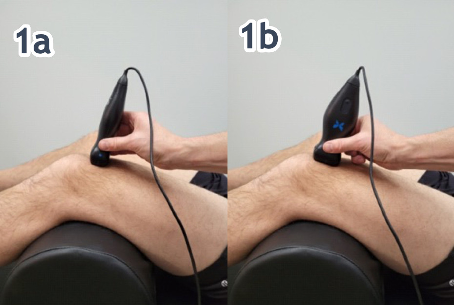
Traditionally, quadricep complex soft tissue injury has been difficult to quantify with the clinical evaluation. Clinical success is always predicated upon an accurate diagnosis and understanding of the pathological process. MSK-US imaging of the has revolutionized the diagnosis of quadriceps muscle and tendon injuries. It provides high-resolution images and animations that enable successful visualization and separation of soft tissues from adjoining bones and ligaments. While a careful history and a thorough physical examination are important steps in the assessment of quadriceps pathology, MSK-US provides high-resolution static of dynamic images that enable successful visualization and separation of soft tissues from adjoining bones and ligaments. MSK-US has become an invaluable component in diagnosing quadriceps muscle and tendon injuries due to its ability to clearly display the affected structures without exposing the patient to radiation or utilizing ionized contrast media. MSK-US can provide precise visualization of edema and can easily distinguish between benign and potentially pathological findings which make it an integral part of any holistic evaluation of quadriceps muscle tendon injury. The speed and accuracy of ultrasound imaging make it a useful tool in differentiating muscle contusion from tendinopathy or strain. In addition to the diagnostic benefits, MSK-US is also beneficial for monitoring progression and recovery of quadriceps muscle and tendon injuries. The accuracy of images helps clinicians evaluate patient progress during physical therapy sessions and make necessary adjustments to treatment plans if needed. This allows for more precise tracking of muscle healing, enabling clinicians to identify any setbacks early on and provide timely intervention if required.
.png)
.png)

Conclusion
In conclusion, MSK-US is an effective diagnostic tool for assessing quadriceps muscle and tendon injuries. MSK-US has minimal risk with no radiation involved and it can provide valuable information about the extent and progression of injury. By understanding MSK-US and its benefits, medical professionals can better diagnose and treat patients with quadriceps muscle and tendon injuries, leading to more successful outcomes. This makes it an invaluable tool for healthcare professionals when treating musculoskeletal disorders. MSK-US should always be used alongside other techniques such as physical examinations, laboratory tests and patient history in order to accurately diagnose quadriceps muscle and tendon injuries. MSK-US is essential for providing insight into the structures affected by injury and helps clinicians create tailored physical therapy plans that lead to optimal outcomes. With its accuracy, precision, and non-invasive nature, MSK-US has become an integral part of diagnosing and treating quadriceps muscle injuries. The goal of this article is to provide a few tips and tricks to assist in using MSK-US as a diagnostic tool for the assessment of quadriceps pathology.