
MSK ultrasound is a non-invasive imaging modality that uses high-frequency sound waves to create real-time images of soft tissues, including muscles, tendons, and ligaments. This non-invasive imaging method provides clear and highly detailed images of the soft tissues in the shoulder area, allowing for a more precise diagnosis. Specifically, MSK ultrasound is a valuable tool in the evaluation of subscapularis injuries by allowing for a detailed assessment of the muscle’s structure and function. This technique can accurately identify the extent and severity of subscapularis damage, which is essential for effective treatment. By using MSK ultrasound, clinicians can assess both the structure and function of the subscapularis muscle and make informed decisions about the most appropriate course of action. With the use of MSK ultrasound, patients can benefit from more accurate evaluations and faster treatment times, improving both the quality of care and the overall outcomes.
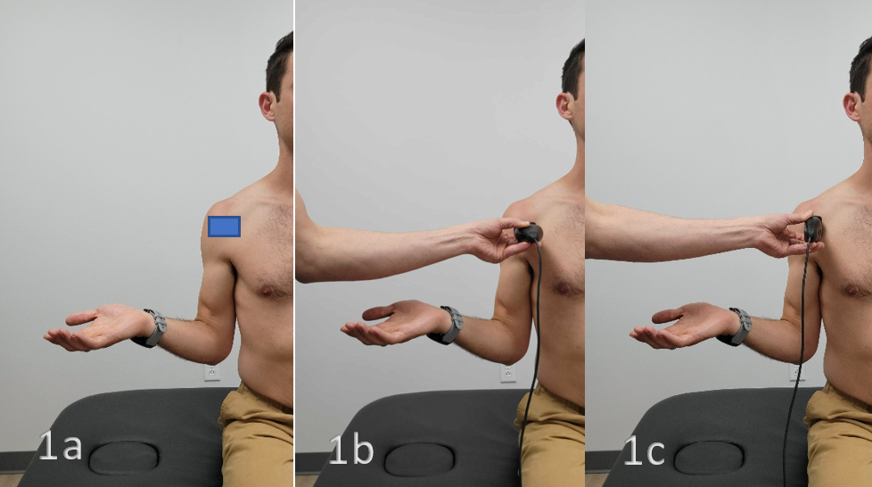
There are several techniques used to obtain accurate images of the subscapularis. One technique involves having the clinician place the patient in a seated position with the arm abducted and externally rotated. This allows the subscapularis muscle to be visualized from multiple angles. The ultrasound probe is then placed over the anterior aspect of the shoulder to visualize the subscapularis muscle (Figure 1).

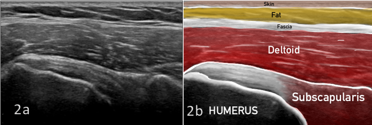
Normal subscapularis anatomy is represented in Figures 2 and 3. As the subscapularis crosses the proximal humerus, multiple hyperechoic tendinous slips can be visualized tapering into an anechoic point (sometimes referred to as a “tendon footprint”) as it inserts on the lesser tuberosity.
_.png)
The linear, anechoic/black area between the lesser tuberosity cortex and the tendon is the normal appearance of the “tendon footprint”. Loss of tendon contour, and an irregular/non-linear tendon footprint are indicative of a compromised, weakened tendon attachment.
_.png)
Some common findings that can be seen on MSK ultrasound images include tears, inflammation, fluid accumulation, and tendinopathy. These issues can be identified with great accuracy, allowing clinicians to better understand a patient’s condition and develop an effective treatment plan. More specifically, the following are some of the key features that an MSK ultrasound can reveal in subscapularis injuries:
-
Muscle thickness: An MSK ultrasound can measure the thickness of the subscapularis muscle, which can be an indicator of muscle atrophy or wasting.
-
Muscle integrity: An MSK ultrasound can detect tears or disruptions in the subscapularis muscle, as well as any associated inflammation or fluid accumulation.
-
Tendon attachment: An MSK ultrasound can assess the attachment of the subscapularis tendon to the humerus at the lesser tuberosity, which can be a site of injury in some cases.
-
Dynamic evaluation: An MSK ultrasound can evaluate the subscapularis muscle in real-time, allowing the clinician to assess its function during active movement of the shoulder joint.
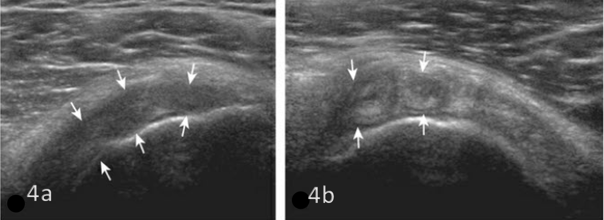
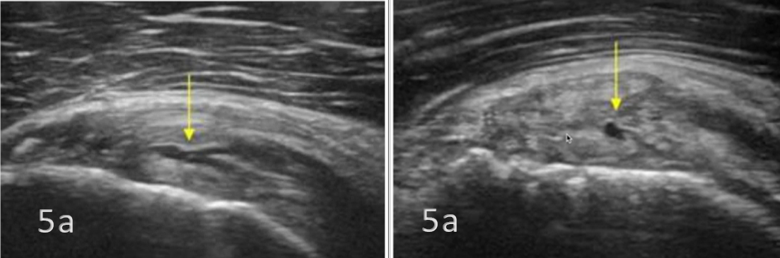
Figures 4 and 5 represent common sonographic findings in pathologies of subscapularis tendinosis as increased thickness and hypoechoic tendon structures. Subscapularis tears and disruptions can also be visualized as hypoechoic or anechoic areas within the muscle or tendon.
_and_long_axis_(lax)_.png)
_and_long_axis_(lax)_.png)
In summary, MSK ultrasound is a valuable tool in the evaluation of subscapularis injuries, providing detailed information on the muscle’s structure and function. Its use offers the benefits of being non-invasive and cost-effective, while providing an accurate diagnosis making it an excellent choice for initial evaluation and monitoring of subscapularis injuries. The techniques employed in an MSK ultrasound scan help to give a clear picture of the injury which helps to differentiate between different types of subscapularis injuries such as tears, tendinosis and strain. Ultimately, when combined with clinical examination, MSK ultrasound proves to be a powerful diagnostic tool in assessing subscapularis pathology.