Introduction
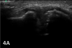
Sports physical therapists often deal with a range of musculoskeletal (MSK) issues, where accurate diagnosis and effective treatment are crucial for an athlete’s recovery and performance. The acromioclavicular joint (ACJ) plays a pivotal role in shoulder stability and movement, being the junction between the acromion process of the scapula and the clavicle. Given its importance, injuries to this joint, commonly resulting from falls, car accidents, or sports activities, can have significant repercussions. These injuries range from mild sprains, where ligaments are partially torn but the joint remains aligned, to severe dislocations with complete ligament tears and joint misalignment. Furthermore, repetitive use or wear and tear can lead to osteoarthritis of the ACJ, characterized by joint pain, stiffness, and swelling. Timely and accurate diagnosis is crucial for effective management. Traditional imaging techniques like radiographs often fall short in adequately assessing these injuries, particularly in visualizing soft tissues and dynamic joint function. MSK ultrasound has become increasingly popular for evaluating ACJ injuries, offering real-time, detailed imaging of soft tissue structures.
Benefits of MSK Ultrasound in ACJ Injury Assessment
MSK ultrasound offers a non-invasive, cost-effective, and dynamic assessment of the ACJ. Its superiority in visualizing soft tissue structures, such as ligaments, articular cartilage, and the joint capsule, allows for a more nuanced diagnosis than what is achievable with radiographs. Its non-invasive nature also allows for repeated assessments, essential in monitoring the healing process or the progression of degenerative changes. This imaging modality is also beneficial for monitoring the healing process and guiding interventions such as injections.
Specific Applications in ACJ Assessment
In acute injury scenarios, MSK ultrasound effectively assesses the extent of ligament and soft tissue damage, aiding in the accurate classification or grading of the injury and facilitating appropriate treatment planning. In chronic conditions, MSK ultrasound aids in identifying degenerative joint changes due to repetitive use or aging. The ability to perform dynamic assessments with MSK ultrasound is particularly useful in evaluating joint stability and function. It also detects concomitant injuries like rotator cuff tears or fractures, often missed by radiographs. In chronic conditions, it helps identify structural changes within the joint due to repetitive stress or aging.
Advantages of MSK Ultrasound for AC Joint Visualization
-
Real-Time Imaging: Unlike static images provided by radiographs or magnetic resonance imaging (MRI)s, MSK ultrasound offers dynamic, real-time imaging. This allows therapists to assess the ACJ under movement, providing a more comprehensive understanding of the injury.
-
High Resolution: Ultrasound provides high-resolution images of soft tissues, making it ideal for detecting subtle changes or injuries in the ligaments and tendons surrounding the ACJ.
-
Patient Comfort and Safety: Being a non-invasive procedure, ultrasound is more comfortable for patients. It also avoids the exposure to radiation that comes with radiographs.
-
Cost-Effectiveness and Accessibility: Ultrasound equipment is generally more affordable and portable compared to other imaging modalities, making it a practical tool in various settings, including clinics and sports fields.
Clinical Applications in AC Joint Injuries
-
Diagnosis: MSK ultrasound can quickly and accurately diagnose ACJ injuries, such as sprains, separations, or arthritis. It can differentiate between types of injuries based on the appearance of soft tissues.
-
Guided Interventions: In some cases, therapeutic interventions like injections are needed. Ultrasound can guide these procedures with precision, ensuring that the treatment is delivered to the exact location.
-
Non-Invasive Monitoring Progress: The non-invasive nature of MSK ultrasound allows for repeated assessments, making it ideal for monitoring injury progression and healing, especially in chronic conditions like osteoarthritis. Rehabilitation providers can use ultrasound to monitor the healing process of ACJ injuries, adjusting treatment plans as needed based on real-time feedback.
Advancements in MSK Ultrasound Technology
Recent advancements have significantly enhanced the utility of MSK ultrasound in ACJ evaluation. High-frequency transducers now offer detailed images of smaller structures like ACJ ligaments with greater resolution and clarity. Additionally, 3D and 4D imaging provide comprehensive evaluations of joint dynamics and movement. The use of contrast agents has also become a game-changer, aiding in distinguishing between active inflammation and structural changes, particularly in diagnosing osteoarthritis.
Conclusion
With advancements in ultrasound technology, there’s potential for even greater use in sports physical therapy. Innovations like 3D imaging and enhanced Doppler capabilities could provide deeper insights into joint health and injury mechanisms. As such, rehabilitation providers should incorporate MSK ultrasound into their diagnostic protocols for ACJ injuries as this represents a significant advancement in the evaluation of ACJ injuries. Its ability to visualize soft tissues, combined with its non-invasive nature, makes it a superior choice for accurate diagnosis and treatment monitoring. Additionally, the ability for repeated assessments makes it a valuable tool in both acute and chronic injury management. Advancements in technology have further improved its capabilities, making it an essential tool for clinicians in the management of ACJ injuries. Early and precise diagnosis is key to successful recovery and preventing complications. Healthcare professionals should actively consider MSK ultrasound in their diagnostic arsenal for ACJ concerns, ensuring proactive and comprehensive care for joint health.
Incorporating MSK ultrasound into their practice requires training and proficiency in ultrasound technology. Understanding the anatomy and common injury patterns of the AC joint is crucial for accurate interpretation of ultrasound images. Continuous education and practice are essential to maintain a high level of skill in ultrasound application.
_joint.png)
.png)


_joint.png)
.png)


