INTRODUCTION
Approximately 200,000 people will injure their anterior cruciate ligament (ACL) in any given year.1 Many of these injuries occur during sporting activities, with injury rates peaking in the second and third decades of life.2 ACL reconstruction (ACLR) is the most common treatment for ACL rupture. The number of reconstructions has increased over the last 30 years, especially in the skeletally immature and middle-aged populations.3–8 Current rehabilitation programs after ACLR are recommended to last 9-12+ months to restore normal knee function and return to sporting activities.9,10 Re-injury of the ACL is common, with re-injury rates as high as 30% to the ipsilateral or contralateral knee.11 Approximately two-thirds of individuals return to their pre-injury level of participation, and approximately three-quarters return to pivoting sports within two years post-ACLR.12,13 Long-term biomechanical deficits also persist,14 which highlights the need to improve rehabilitation practices.
Neuromuscular dysfunction of the quadriceps, particularly arthrogenic muscle inhibition (AMI), and muscle atrophy are common following anterior cruciate ligament injury and reconstruction.15,16 Persistent weakness of the quadriceps is associated with ACL graft rupture,17 worse patient-reported outcome (PRO) measures,18–20 increased risk for osteoarthritis,21 and altered biomechanics of the lower extremity.14,22 Achieving a quadriceps limb symmetry index (LSI) of >90% when compared to the nonsurgical limb improves PROs, knee joint biomechanics during movement, and the likelihood of reaching a patient acceptable symptom state (PASS).16–21 LSI may not provide the best benchmark for return to sport based on age. Pediatric athletes undergoing physeal-sparing grafts do not have disruption of the extensor mechanism and tend to reach LSI targets more easily than older athletes,23,24 yet reinjury risks in younger populations remain high.25,26 On the other end of the spectrum, older and/or less active individuals may be more likely to undergo hamstring autograft or allograft options,27,28 thus avoiding disruption of the extensor mechanism. Although specific evidence is limited, these individuals could theoretically meet LSI targets while maintaining low peak torque-to-bodyweight ratios, suggesting that normalized peak torque may be a better benchmark than LSI.
Current research suggests reaching a normalized peak isometric torque of 3-3.1 newton-meters per kilogram of body mass (Nm/kg) is correlated with better PROs.29,30 A large number of individuals do not meet this metric even after ~8-12+ months of rehabilitation post-surgery.29,31,32 Further, this metric may not reflect variation in quadriceps peak torque normalized to body mass across the lifespan or account for sex differences in strength.
The hamstring muscle group plays an important role as an agonist to the ACL.33,34 Lower hamstring peak torque compared to quadriceps peak torque has been linked to a higher reinjury risk post-ACLR.17,35 Because of this relationship, hamstring peak torque is evaluated relative to quadriceps peak torque using the hamstring-to-quadriceps ratio (H:Q ratio). Historically, it has been suggested that the H:Q ratio should reach at least 60% when tested on an isokinetic dynamometer at 60°/second.36 Evidence indicates that healthy individuals do not consistently meet this threshold.37,38 Similar to recommendations for normalized quadriceps peak torque, this benchmark may not account for normal variation across the lifespan nor for sex differences in muscle strength.
Current evidence suggests that chronological age should no longer be considered a contraindication for ACLR surgery, therefore it is important to understand how quadriceps and hamstring strength change with age.39 The purpose of this study was to examine the relationship between age and isokinetic quadriceps and hamstring peak torque normalized to body mass, as well as the H:Q ratio, and to evaluate differences between sexes, using the uninvolved limb from pre-ACLR testing. It was hypothesized that isokinetic quadriceps and hamstring peak torque, normalized to body mass, would vary with age and that females would demonstrate lower normalized peak torque than males. Improved characterization of peak torque values across the lifespan may help inform clinical expectations following ACLR.
METHODS
Strength Testing
Quadriceps and hamstring peak torque (a proxy for “strength”) was assessed using an isokinetic dynamometer (Biodex System 4 Pro; Biodex Medical Systems) with hips and knees flexed to 90°, the femoral condyles aligned with the axis of rotation, and the distal pad affixed to the distal shank about 5cm proximal to the medial malleolus. As a part of the UW Health post-operative protocol, pre-operative testing is performed to determine an athlete’s baseline strength. Prior to testing, athletes are weighed on a standard scale. The uninvolved limb was completed first, with three submaximal trials performed as a warm-up at 50%, 75%, and maximum effort. Pre-operative testing was performed bilaterally if there were no contraindications to perform testing on the involved limb. Testing was performed unilaterally (uninvolved only) when contraindications were present on the involved limb, which included: 1) a meniscal root tear, 2) a bucket-handle meniscal tear, 3) significant pain during warm-up, and 4) if the range of motion was not between 0 and 120 degrees on the involved limb. Testing then consisted of five consecutive maximum efforts of extension and flexion at 60° per second in a concentric manner. Participants were provided with standardized verbal encouragement (“Kick as hard as you can, pull as hard as you can”) once per rep. The peak torque normalized to body mass (Nm/kg) across the five repetitions on the uninvolved leg was determined for the quadriceps and hamstrings, and this was used to calculate the H:Q ratio.
Participants
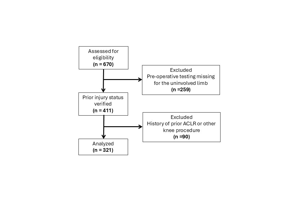
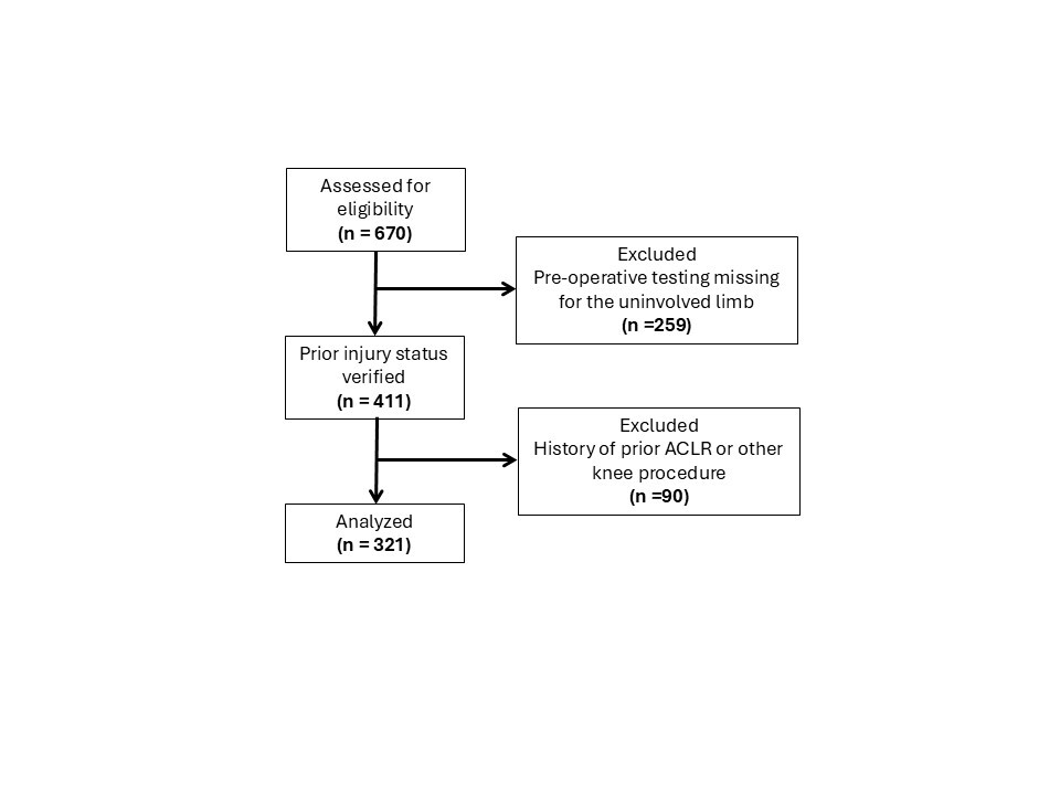
Three hundred twenty-one athletes were identified in the UW Health Sports Rehabilitation Database who had planned ACLR and underwent pre-operative testing at one of two hospital-based locations between July of 2021 and January of 2026 (Figure 1). The mean time in days from injury to testing is reported in Table 1. Participants were included if they met the following criteria: 1) they had a documented athletic activity to return to after surgery, and 2) they had pre-operative isokinetic testing on their uninvolved limb. Participants were excluded from data analysis if they met the following criteria: 1) this was not their primary ACL injury, and 2) they had a history of any prior knee surgery (involved or uninvolved). This retrospective records review was determined to be exempt by the University’s Health Sciences Institutional Review Board (IRB# 2018-1006), with a waiver of informed consent. Testing was performed as part of standard clinical care. The study received no external funding.
Statistical Analysis
Linear regression models were used to examine the relationships between age, sex, and their interaction on three outcomes: extension strength (Nm/kg, quadriceps), flexion strength (Nm/kg, hamstring), and hamstring-to-quadriceps ratio (H:Q ratio). For each outcome, the model included age (continuous variable), sex (binary), and age by sex as predictors.
Linear regression assumptions were assessed using visual and quantitative diagnostic methods. Residuals-versus-fitted plots supported assumptions of linearity and homoscedasticity, while Q–Q plots demonstrated approximate normality with only minor tail deviations. Influence diagnostics (Cook’s distance, leverage, and studentized residuals) identified a small number of outlying observations. Cook’s distance values remained low across all models (maximum = 0.087), indicating no single observation exerted undue influence on model estimates. Although the hamstring-to-quadriceps ratio model demonstrated the greatest departure from model assumptions, including three observations with studentized residuals exceeding the conventional ±3 threshold, diagnostic plots revealed no meaningful violations of regression assumptions. Accordingly, no observations were excluded from the primary analyses.
Potential sources of bias were also considered. Selection bias may be present due to the single-institution nature of the cohort, which consisted primarily of individuals undergoing sport-related rehabilitation; therefore, findings may not be fully generalizable to broader or non-athletic populations. Measurement bias is also possible because testing was conducted across multiple clinical sites. Although standardized protocols and equipment were used, formal inter-rater and inter-site reliability data were not available. Any resulting measurement variability is expected to be non-differential with respect to age and sex and would likely attenuate observed associations. These potential biases were considered when interpreting the findings by emphasizing effect estimates and 95% confidence intervals, evaluating consistency across outcomes, and recognizing the likely attenuating effect of non-differential measurement error.
All statistical analyses were conducted in R (version 4.5.3, R Foundation for Statistical Computing, Vienna, Austria). Predicted values with 95% confidence intervals were generated for each outcome and visualized as smooth curves stratified by sex using ggplot2.40 Statistical significance was set at α < 0.05. Because the primary analyses were pre-specified and focused on closely related biomechanical outcomes, adjustment for multiple comparisons was not applied to the primary models. However, Bonferroni-adjusted sensitivity analyses were performed and did not alter the study conclusions.
RESULTS
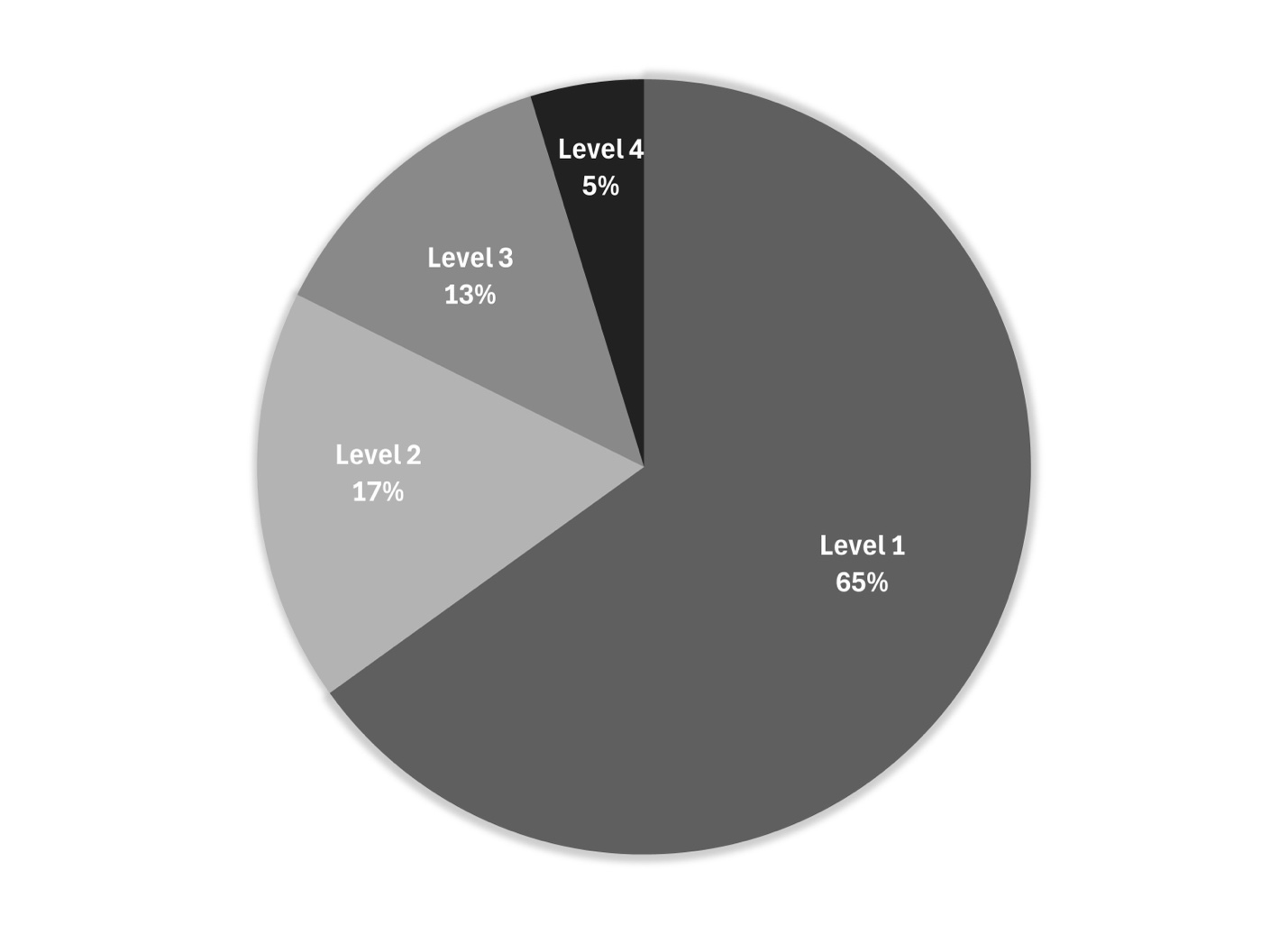
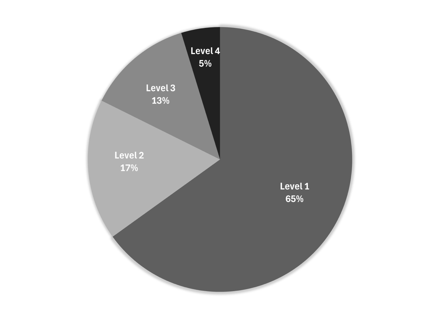
Three hundred twenty-one athletes (n=171 female; 27.5±11.2 years, n=150 male; 25.0±10.4 years) with uninvolved limb pre-ACLR testing were included in the analysis. Descriptive statistics were used to report demographic data (Table 1). Sport participation is self-reported at intake, with 65% partaking in Level 1 sports (Figure 2).41
Extension (Quadriceps) Strength
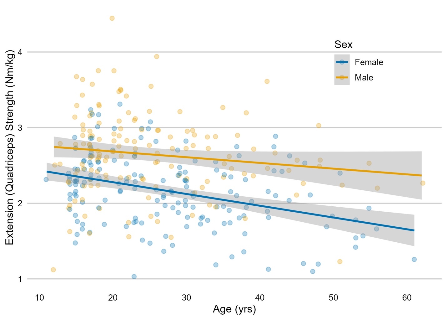
Age was negatively associated with extension strength (β = –0.016 Nm/kg per year, 95% CI: –0.022 to –0.009, p < 0.001), with a moderate standardized effect size (standardized β = –0.30). Males demonstrated higher mean strength than females, although the sex effect did not reach statistical significance (β = 0.246 Nm/kg, 95% CI: –0.034 to 0.525, p = 0.085). The age-by-sex interaction was not significant (β = 0.008, 95% CI: –0.002 to 0.018, p = 0.113).
This model accounted for approximately 25% of the variance in extension strength (R2 = 0.253). Predicted values plotted across the age range demonstrated a gradual decline in strength for both sexes (Figure 3).
_in_relation_to_age._males_and_females_b.jpeg)
Flexion (Hamstring) Strength
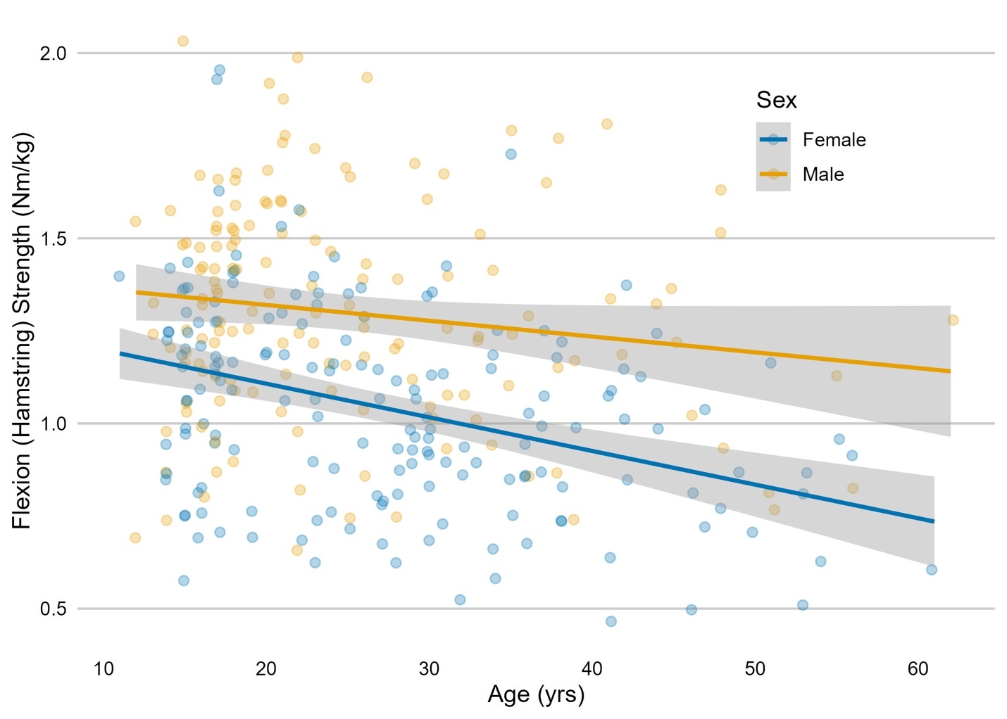
Age was similarly negatively associated with flexion strength (β = –0.009 Nm/kg per year, 95% CI: –0.013 to –0.005, p < 0.001), with a moderate standardized effect size (standardized β = –0.32). Males demonstrated slightly higher flexion strength than females, although this effect was not statistically significant (β = 0.116 Nm/kg, 95% CI: –0.043 to 0.275, p = 0.151). The age-by-sex interaction was also non-significant (β = 0.005, 95% CI: –0.001 to 0.010, p = 0.094).
This model accounted for approximately 24% of the variance in flexion strength (R2 = 0.238). Predicted curves showed a modest decline in flexion strength with increasing age across both sexes (Figure 4).
_in_relation_to_age._males_and_females_hav.jpeg)
Hamstring-to-Quadriceps Ratio (H:Q Ratio)
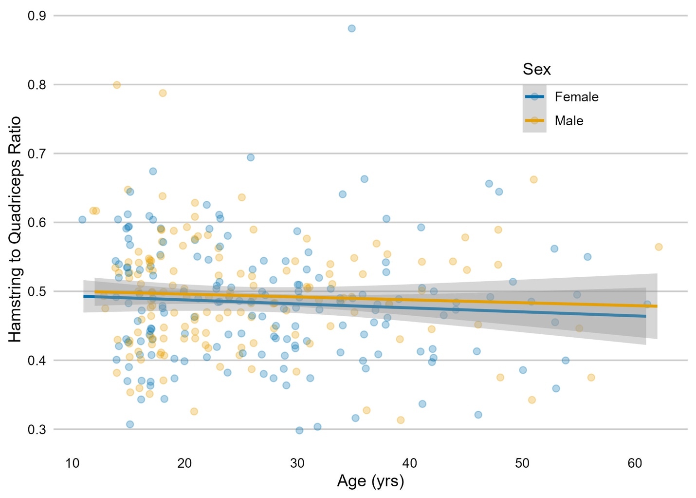
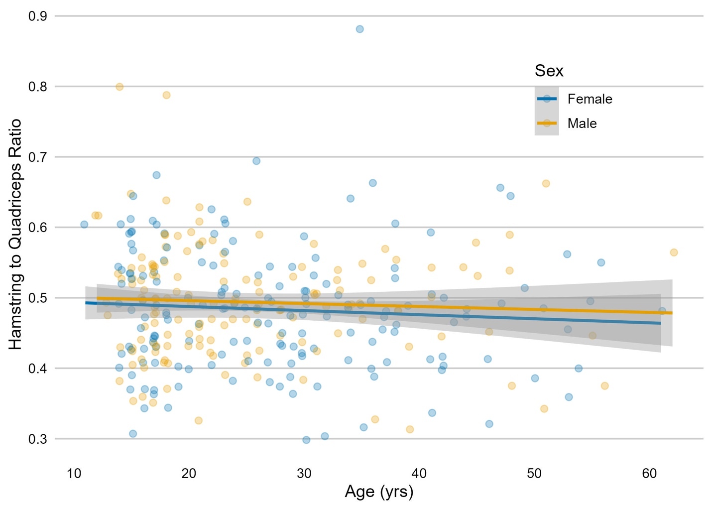
Neither age, sex, nor their interaction significantly predicted the H:Q ratio (age: β = –0.001 per year, 95% CI: –0.002 to 0.001; sex: β = 0.005, 95% CI: –0.043 to 0.054; interaction: β = 0.0002, 95% CI: –0.0016 to 0.0019; all p > 0.30).
This model explained minimal variance in the outcome (R2 = 0.009). Predicted H:Q ratio curves were essentially flat across the age range, with overlapping confidence intervals between males and females (Figure 5).
DISCUSSION
This study aimed to provide isokinetic peak torque values for the quadriceps and hamstrings normalized to body mass across different life stages in active individuals undergoing ACLR. The findings demonstrate differences across age groups in normalized peak torque, with a general decline as athletes age. This study provides clinical reference data to establish age- and sex-related norms for peak torque measures (Table 1). Clinicians can utilize Table 1, Figure 3, and Figure 4 as comparison values for their athletes based on age and biological sex.
Quadriceps and hamstring peak torque normalized to body mass linearly decrease with age. Mean values for males were higher than those for females across all age groups; however, this finding was not statistically significant and may require further study. It was originally hypothesized that females would have lower peak torque normalized to body mass compared to males due to several factors, namely lower testosterone,42 increased fat mass relative to muscle mass,43 and, historically and still not uncommonly, decreased strength-training exposure compared to males.44 As hypothesized, both groups experienced a general decline throughout the decades of life, which could be attributed to natural declines in muscle mass and strength, with relative inactivity compounding this effect.45 Age explains 25% and 24% of the variability in quadriceps and hamstring peak torque normalized to body mass across age groups, respectively. The remaining 75% could be attributed to individual physiological factors,46 exposure to strength training,44,45 activity levels,45 and comorbidities.47
When comparing these study results to published data on isokinetic strength in healthy athletes, the relationship varies with age and level of activity. In the 10-14 age group, both males and females in this study showed higher quadriceps and hamstring peak torque normalized to body mass than published norms.48–50 This could be attributed to the smaller sample size in this age group, which makes it more sensitive to high performers. In the 15-19 age group, both male and female quadriceps and hamstring normalized peak torque are similar to those in this study.48–50 In the 20-29 age group, athletes demonstrated normalized peak torque similar to published norms for non-elite athletes but were weaker than their elite and professional athlete counterparts.38,51 It is reasonable to expect that elite and professional athletes will have higher normalized peak torque at baseline and, therefore, require restoration of higher normalized peak torque after surgery.38,51
A commonly reported recommendation for normalized peak torque of the quadriceps is 3.0-3.1 Nm/kg, as this correlates with improved patient-reported outcomes.29,30 It is important to note that both studies that determined these values used a subject pool in the third decade of life and measured isometric quadriceps torque.29,30 These recommendations may not apply to younger and older individuals and require further study. Isokinetic-derived torque may provide lower absolute values than isometric-derived, thus producing lower normalized torque outputs.52 Therefore, these recommendations should not be extrapolated to isokinetic testing. There is a paucity of literature on the relationship between normalized quadriceps peak torque tested at 60º/sec and patient-reported outcomes. Using isokinetic testing at 90º/sec, Ashnai et al. found that males achieving ≥2.6 Nm/kg and females achieving ≥2.1 Nm/kg had higher odds of reaching a patient-acceptable symptom state at 1-year post-reconstruction53; however, this faster isokinetic speed yields smaller torque values than at 60º/sec in relation to the force-velocity curve. A desirable quadriceps cut-off score tested isokinetically at 60º/sec likely sits between the values from 90º/sec and isometric tests29,30,53; however, this was not investigated in the current study. An individual’s starting point for normalized peak torque likely influences expectations for the timeline to regain strength or meet a standard goal. Education of patients on expectations of this timeline should align with this. This data can be used to better inform specific recommendations for restoration of quadriceps and hamstring strength based on patient demographics.
H:Q ratio is another measurement criterion often used after ACLR due to the agonistic role the hamstrings play with the ACL.33,34,36,37 The literature remains inconclusive on whether the H:Q ratio is an independent risk factor for ACL injury.54 There is evidence of the H:Q ratio being a contributing factor. Almeida et al. used a classification and regression tree analysis that included an H:Q ratio cut-off of 46.2% in the model, achieving 90.9% accuracy in correctly classifying those who did and did not sustain a second ACL injury.35 Kyritsis et al. found a hazard ratio of 10.6 per 10% decrease in the H:Q ratio at 60º/sec.17 These findings indicate that aiming for a higher H:Q ratio is beneficial and that restoring the ratio as close to the commonly recommended 60% (tested isokinetically at 60º/sec) may still be the best target.17,35,36 This study’s cohort had an average H:Q ratio below 60%, indicating that many individuals enter surgery with subpar hamstring strength relative to quadriceps strength. It is important to note that higher H:Q ratios (0.8-1.0) are found when testing at higher isokinetic speeds or when eccentric hamstring strength is compared to concentric quadriceps strength at varying speeds.36–38,54 It is imperative that clinicians recognize the differences in H:Q ratio recommendations across measurement speeds and modes. There is some evidence of a disproportionate increase in quadriceps strength relative to hamstring strength during puberty in females,55 which altered movement mechanics may partially explain.56 This would naturally lead to a lower H:Q ratio in those in the second decade of life, requiring more work after ACLR to reach recommended values. The current study did not support this theory, with H:Q ratio staying relatively stable across the lifespan.
There are a few limitations of this study. The cohort underwent strength testing after an injury, with variable time since injury to testing and activity modification. Further, it could be argued that the uninvolved side may have also been impacted,38 thereby yielding lower-than-normal normalized peak torque values. However, the results are comparable to other published data, mitigating this limitation. Second, all participants had sustained an ACL injury. This raises the question of whether there would be differences in strength values compared with those who have not injured their ACL in an age-, sex-, and activity-matched cohort. While all participants were from a healthy and active population prior to injury, the levels of sport participation, positions, and exposure rates were not included in the analysis. External validity is limited by the single-institution design and the sport-rehabilitation–skewed cohort. Additionally, fewer subjects represented in the youngest (10–14 years) and oldest (50+ years) ages may reduce the precision of estimates within these groups. Therefore, findings should be interpreted primarily as applicable to a physically active clinical rehabilitation population rather than the general population. External validation in independent cohorts, particularly those with broader age and activity distributions, is warranted to establish broader generalizability.
CONCLUSION
The results of the current study demonstrate that age explains 25% and 24% of the variability in quadriceps and hamstring peak torque normalized to body mass across age groups, respectively, whereas the H:Q ratio remains constant. Recommendations for normalized quadriceps and hamstring peak torque after ACLR should consider the influence of age-related biological and lifestyle constraints on attainable strength levels. The data from this study can serve as a reference for clinicians to compare patients’ isokinetic data. Clinicians should understand that age and the mode of testing may affect reported peak torque data. Future research should explore the effects of age and testing mode on normalized peak torque after ACLR and reinjury rates to better inform return-to-sport recommendations for those recovering from ACLR. Further, the results of the current study should be explored in relation to an uninjured population.