INTRODUCTION
Athletic groin pain is a common condition in athletes participating in multidirectional sports and can be challenging to accurately diagnose and manage. The complex regional anatomy and the presence of multiple overlapping pathologies have historically resulted in inconsistent terminology and variability in diagnostic approaches across the literature and clinical practice.1 In response, the 2015 Doha agreement established consensus-based terminology and clinical classification criteria to improve diagnostic clarity. Although this framework has improved consistency in identifying subtypes of groin pain, uncertainty remains regarding optimal management strategies.
Adductor-related athletic groin pain (ARAGP) represents the most frequently reported subtype of athletic groin pain and contributes substantially to time loss and performance limitation.2–4 According to the Doha Agreement, adductor related athletic groin pain is distinguishable from other similar clinical entities (e.g., iliopsoas related athletic groin pain, inguinal related athletic groin pain, pubic related athletic groin pain or hip related athletic groin pain) by the presence of pain localized to the adductor region and reproducible symptoms with resisted hip adduction or adductor palpation.1,5,6 Multidirectional sports such as soccer and ice hockey demonstrate particularly high rates of groin injury. In NCAA athletics, hip and groin injuries have been reported at a rate of approximately 53 per 100,000 athlete exposures, with adductor tears representing the most common diagnosis.3 Similarly, in professional men’s ice hockey, non-contact adductor strains account for approximately 10-11% of all injuries.7 Although prevalence appears highest in field and ice sports, ARAGP and related groin injuries also occur in basketball and dance populations, underscoring the broad relevance of this condition across athletic disciplines.8,9
Establishing accurate incidence remains challenging, as many studies rely on self-reported or time-loss data, and minor groin symptoms may go unreported.10,11 Additionally, male athletes consistently demonstrate higher rates of ARAGP and related groin injury compared to female athletes, with some studies reporting a relative risk more than twice as high in multidirectional sports.10,12 The underlying reasons for this disparity remain incompletely understood.
Despite its prevalence and potential impact on athlete availability and performance, debate persists regarding best practices for risk identification, prevention, and rehabilitation of ARAGP.13–16 The purpose of this clinical commentary is to provide clinicians and sports performance professionals with an evidence informed framework for the recognition, management, and risk reduction of adductor related athletic groin pain (ARAGP).
ANATOMY
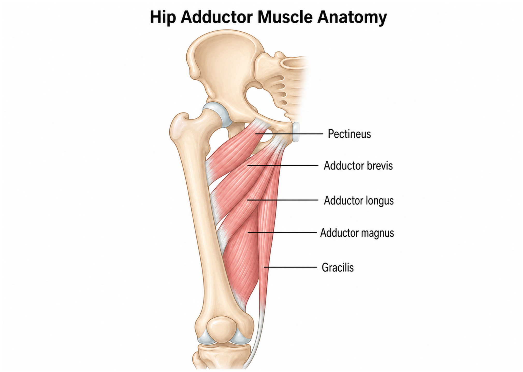
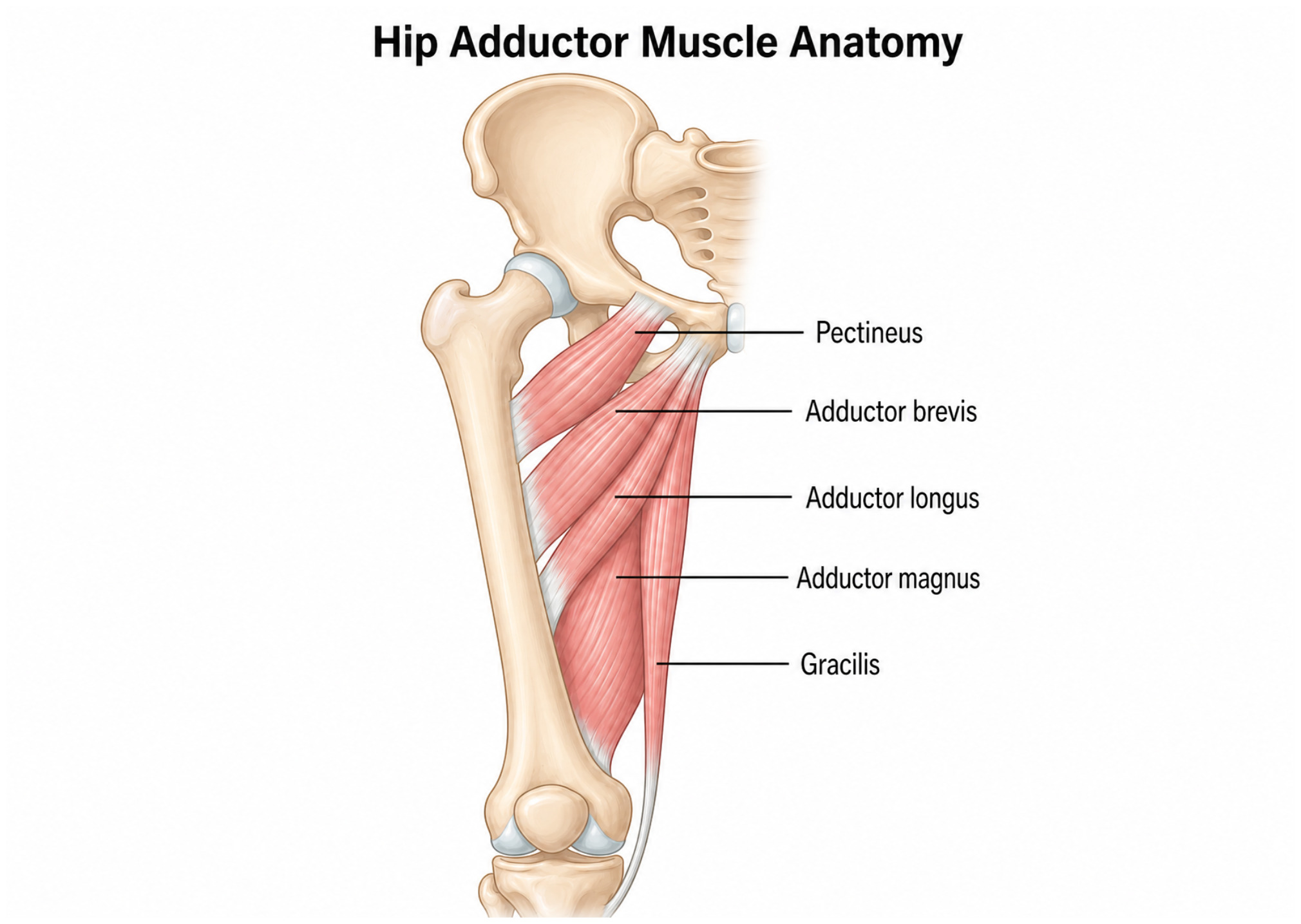
A foundational understanding of regional anatomy is essential for the accurate diagnosis and management of adductor-related athletic groin pain. The adductor musculature consists of six muscles located along the medial thigh that contribute primarily or secondarily to hip adduction: the adductor longus, adductor brevis, adductor magnus, pectineus, gracilis, and obturator externus.17 These muscles originate primarily from the anteroinferior pubis and are predominantly innervated by the obturator nerve. Notable exceptions include the pectineus, which receives femoral nerve innervation, and the hamstring portion of the adductor magnus, which is innervated by the sciatic nerve.
Anatomically, the adductors are positioned to generate substantial torque during hip adduction in both pelvis-on-femur and femur-on-pelvis movements.18 Beyond their primary role in hip adduction, the adductor complex contributes to multiplanar hip function, including internal and external rotation, as well as flexion and extension depending on hip position. When the hip is near neutral or extended, many of the adductor muscles assist with hip flexion; however, as the hip moves into approximately 40–70 degrees of flexion, their line of pull shifts relative to the axis of rotation, enabling contribution to hip extension.18 The adductor magnus, in particular, plays a substantial role in hip extension across a range of positions and is often described functionally as a secondary hip extensor. Collectively, these anatomical and functional characteristics underscore the integral role of the adductor complex in dynamic lower extremity tasks common in sport. Beyond their contributions to isolated hip movement, the adductor complex serves as a dynamic stabilizer of the pelvis during rapid multiplanar loading. During cutting and change of direction tasks, the hip adductor muscles must simultaneously resist hip abduction forces generated by ground reaction forces while contracting eccentrically as the limb is driven into further abduction. Electromyographic analyses show the adductor longus is active throughout the entire ground contact period of cutting maneuvers, not only at push off, and show peak activation at approximately 53% of the cutting stance phase during an eccentric contraction phase.19,20 This constant eccentric demand during high velocity directional change represents the primary mechanical basis for adductor injury risk and underscores why multiplanar sport tasks place increased stress on this muscle group.
RISK FACTORS
Several risk factors for ARAGP have been identified in the literature. Established evidence supports an increased risk in athletes with a prior history of groin injury, those participating at an elite or professional level compared to lower levels of competition, and in those demonstrating imbalances in hip adduction and abduction strength.21–23 Previous injury to the adductor complex has consistently been reported as one of the strongest predictors of future ARAGP.17,23 Residual strength deficits, altered neuromuscular control, and persistent side-to-side imbalances have been proposed as contributing factors following initial injury.12 A summary of commonly reported risk factors and their clinical implications is presented in Table 1.
Strength profiling, particularly the hip adduction-to-abduction strength ratio, has received considerable attention in the literature. In a prospective study of professional ice hockey players, uninjured athletes demonstrated adduction strength values approximately 95% of abduction strength, whereas athletes who later sustained groin injuries demonstrated adduction strength values closer to 78% of abduction strength.22 However, a subsequent prospective cohort study including 163 male ice hockey athletes found that preseason isometric adduction and abduction strength were not significantly associated with subsequent hip and groin injuries, whereas previous non-time-loss groin symptoms was the strongest independent predictor of future injury (OR; 3.3).24 These conflicting results suggest that isometric strength ratio thresholds derived from a single sport population should not be applied as universal screening benchmarks across all groups of athletes. More recent findings suggest that the hip adduction to abduction strength ratio itself may not represent a robust independent predictor. A 2025 systematic review and meta-analysis of healthy athletes reported that lower hip adduction strength demonstrated a moderate association with future groin injuries, whereas the adductor to abductor strength ratio showed only trivial association with subsequent groin pain or time loss groin injuries.25
A recent systematic review further identifies reduced eccentric hip adduction strength as one of the most consistent intrinsic risk factors associated with groin pain in athletes.26 Emerging evidence also suggests that the presence of subclinical groin symptoms, even in the absence of time-loss injury, may increase the likelihood of subsequent clinically significant groin pain.26 Collectively, these findings emphasize the importance of eccentric strength capacity and early symptom monitoring when designing targeted prevention strategies.
Playing surface has also been investigated as a potential extrinsic risk factor for ARAGP. Current evidence does not support a clinically meaningful difference in adductor injury risk between artificial turf and natural grass, with large-scale prospective data indicating no significant surface effect on adductor-related injury incidence after controlling for relevant covariates.27 These findings suggest that surface type alone should not be a primary focus of risk mitigation for ARAGP, and that emphasis should remain on modifiable intrinsic factors such as adductor eccentric strength capacity, prior injury history, and training load management.
Additional factors associated with groin injury risk include reduced hip range of motion, higher body mass index, greater body mass, and increased height.12 While these variables demonstrate associations with injury risk, their individual contribution to injury development remains multifactorial and likely sport specific.23 Reduced hip internal rotation and abduction range of motion have been associated with increased groin injury risk across multiple athlete cohorts, potentially reflecting underlying morphological factors that alter hip loading mechanics during sport.12 Increased competitive level and prolonged exposure have also been identified as contextual risk factors, consistent with the dose-response relationship between mechanical loading and tissue tolerance.
Sex is another clinically relevant risk factor, with male athletes consistently demonstrating higher rates of adductor related athletic groin pain compared to females, a disparity that appears to reflect fundamental differences in pelvic and hip morphology rather than exposure alone.10,12 Male athletes typically exhibit a narrower subpubic angle and greater stress across the pubic symphysis during athletic loading, while female athletes exhibit greater acetabular coverage, greater femoral anteversion, and wider pelvic width to femoral shaft length ratios, characteristics that may reduce adductor origin loading during cutting and other change of direction tasks.28 When female athletes do present with hip and groin pain, the distribution among Doha categories differs substantially from males. Evidence from female team sport athletes suggests that iliopsoas-related athletic groin pain is substantially more common than adductor related athletic groin pain accounting for 58.8% and 11.8% of cases, a pattern that is essentially the inverse of male athletes. From a clinical perspective, these results suggest that assessment strategies validated largely in male athletes, including adductor palpation and resisted adduction testing, may not directly translate to female, for whom iliopsoas palpation and resisted hip flexion testing require an equal emphasis.
CLINICAL DIAGNOSIS AND EXAMINATION
Accurate clinical diagnosis of ARAGP requires a structured examination approach that incorporates symptom history, palpation, and provocation testing within the framework established by the Doha Agreement.14 According to the Doha classification, adductor-related athletic groin pain is distinguished from other groin pain entities by two primary clinical criteria: tenderness on palpation of the adductor muscle origin and reproduction of familiar pain with resisted hip adduction.29 These criteria form the diagnostic foundation upon which clinical examination should be structured.
Clinical assessment begins with a comprehensive history of groin injury, including symptom onset, location, and aggravating factors during sport participation.
Athletes with ARAGP typically report medial thigh or pubic region pain that is provoked by cutting, kicking, and change-of-direction tasks and may describe a gradual onset rather than a discrete traumatic event.30 Pain distribution can assist in directing examination focus: adductor-related pain localizes primarily to the medial thigh and pubic region, while inguinal-related pain tends to distribute in more of the superiorly and anteriorly groin.31
Systematic palpation of the adductor complex follows history-taking, beginning at the proximal adductor origin on the anteroinferior pubis and progressing distally along the muscle belly. The proximal adductor origin palpation is the single most powerful test for excluding adductor pathology: a pain-free adductor origin has a negative likelihood ratio of 0.11, meaning that the absence of tenderness substantially reduces the post-test probability of adductor-related pathology.32 However, positive palpation findings alone lack specificity and must be combined with resisted testing for reliable classification.5
Resisted hip adduction in the neutral position (0° hip flexion) has been shown to most reliably provoke adductor longus origin pain compared to other test positions.33 Testing in neutral maximizes tensile load at the proximal adductor longus tendon and pubic attachment; hip-flexed positions reduce mechanical specificity at the proximal origin. Reliable Doha adductor-related classification requires both palpation tenderness and pain on resisted adduction to be present, applying both criteria in combination improves inter-examiner reliability from fair (k = 0.40) to moderate (k = 0.49) and achieves 100% agreement in athletes with unilateral single-entity presentations.5 Resisted adduction is performed with the athlete supine, hips in neutral, and the examiner providing manual resistance at the medial knee bilaterally. A positive test reproduces the athlete’s familiar groin pain rather than simply producing discomfort.
The adductor squeeze test at 90° of hip flexion provides a broad screening function with 85.4% sensitivity for athletic groin pain, making it a useful first-step screen; however, a negative likelihood ratio of 1.95 indicates that a negative result increases rather than decreases post-test probability, confirming the test has no value as a rule-out instrument and must not be used in isolation for diagnostic classification.32 Squeeze test positions at 0° preferentially load the pubic symphysis and aponeurosis and may be more useful when pubic-related pathology is suspected. All squeeze test positions function as screening instruments rather than confirmatory tests, and serial squeeze testing at 45° or 90° demonstrates acceptable intrarater reliability for longitudinal load tolerance monitoring.34
DIFFERENTIAL DIAGNOSIS
A structured clinical examination must also systematically consider overlapping entities that can mimic or co-exist with ARAGP. The Doha Agreement identifies five groin pain entities that may present in isolation or combination: adductor-related, iliopsoas-related, inguinal-related, pubic-related, and hip-related pain, each with distinct clinical examination findings that guide differential diagnosis.5,29 Accurate classification requires applying criteria across all five categories rather than anchoring prematurely to a single diagnosis, as multiple entities frequently co-exist in the same athlete.
Iliopsoas-related groin pain is identified through iliopsoas palpation tenderness and reproduction of pain with resisted hip flexion or the Thomas test. In female athletes specifically, iliopsoas-related pain is the predominant groin pain category and should receive priority in the examination sequence.35 Inguinal-related groin pain is differentiated by the absence of adductor palpation tenderness and reproduction of pain with abdominal compression testing or Valsalva maneuver rather than resisted adduction.14 Pubic-related groin pain is characterized by pubic symphysis tenderness; notably, pubic-related classification carries poor inter-examiner reliability (k = 0.12) and should not be assigned as a primary diagnosis without confirmatory imaging (Heijboer et al., 2023).5
Femoroacetabular impingement syndrome (FAIS) represents the primary hip-related entity to consider in the differential, as cam morphology is highly prevalent in elite contact and multidirectional sport athletes and frequently co-occurs with adductor-related pain.36 FAIS typically presents with anterior hip or groin pain reproduced by the FADIR test (flexion, adduction, internal rotation), with associated hip internal rotation restriction on examination. The FADIR test has high sensitivity but low specificity in athletic populations and should be interpreted alongside symptom history and imaging when FAIS is suspected. Critically, FAIS and ARAGP are not mutually exclusive. The clinician’s task is not simply to choose between them but to identify which entities are contributing and to what degree. Applying the Doha criteria systematically across all five categories represents the most reliable clinical approach to groin pain classification.5 Treatment planning should reflect the full clinical picture, as unrecognized co-existing entities are a common source of incomplete recovery and delayed return to sport.
IMAGING
The athletic groin represents a complex anatomical region, and both magnetic resonance imaging (MRI) and diagnostic ultrasound are commonly utilized to support evaluation.37–40 Given the overlap in clinical presentation among various causes of groin pain, imaging can assist in clarifying diagnosis when physical examination findings are inconclusive.37 MRI and ultrasound have demonstrated validity and reliability in the assessment of adductor-related pathology.41,42
Imaging is most frequently used to evaluate the presence, location, and severity of acute adductor strains, including involvement at the muscle belly, myotendinous junction, or proximal tendon origin.14,41,43 MRI remains the reference standard for detailed assessment of soft tissue injury, providing high resolution visualization of edema patterns, tendon integrity, and associated pelvic findings. Ultrasound offers advantages including accessibility, dynamic assessment, and the ability to evaluate tissue behavior during functional contraction.
Ultrasound has also been used in research settings to examine muscle morphology and architectural characteristics. Although architectural adaptations have been described in other muscle groups following strength training or injury, limited evidence currently exists evaluating longitudinal architectural changes in the hip adductors in relation to rehabilitation, performance, or reinjury risk.44 Importantly, imaging findings must be interpreted in conjunction with clinical presentation, as structural abnormalities may be present in asymptomatic athletes and do not necessarily correlate with pain severity or functional limitation. Imaging should therefore serve as an adjunct to, rather than a replacement for comprehensive clinical examination and functional assessment.
PATHOMECHANICS
Clinical movement assessments have demonstrated limited ability to reliably predict future athletic groin injury, despite theoretical rationale supporting their use in identifying altered hip and pelvic mechanics.16 Systematic reviews indicate that while movement quality screening may reveal biomechanical differences, the evidence linking specific movement patterns to prospective injury risk remains inconclusive.45 However, biomechanical investigations have demonstrated that athletes with ARAGP exhibit altered movement and loading strategies. Athletes with ARAGP have been shown to display longer ground contact times, reduced peak vertical ground reaction force, and diminished rates of force development during hopping tasks, suggesting modified force attenuation and propulsion characteristics.46 Importantly, these loading parameters appear responsive to rehabilitation, with normalization of vertical ground reaction force measures and concurrent changes in trunk and pelvic kinematics. Similarly, a study evaluating cutting mechanics showed post-rehabilitation adaptations characterized by increased pelvic rotation toward the direction of travel, a more anterior center of mass relative to the center of pressure, and greater ankle plantarflexion moments and power, reflecting reoptimized movement strategies following recovery.47
These biomechanical adaptations following rehabilitation reflect meaningful functional recovery that extends beyond simple pain resolution. Increased pelvic rotation toward the direction of travel reflects improved hip dissociation and reduced reactive loading on the adductor complex during directional change.48 A more anterior center of mass relative to the center of pressure indicates improved force transfer through the stance limb, reducing the compensatory lateral trunk shift commonly observed in symptomatic athletes. Greater ankle plantarflexion moments and power reflect restored push-off propulsion and improved force transmission from the proximal to distal kinetic chain, changes that are clinically observable as a more fluid, powerful cutting stride.48,49 Taken together, these adaptations suggest that successful rehabilitation restores not only tissue tolerance but the whole-body movement efficiency required for high-intensity sport participation.50
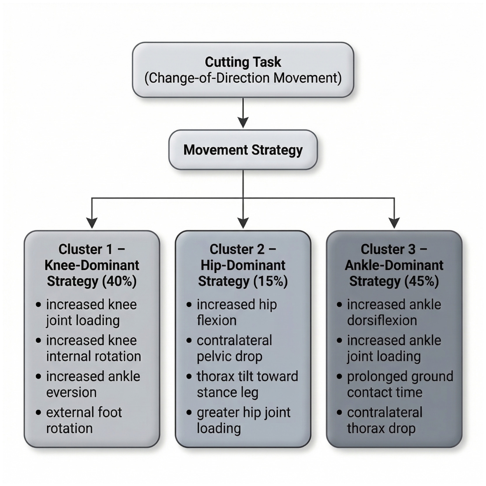
Similar biomechanical work demonstrated that athletes with long-standing ARAGP adopt varied movement strategies. Specifically, three distinct movement clusters have been observed: Cluster 1 (40%) characterized by increased ankle eversion and external rotation, knee internal rotation, and greater knee joint moments; Cluster 2 (15%) characterized by increased hip flexion, contralateral pelvic drop, thorax tilt, and greater hip joint moments; and Cluster 3 (45%) characterized by increased ankle dorsiflexion, contralateral thorax drop, greater ankle joint loading, and prolonged ground contact time (Figure 2).45 Collectively, current evidence suggests that movement assessments may be valuable for characterizing symptom presentation and functional limitation, although its role in injury prediction remains unclear. The clinical utility of this cluster framework extends beyond biomechanical characterization. Evidence suggests that individual athletes consistently reproduce their cluster-specific movement patterns across multiple repetitions, supporting the use of brief change-of-direction assessments for biomechanical subtyping in clinical practice.48
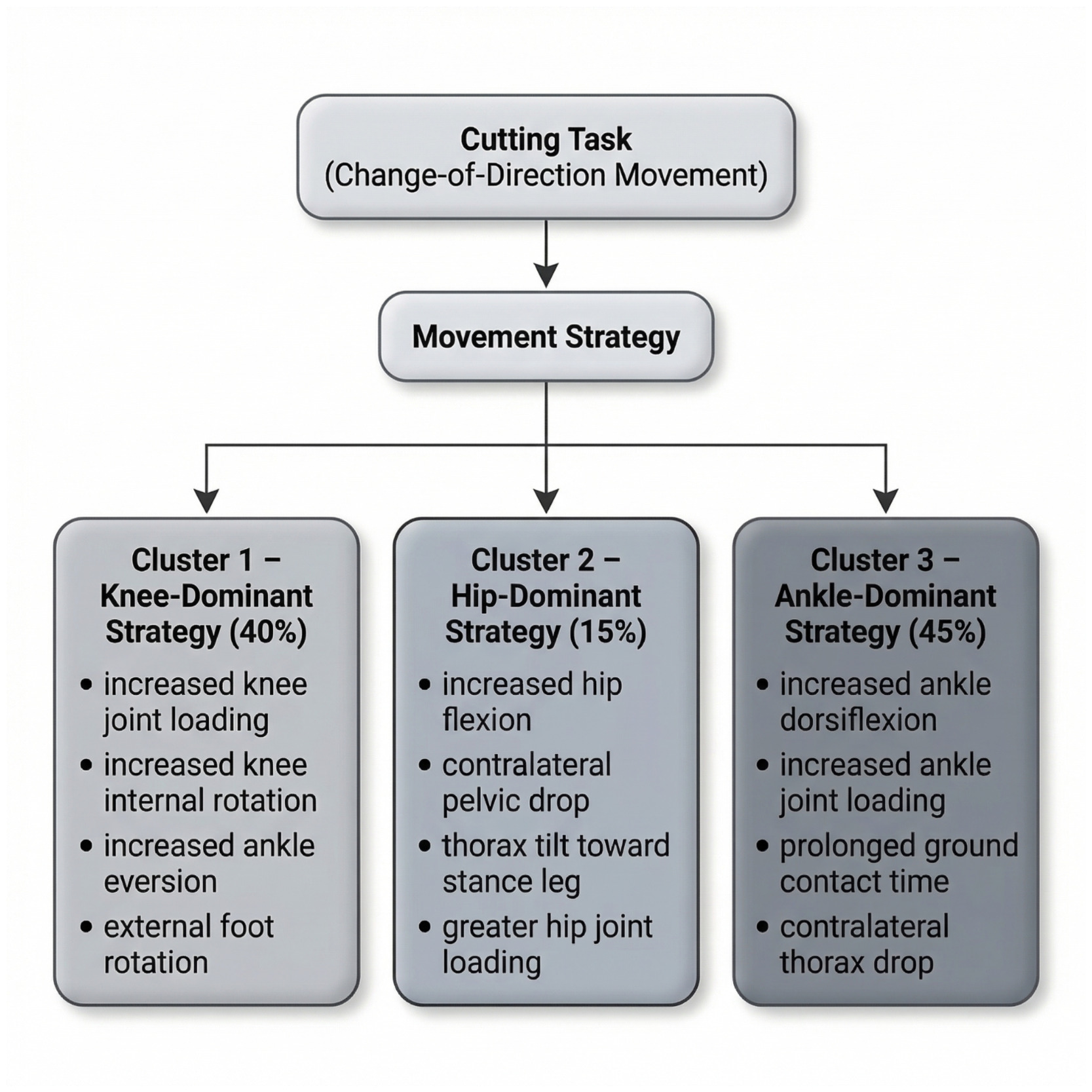
Each cluster carries distinct implications for exercise prescription. Cluster 1 athletes, characterized by reduced hip adductor moment and increased contralateral trunk lean, may benefit most from progressive adductor loading combined with concurrent trunk stability demands. Cluster 2 athletes, characterized by elevated hip adductor moment, may require movement redistribution strategies aimed at reducing tissue loading through improved lumbopelvic control and hip abductor co-activation. Cluster 3 athletes, characterized by generalized reductions in force output across all planes, typically require global strength development and neuromuscular re-education before task-specific change-of-direction training is introduced.48 Identification of an athlete’s cluster pattern early in rehabilitation may help guide exercise selection and provide insight into why a standardized adductor strengthening program alone may be insufficient, particularly in athletes whose primary impairment is movement control rather than isolated strength deficits.
In contrast to the limited predictive value of movement screening for prospective injury risk, individuals currently experiencing hip and groin pain often demonstrate distinct movement strategies compared to asymptomatic controls. Altered performance has been observed during functional tasks such as the Star Excursion Balance Test, particularly in the posterolateral reach direction,16,51 as well as during cutting and change-of-direction tasks where differences in intersegmental coordination and trunk–pelvic mechanics have been reported.45,46,48,52 Whether these altered movement strategies represent a predisposing factor, a response to pain, or a protective adaptation remains unclear.14,53 Although rehabilitation programs frequently emphasize intersegmental control and adductor strengthening45,54 the extent to which restoration of movement patterns reduces future injury risk has not been definitively established.
Video-based analyses of acute adductor injuries provide additional insight into common injury scenarios. Change-of-direction tasks and kicking actions represent the most frequently reported mechanisms, with a substantial proportion of injuries occurring during non-contact events.14,30,41 In professional soccer players, change of direction accounted for approximately one-third of adductor longus injuries, followed by kicking and reaching tasks.30 These findings suggest that rapid multiplanar loading during high-intensity sport places considerable mechanical demand on the adductor complex. Collectively, current evidence suggests that while movement assessment may help characterize symptom presentation and functional limitation, its role in injury prediction remains uncertain.
The adductor complex does not operate in biomechanical isolation during sport. Through shared fascial and osseoligamentous connections at the pubic symphysis, the adductors interact continuously with the lumbopelvic stabilizers.55,56 During cutting tasks, the adductor longus generates an anterior pelvic shear force that must be counteracted by transversus abdominis and pelvic floor co-activation.55,57 Athletes with ARAGP demonstrate disrupted neuromuscular recruitment sequences across this kinetic chain, including delayed gluteus medius onset and advanced rectus abdominis onset during cutting maneuvers, suggesting a compensatory global stabilization strategy substituting for impaired local stabilizer function.58 Importantly, residual lumbopelvic control deficits have been observed even after clinical return to sport, with athletes demonstrating altered pelvic kinematics and reduced hip extension velocity during kicking tasks compared to uninjured controls.59 These findings reinforce that lumbopelvic stability assessment should be incorporated into both rehabilitation programming and return-to-sport evaluation rather than treated as secondary to isolated adductor strength restoration.
PREVENTION
Given that ARAGP most commonly occurs during high-intensity change-of-direction and kicking tasks, prevention strategies should consider the demands of rapid multiplanar loading and deceleration commonly encountered in sport. Reduced eccentric hip adduction strength represents the most consistently modifiable intrinsic risk factor, and targeted eccentric loading has become the central focus of evidence-based prevention programming. The Copenhagen adduction exercise has demonstrated high activation of the adductor longus and has been shown to significantly increase eccentric hip adduction strength following structured implementation.60,61 In a cluster-randomized trial involving 35 semiprofessional Norwegian soccer teams, inclusion of a Copenhagen-based strengthening program resulted in a 41% reduction in groin injuries and a 20% reduction in time lost from sport compared to controls.61 The Copenhagen adduction exercise therefore represents the most evidence-supported single intervention currently available for adductor-related groin injury prevention in team sport populations.
Comprehensive warm-up programs may also contribute to groin injury reduction. The FIFA 11+ program, which incorporates neuromuscular control, strength training, balance, plyometrics, and agility components, has demonstrated reductions in lower extremity injuries, including hip and groin injuries.62 A systematic review and meta-analysis reported a 39% reduction in overall soccer-related injuries and a 41% reduction in hip and groin injuries among recreational players implementing the program. A recent scoping review of randomized trials highlighted that the Copenhagen adduction exercise remains the most frequently studied preventive intervention, with comparatively limited investigation of alternative or multifaceted exercise strategies.63 Emerging evidence supports the integration of trunk-hip coordination training alongside isolated adductor strengthening. Fujisaki et al. demonstrated reduced groin injury incidence in male high school soccer players when the Copenhagen adduction exercise was combined with hip and core coordination exercises in a cluster-randomized trial.64 A registered RCT protocol in futsal athletes proposes a multimodal approach combining core stability exercises with Copenhagen and sliding hip abduction exercises, targeting the lumbopelvic-hip complex rather than adductor strength in isolation, a design that reflects the growing clinical consensus that prevention programming should address the full kinetic chain.65 An 8-week adductor strengthening RCT by Wang et al. in youth players similarly demonstrated improved adductor-to-abductor strength ratios and reduced groin injury risk factors, with the inclusion of female athletes representing an important methodological advance given their historical underrepresentation in groin injury prevention trials.66 Collectively, current evidence supports the inclusion of structured eccentric strengthening and neuromuscular training within prevention strategies for athletes at risk of adductor-related groin injury, with multimodal trunk-hip approaches offering additional benefit beyond adductor-isolated loading alone.
REHABILITATION
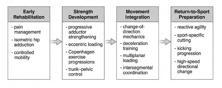
Early rehabilitation approaches for ARAGP emphasized progressive strengthening of the adductor musculature. Hölmich et al. described an active rehabilitation program centered on targeted adductor strengthening, which demonstrated favorable outcomes in athletes with long-standing groin pain.67 Since that time, rehabilitation strategies have evolved to incorporate a broader range of exercises supported by surface electromyography (sEMG) analyses to guide exercise selection and optimize adductor activation.60 While the adductor longus receives particular emphasis in the sEMG literature given its status as the most commonly injured adductor muscle and primary focus of provocation testing, the broader adductor complex, including the adductor brevis, adductor magnus, and gracilis, contributes to pelvic stabilization and sport-specific loading and should be considered within a comprehensive rehabilitation program. Exercises demonstrating high adductor longus activation include isometric hip adduction, the Copenhagen adduction exercise, and resisted hip adduction variations, incorporating isometric, concentric, and eccentric loading strategies. Successful rehabilitation requires coordination of multiple interacting components including progressive adductor strengthening, restoration of intersegmental movement control, appropriate load management, and gradual reintegration of sport-specific tasks. Rehabilitation models for ARAGP typically progress loading intensity and exercise complexity over time, with gradual advancement from lower-load isometric and concentric exercises toward higher-intensity strengthening as tolerated (Figure 3).44,68–71 The Copenhagen adduction exercise, in addition to its role in injury prevention, has been incorporated into post-injury rehabilitation programs to restore adductor strength and neuromuscular control.69–71
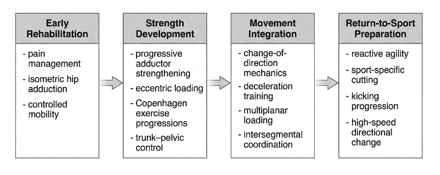
Rehabilitation progresses from early pain management and low-load strengthening toward progressive adductor strengthening, movement integration, and sport-specific exposure prior to unrestricted return to sport.
The progressive rehabilitation model illustrated in Figure 3 provides a clinically useful framework for structuring intervention across four broad phases. In the early phase, isometric hip adduction serves as the primary exercise modality, offering adductor activation with minimal joint loading, a strategy supported by evidence for pain modulation in tendinopathic presentations. Pain-free isometric loading during the initial days to weeks following injury facilitates neuromuscular re-engagement and provides athletes with a manageable entry point into structured rehabilitation. The second phase introduces progressive concentric and eccentric loading through resisted hip adduction variations and short-lever Copenhagen adduction progressions, with load and range of motion advanced systematically as pain tolerance improves. The third phase targets intersegmental movement control through lumbopelvic stability exercises, trunk-hip coordination tasks, and movement pattern retraining, reflecting evidence that disrupted co-activation between the adductor complex and lumbopelvic stabilizers persists beyond pain resolution and should be specifically addressed during rehabilitation.58 The fourth and final phase integrates progressive sport-specific loading through change-of-direction drills, deceleration training, and reactive cutting tasks performed at increasing speeds and levels of complexity. Successful progression to sport-specific loading depends on the restoration of adequate intersegmental control, as unresolved deficits in trunk, pelvic, and hip coordination contribute to persistent symptoms, delayed return to sport, and increased reinjury risk.
Biomechanical analyses of athletes with long-standing ARAGP have identified distinct movement strategy patterns during dynamic tasks such as cutting.48 Although these movement classifications did not correlate directly with localized pain findings, rehabilitation programs targeting intersegmental control and strength have been associated with successful return-to-sport outcomes. Following rehabilitation, athletes demonstrated movement adaptations consistent with a more mechanically efficient cutting strategy, including reduced ipsilateral trunk side flexion over the stance limb, increased pelvic rotation toward the direction of travel, and greater translation of the center of mass relative to the center of pressure. These changes were accompanied by alterations in lower extremity mechanics, such as reduced knee flexion during stance and increased ankle plantarflexor moments and power, suggesting improved force transmission and propulsion during directional changes. In that cohort, 73% of participants returned to sport pain-free at a mean of 9.9 weeks, alongside improvements in patient-reported outcomes and cutting biomechanics. Collectively, contemporary rehabilitation approaches emphasize progressive strengthening, restoration of intersegmental control, and the development of movement strategies that support efficient sport specific loading.
RETURN TO SPORT
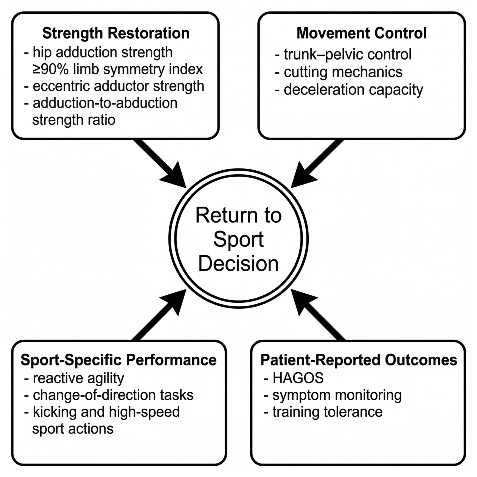
Return to sport following ARAGP is a multifactorial process that should incorporate strength, agility, and sport-specific performance testing to ensure safe progression (Figure 4).5,17 Objective assessment of isolated hip adductor strength is commonly included to quantify force-generating capacity and identify residual deficits.17
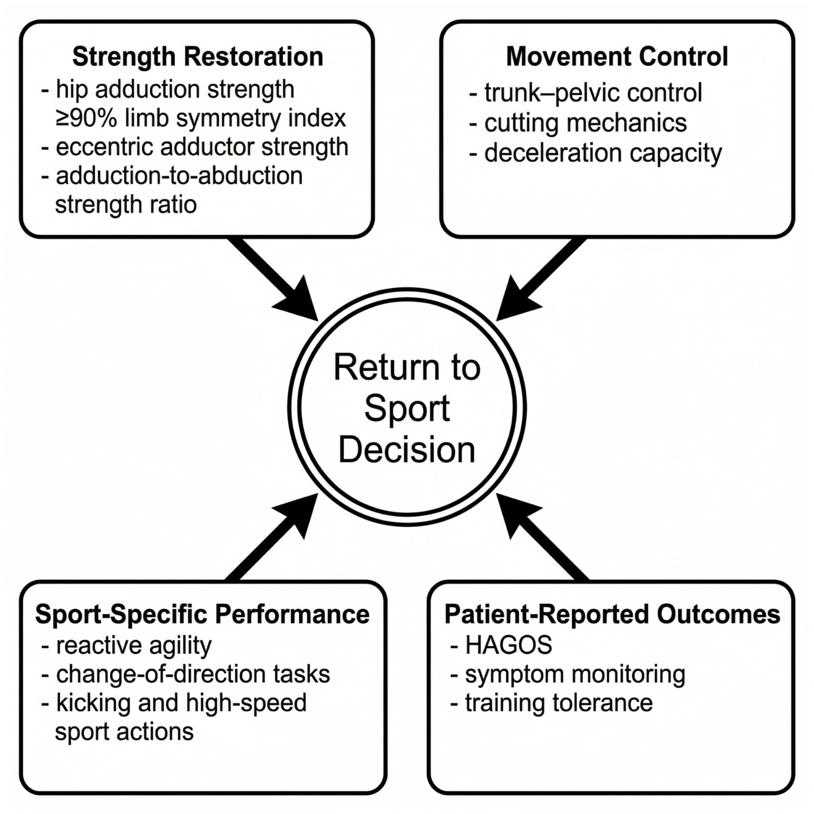
Hip adduction strength within approximately 10% of the contralateral limb (limb symmetry index ≥ 90%) is often used as a clinical benchmark prior to unrestricted return to sport.72 In elite soccer players utilizing criteria-based rehabilitation, eccentric hip adduction strength limb symmetry approaching 95-100% has been reported at discharge, coinciding with successful return to sport. Evaluation of the hip abduction-to-adduction strength ratio may provide additional context, as ratios below 80% have been associated with increased groin injury risk in prospective and cohort studies of field sport athletes.54,73
Side-to-side strength asymmetries, particularly in the dominant limb, have been described in relation to groin injury history.23,54 While these measures should not be interpreted in isolation, they may assist clinicians in identifying persistent deficits prior to return to full participation. The adductor squeeze test has demonstrated validity and reliability in athletes with groin pain and may serve as a useful adjunct measure during return-to-sport decision-making.34,74,75
Recent data demonstrate strong week-to-week reliability of long-lever hip adduction squeeze testing in elite youth athletes (ICC >0.90), with a minimal detectable change of approximately 20–24%, supporting its utility in serial monitoring frameworks.76 Strong test-retest reliability is clinically meaningful because it confirms that score changes reflect true physiological change rather than measurement noise, which is essential for using any monitoring tool to guide clinical decisions. The minimal detectable change of 20–24% establishes the threshold above which a strength change can be confidently attributed to real recovery rather than test variability. This means that a 10–15% improvement in squeeze test score between sessions should not be interpreted as meaningful progress. Ongoing monitoring with a reliable adductor squeeze test allows clinicians to track genuine recovery trajectories, avoid premature progression based on measurement noise, and communicate objective benchmarks clearly to athletes and coaching staff.
Unilateral assessment may provide greater sensitivity to side-specific deficits compared to bilateral testing.17 Comprehensive return-to-sport assessment should include evaluation of strength across multiple planes of motion, as well as functional testing reflective of sport demands. In addition to hip adduction, clinicians should evaluate hip abduction to contextualize the adduction-to-abduction ratio, hip flexion to assess iliopsoas capacity relevant to kicking athletes and to differentiate from iliopsoas-related pain, hip extension to evaluate gluteal and adductor magnus contribution to propulsion and deceleration, and hip external and internal rotation particularly in athletes with rotational sport demands. Normative hip strength values across competitive levels in field sport athletes provide a reference framework for interpreting these measures, though sport- and position-specific benchmarks remain limited in the current literature.77
These measures should be interpreted within a broader performance framework that includes multiplanar strength, reactive agility, and sport specific tolerance to high-speed directional change. Comprehensive return to sport assessment should therefore extend beyond isolated strength metrics to include evaluation of movement efficiency, deceleration capacity, and progressive exposure to sport specific loading demands. Incorporating patient-reported outcome measures such as the Hip and Groin Outcome Score (HAGOS) may further support clinical decision-making and progression toward unrestricted participation. Expert consensus further reinforces a multidimensional approach to return-to-sport decision-making. A three-round Delphi study involving 32 sports medicine experts reached consensus that return-to-sport criteria should include strength assessment, performance on sport-specific functional tests, and demonstrated capacity for sport-specific change-of-direction tasks.78 Notably, experts reached strong negative consensus that imaging findings should not serve as a return-to-sport criterion, directly challenging imaging-based clearance practices that persist in some clinical settings. Performance tests analysis was endorsed by 93.7% of experts as a component of the return-to-sport process, with planned and unplanned COD tasks at varying angles (45°–180°) achieving 96.9% consensus as appropriate performance criteria. Additionally, emerging evidence suggests that peak isometric adductor force alone may be insufficient as a return-to-sport criterion. Deceleration capacity during change-of-direction tasks correlates more strongly with adductor rate of torque development than with peak torque, suggesting that the speed of force production, rather than maximal strength, may be the adductor quality most directly relevant to the cutting and deceleration demands of multidirectional sport.79 Collectively, these findings support a return-to-sport framework that extends beyond isolated strength measures and incorporates movement quality, sport-specific performance, and athlete-reported readiness.
CONCLUSION
ARAGP is a common and multifactorial condition affecting athletes participating in multidirectional sports. Accurate diagnosis supported by an understanding of regional anatomy, recognized risk factors, and appropriate use of imaging is essential for effective management. Current evidence supports the role of progressive strengthening, neuromuscular training, and structured prevention programs in reducing injury incidence and facilitating return to sport.
Although movement assessment and strength profiling may assist in identifying deficits and guiding rehabilitation, the predictive value of specific screening tools remains uncertain. Collectively, current evidence suggests that eccentric hip adduction capacity and intersegmental trunk-pelvic control represent modifiable targets within both prevention and rehabilitation frameworks. Continued research is needed to further clarify mechanisms of injury and optimize evidence-informed prevention and return-to-sport strategies. Overall, successful management of ARAGP requires an individualized, comprehensive approach that integrates strength restoration, functional progression, and sport-specific demands to support safe and durable return to participation. Future research should continue to examine sport-specific biomechanical demands and longitudinal rehabilitation outcomes to better inform evidence-based return-to-sport decision making.