Introduction
Diagnostic musculoskeletal ultrasound (MSKUS) has become an essential tool in the evaluation of musculoskeletal structures. Glenoid labral tears are common sports and orthopedic settings. Taljanovic and colleagues performed MSKUS on cadaveric specimens and then followed with arthroscopy.1 They found concordance between sonography and arthroscopy to be 86%. In the ability to differentiate abnormal labral tissue from normal, they found sensitivity of 63%, specificity of 98%, positive predictive values or 94%, negative predictive value of 86% and accuracy of 88%. This paper aims to provide a comprehensive review of diagnostic MSKUS of the glenoid labrum, including relevant anatomy, scanning technique, normal and abnormal imaging characteristics and common pathological findings.
Anatomy of the Glenoid Labrum
The glenoid labrum is a fibrocartilage ring that surrounds the outer periphery of the glenoid. The labrum deepens and enlarges the joint cavity by creating increased area of contact between the tissue and the humeral head, which in turn helps increase shoulder stability. Labral tissue is composed mainly of fibrocartilage tissue with various collagen fingers arranged in a circular fashion. In addition to collagen there appear to be some elastin fibers within the labrum which allow some degree of mobility and deformation of the tissue.2 The labrum itself is not uniform around the circumference of the glenoid. The superior labral tissue merges with the long head of the biceps to attach via a cojoined tendon onto the supraglenoid tubercle. The first of two sublabral recesses is seen beneath the superior labrum,3 while the second is located slightly more anterior. When there is a complete loss of continuity of the labral tissue and the glenoid it is considered a sub-labral foramen. A final variant, termed the Buford complex, is when there is a lack of anterosuperior labrum combined with a cordlike thickening of the medial glenohumeral ligament. All of these are normal variants that present as a gap between the labrum and the glenoid, and have in the past been confused with tears. The superior attachment of the labrum is much more loose compared to the inferior counterpart. The labrum continues anteriorly and inferiorly and attached deep to the glenohumeral ligaments. Finally, the inferior and posterior labrum is somewhat thinner than the superior and anterior labrum and is not reinformed by ligament thickenings like the superior and anterior portions.4 The labral tissue is formed from a continuation of the joint cartilage of the glenoid inferiorly with a transitional fibrocartilage zone, giving it significantly less mobility in this region.
The Role of MSK Ultrasound in Evaluation of the Glenoid Labrum
Advantages
-
Real-Time Imaging: MSKUS allows dynamic evaluation of labrum while the shoulder can be moved through available range of motion. It provides real-time assessments of the soft tissue, their motion and provides information beyond static morphology.
-
High-Resolution Visualization: Provides detailed images of the labrum and its attachment to the glenoid fossa.
-
Accessibility and Cost-Effectiveness: Magnetic resonance imaging (MRI) is the gold standard reference for imaging musculoskeletal soft tissues, such as the labrum.3,5,6 However, it is extremely expensive, time consuming, and may be contraindicated in certain patients. MRI without contrast may also have limitations in detecting smaller tears or partial labral tears. MSKUS is portable, widely available, and less expensive than MRI.
Limitations
-
Operator Dependency:MSKUS requires skill and experience for accurate interpretation of findings. The ability to sonograph labral tissue and surrounding structures is to a large extent influenced by the operator and the availability and technical considerations of state-of-the-art equipment, and the position of the shoulder during the assessment.
-
Depth Limitations: Visualization is usually not a problem as the labrum is within the appropriate depth limitations of MSKUS.
-
Artifacts and Shadows: Bone shadowing from the glenoid may create image artifacts, requiring adjustments in probe positioning and frequency.
Sonographic Technique for Evaluating the Labrum
Equipment Setup
-
Probe Type: The depth of the labrum allows for a standard high-frequency 3-13 MHz, linear array probe; however, deeper depths may require a curvilinear 1-6 MHz probe.
-
Patient Position: The patient is seated with the shoulder in neutral.
-
Dynamic Assessment: A passive or active movement of shoulder can create dynamic movements of the humerus that allows stress to be applied to labral tissue, helping to accentuate issues seen during ultrasonography.
Examination Protocol
Normal Sonographic Appearance of the Glenoid Labrum
In a normal healthy glenoid labrum, the tissue appears as a thin, triangular shaped, fibrocartilaginous ring that is continuous and smooth, with well-defined margins and uniform homogeneous echogenicity throughout, that is similar to surrounding soft tissues with no focal hyper or hypo echogenicity.7 The tissue should be smooth and free of any irregularities, clefts, or fraying. Acoustic shadowing is rare and only occurs if there is a bony spur or calcification of some type in the shoulder. Like many locations in which MSKUS is used, various views may require some toggling, or heel toe movements to reduce anisotropy.
One way to view the labrum is to visualize it similar to a clock-face, dividing the labrum into quadrants for assessment. The best locations for viewing include:
Superior Labrum: Due to the bony acromion, visualization is less optimal here. The superior labrum will be partially visible and appears as a thin, continuous band of labral tissue with smooth margins. To see this portion of the labrum the patient will be seated, arm in neutral or externally rotated.
Anterior Labrum: The anterior labrum is easier to see at the edge of the glenoid directly under the subscapularis tendon. It’s slightly thicker than the superior labrum, but still smooth and continuous band of labral tissue.
Inferior Labrum: The position to view the inferior labrum is from an axillary approach. The normal tissue should appear as a smooth, continuous band of labral tissue. To view the inferior labrum the patient should be supine, in maximal abduction and external shoulder rotation.
Posterior Labrum: The posterior labrum is best seen from a posterior approach while also evaluating the infraspinatus and teres minor. The labral tissue should be thinner again, but still smooth, continuous labral tissue. To view the posterior labrum the patient will be seated.
Pathologic Findings for the Glenoid Labrum
-
Irregular margins, clefts, or fraying.
-
Hyperechogenic foci, which would include possible calcifications, or fluid or degeneration of labral tissue.
-
Displacement may be seen during dynamic movement of the humerus on the glenoid.
Clinical Implications for Rehabilitation Providers
MSKUS provides real-time feedback for rehabilitation professionals, facilitating early diagnosis and intervention. Key applications include:
-
Early Detection of Injury / Accurate Injury Grading: MSKUS can differentiate normal labral tissue from pathologic. It should be remembered that slight fraying or even some slight superior labral disruption from the glenoid may be very common and non-pathologic in overhead athletes.
-
Dynamic Functional Testing: Rehabilitation professionals can use MSKUS during physical therapy sessions to monitor recovery and assess labral tissue function dynamically. Serial MSKUS imaging aids in assessing labral healing and remodeling to help with readiness for rehabilitation progression.
-
Guided Interventions: Ultrasound imaging assists in precision-guided procedures such as dry needling or injections.
-
Patient Education: Real-time imaging serves as a visual aid to explain the nature of the injury and set realistic expectations for recovery.
Limitations and Challenges
Despite its advantages, MSKUS cannot entirely replace MRI with or without intraarticular contrast for complex cases of labral tears.8 Additionally, the expertise required for optimal imaging technique limits its immediate adoption across all rehabilitation settings.
Conclusion
In summary, diagnostic MSKUS offers rehabilitation professionals a powerful, clinically relevant extension of the physical examination when evaluating the glenoid labrum and labral tissue. Its ability to provide high-resolution, real-time, and dynamic visualization of the labral tissue architecture enhances diagnostic precision, supports early clinical decision-making, and allows serial monitoring of tissue healing and load tolerance. When integrated with a thorough understanding of anatomy, injury mechanisms, and functional biomechanics, MSKUS becomes more than an imaging modality—it becomes a performance-informed clinical tool. While operator skill and certain anatomic limitations must be acknowledged, the thoughtful incorporation of MSKUS into sports and orthopedic practice has the potential to elevate assessment accuracy, refine rehabilitation progression, and ultimately improve return-to-play outcomes for athletes with shoulder injuries and in particular glenoid labral injuries.
Patient Positioning for Viewing Glenoid Labrum
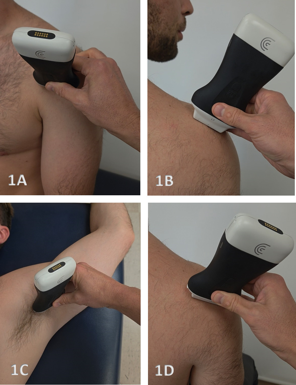
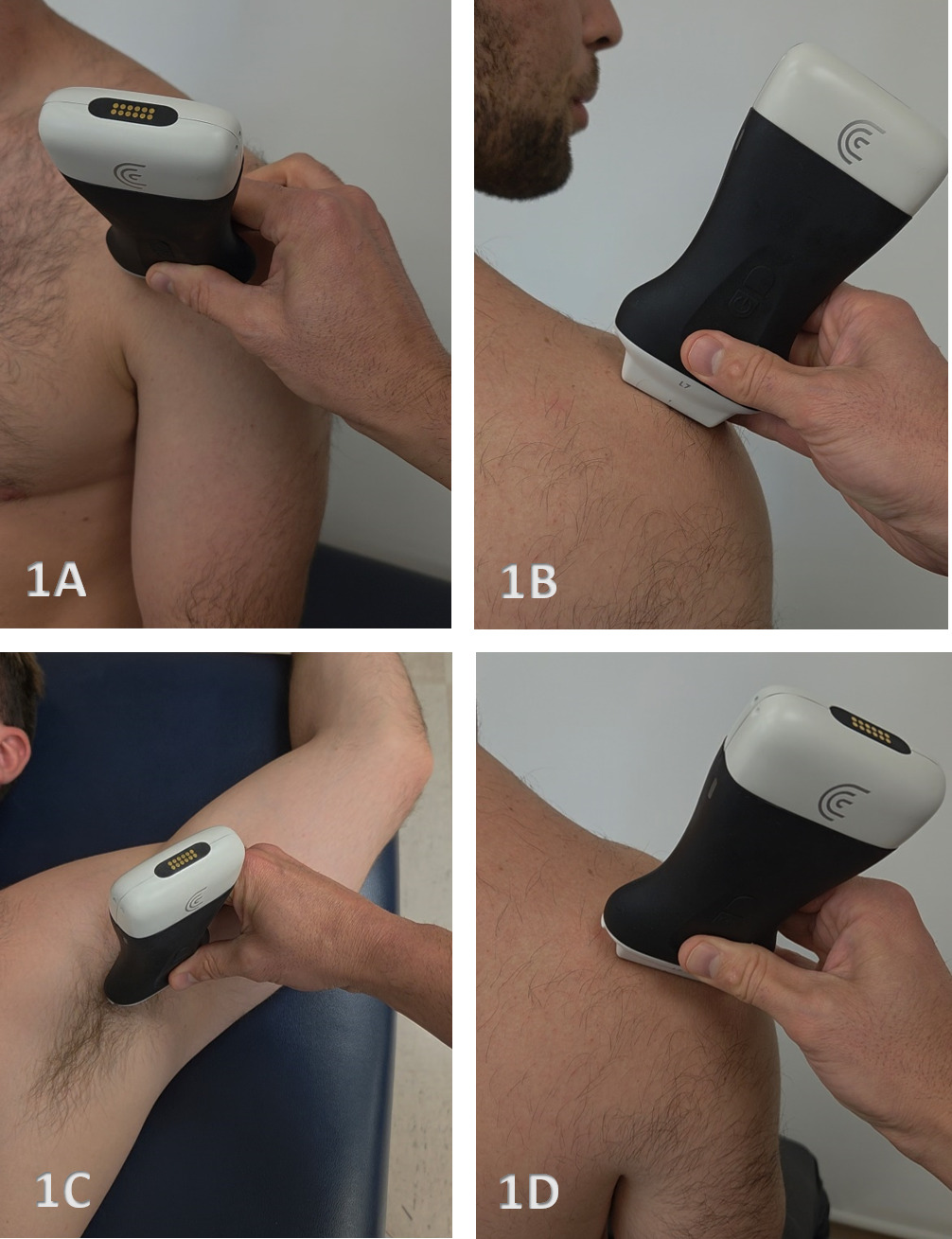
Evaluation of the glenoid labrum requires the patient to transition between seated and supine positions to optimize visualization of the different labral regions. The posterior and superior labrum are primarily assessed with the patient seated, while the anterior and inferior labrum may require transition to a supine position to stabilize the scapula and provide improved access to these regions. The arm position is adjusted throughout the examination to facilitate both static and dynamic assessment of the labrum.
Figure 1A: Transducer Placement for the Anterior Labrum: 1–4 o’clock
The following clock faces refer to the position of the left side of the transducer. The anterior labrum includes the anterosuperior 1–2 o’clock region and the anterior 3–4 o’clock region. The patient may be examined seated; however, a supine position can improve visualization of the 3–4 o’clock region by stabilizing the scapula against the examination table. The arm is positioned in slight external rotation, and the transducer is aligned with the anterior glenoid rim. Trapezoidal imaging, positioning the labrum near the edge of the field of view, and gentle pressure along the medial edge of the transducer may improve visualization. Dynamic internal and external rotation is particularly important because an unstable anterior labrum may demonstrate medial displacement during internal rotation and reduction toward the glenoid rim during external rotation.
Figure 1B: Transducer Placement for the Superior Labrum: 11–12 o’clock
The superior labrum is evaluated with the patient seated. At the 11 o’clock position, visualization may be limited by acoustic shadowing from the posterior acromial angle. At 12 o’clock, the transducer is positioned over the supraspinatus fossa within the soft tissue window medial to the acromion, anterior to the scapular spine, and posterior to the clavicle. Trapezoidal imaging or a convex transducer may improve visualization by allowing obliquely directed ultrasound beams to pass beneath the acromion. The superior labrum and biceps–labral complex should be evaluated dynamically with shoulder rotation, abduction, and adduction.
Figure 1C: Transducer Placement for the Inferior Labrum: 5–7 o’clock
The inferior labrum is evaluated with the patient supine and the shoulder placed in maximal abduction and external rotation with the arm elevated overhead. The 6 o’clock position serves as the primary starting point. From this position, the transducer is rotated approximately 30° anteriorly to visualize the 5 o’clock region and approximately 30° posteriorly to visualize the 7 o’clock region. The inferior labrum is generally well visualized from this approach, with the humeral head, glenoid rim, labrum, and overlying joint capsule serving as the primary sonographic landmarks.
Figure 1D: Transducer Placement for the Posterior Labrum: 8–10 o’clock
The posterior labrum is evaluated with the patient seated and the examiner positioned behind the patient. The transducer is placed horizontally just inferior to the scapular spine and aligned with the long axis of the scapula. The 9 o’clock position generally provides the easiest and most reliable starting point. From this position, the transducer can be shifted slightly inferiorly and rotated approximately 30° caudally to visualize the 8 o’clock region or shifted superiorly and rotated approximately 30° cranially to visualize the 10 o’clock region. Internal and external rotation of the shoulder should be incorporated dynamically to assess labral contour, attachment, and stability.
NORMAL GLENOID LABRUM
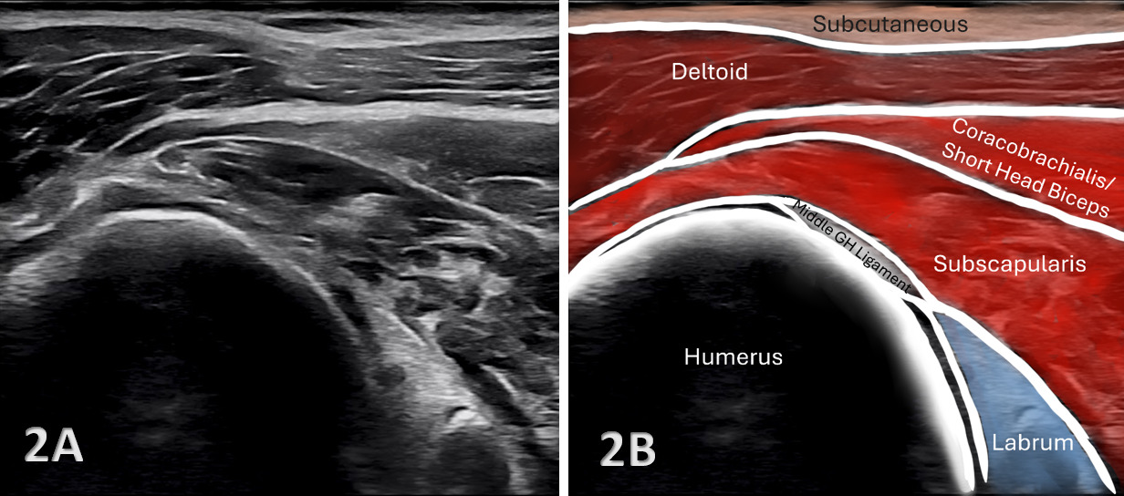
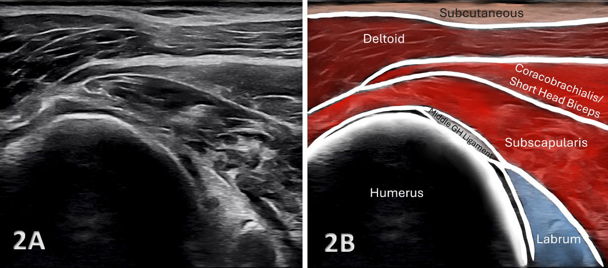
Figures 2A and 2B: Anterior Labrum: 1–4 o’clock (Figure 1A)
In the anterior view, the cortical margin of the glenoid appears as a bright hyperechoic interface, with the anterior labrum visualized superficially as a triangular echogenic structure extending from the glenoid rim. The humeral head lies lateral to the glenoid and provides an additional osseous landmark. Care should be taken when defining the anterior labral margins because the overlying glenohumeral ligaments, particularly the middle glenohumeral ligament, may contribute to the sonographic appearance of the anterior capsulolabral complex. Dynamic internal and external rotation is particularly important in this region. A normal anterior labrum should maintain its attachment and relationship to the glenoid throughout rotation without abnormal medial displacement, deformation, or development of a hypoechoic cleft.
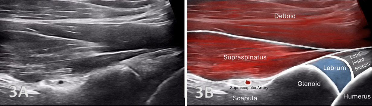
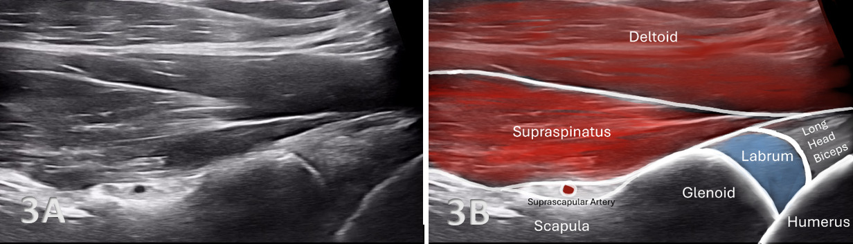
Figures 3A and 3B: Superior Labrum: 11–12 o’clock (Figure 1B)
The superior labrum is identified as an echogenic triangular structure extending from the superior glenoid rim. The long head of the biceps tendon may also be visualized as it approaches the superior labrum and forms the biceps–labral complex. External rotation may improve visualization of the biceps anchor. The superior labrum should demonstrate a smooth contour and remain appropriately positioned against the glenoid margin during dynamic rotation, abduction, and adduction. Care should be taken not to mistake a normal sublabral recess for labral pathology. When acoustic shadowing limits visualization, trapezoidal imaging or a convex transducer may improve assessment by allowing the ultrasound beam to pass obliquely beneath the acromion.
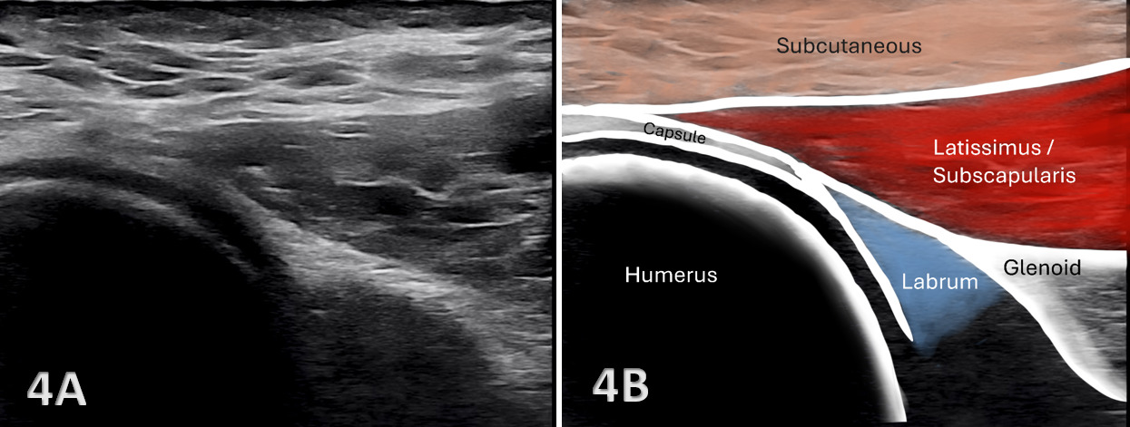
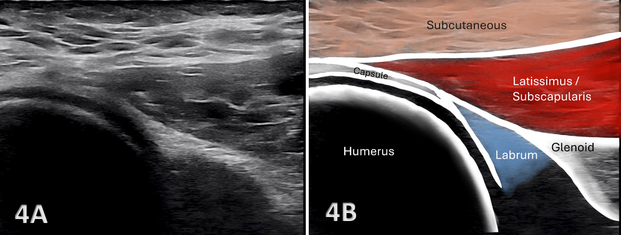
Figures 4A and 4B: Inferior Labrum: 5–7 o’clock (Figure 1C)
In the inferior view, identify the humeral head on one side of the image and the hyperechoic cortical margin of the inferior glenoid on the other. The inferior labrum appears as a triangular echogenic structure extending from the glenoid rim. At the 6 o’clock position, a narrow portion of the scapular body may also be visualized and serves as a useful landmark confirming transducer orientation. The joint capsule is relatively thin in this region because it is not reinforced by the glenohumeral ligaments at the direct inferior position. Dynamic rotation may be incorporated to confirm stability of the labrum and evaluate for subtle contour abnormalities or displacement.
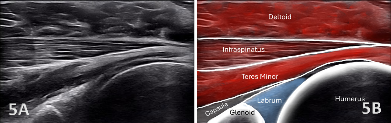
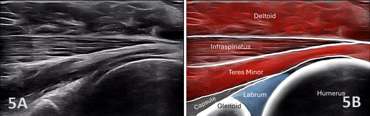
Figures 5A and 5B: Posterior Labrum: 8–10 o’clock (Figure 1D)
To visualize the posterior labrum, place a high-frequency linear transducer horizontally just inferior to the scapular spine and align the transducer along the long axis of the scapula over the posterior glenohumeral joint. The 9 o’clock position generally provides the most accessible acoustic window and serves as a useful starting point for examination of the posterior labrum. From this position, the transducer may be shifted slightly inferiorly and rotated approximately 30° caudally to evaluate the 8 o’clock region or shifted superiorly and rotated approximately 30° cranially to evaluate the 10 o’clock region. Maintain consistent transducer pressure and alignment with the scapular plane while dynamically rotating the shoulder.
Pathology of the Glenoid Labrum
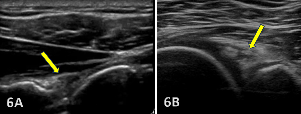
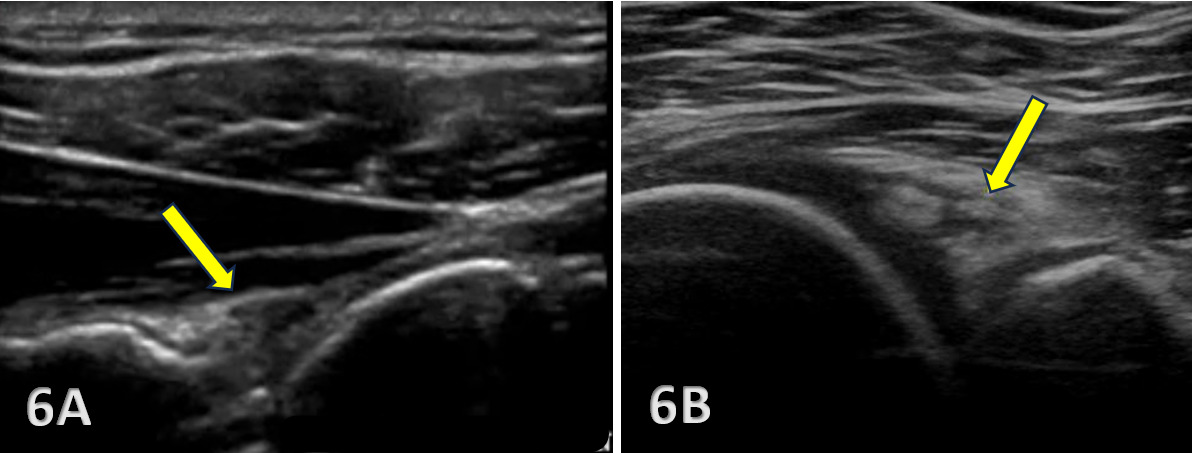
Figures 6A and 6B: Posterior Glenoid Labral Tears
Shown above in Figures 6A and 6B, evaluation of a posterior glenoid labral tear demonstrating irregularity and disruption of the normally triangular labral contour, focal hypoechoic cleft formation, and mild separation of the labrum from the posterior glenoid rim. Dynamic internal and external rotation is particularly important, as subtle labral displacement or abnormal mobility may become more apparent with movement. Comparison with the contralateral side may further assist in identifying abnormal labral morphology or motion.