INTRODUCTION
Lateral ankle sprains (LAS) are the most common orthopedic injury with an annual health care cost of $4.2 billion.1,2 Many who suffer an ankle sprain do not regard it as a substantial injury and avoid seeking formal medical care.3 This may account for the re-injury rate of ankle sprain being as high as 73%, with a history of previous LAS being the most important risk factor for incurring another sprain injury.4 Consequently, after an initial ankle sprain, 40% of individuals eventually develop CAI.5 CAI is characterized as a feeling of instability accompanied by repetitive bouts of giving way with self-reported decrease in function and recurrent ankle sprains. Commonly observed deficits in individuals with CAI include decreased range of motion, poor postural control, reduced strength, and dysfunctional neuromuscular control.5–8 LAS often result from an inversion and plantar flexion mechanism.1,2 The peroneal muscles constitute the primary muscular defense against the inversion movement and provide antagonizing forces to the sudden inversion thrust.9 Weakness of the peroneal muscles is considered a risk factor for ankle sprains,10 and pain and tenderness in the peroneus longus muscle are frequently reported after LAS.
Peroneal dysfunction is associated with impairment in postural control in subjects with CAI.11 Decreased reaction times have been documented in the peroneal muscle group in the CAI population.12 It is believed that impairment in peroneal muscles may also result indirectly from arthrogenic muscle inhibition.13 Simon and Docherty9 found that individuals with CAI had significantly slower nerve conduction velocity (NCV) in the superficial peroneal nerve. The slowness in NCV may contribute to chronic symptoms of giving way and instability in CAI. Peroneal strength deficits in the CAI population have been reported previously,14 with peroneal weakness of approximately 39% compared with the contralateral healthy side.14 Eversion strength deficits of 0.46 N-m/kg compared to a healthy group have been similarly reported, without concomitant differences in EMG-based muscle activation. These conflicting EMG findings are attributable to inherent limitations of EMG including cross talk from surrounding muscles, electromechanical delays, and discomfort associated with fine-wire EMG.15,16
Ultrasound (US) imaging provides an opportunity to investigate multiple facets of muscle function including muscle size and quality.17,18 As a non-invasive technique, US imaging offers enhanced visualization compared to EMG measurements and allows assessment of muscle size through CSA and muscle quality through echogenicity analysis.18,19 Muscle size and tissue quality are both considered independent measures of muscle strength and function.20 Lobo et al.21 found significant differences in peroneal muscle CSA between LAS and healthy groups. While deficits in peroneal strength and CSA in patients with CAI compared with healthy controls are established,21 no study has analyzed the change in peroneal muscle size and quality before and after an impairment-based rehabilitation program using US imaging. Therefore, the objective of this study was to determine peroneal muscle size and quality changes using US imaging following impairment-based rehabilitation in patients with CAI. It was hypothesized that there would be an increase in size and improvement in quality of the peroneal muscles in patients with CAI following rehabilitation.
METHODS
Study Design
This prospective cohort study was embedded within a larger randomized controlled trial conducted in a university setting, designed to compare clinical and patient-reported outcomes between groups receiving gait training and those not receiving gait training.22 This study represents a pre-post analysis focused specifically on evaluating peroneal muscle quality and size, with data pooled across all participants. This approach was taken because the larger randomized trial found no significant differences in peroneal muscle size or quality between the gait training and non-gait training groups. Data collection occurred at two time points: baseline and after completion of a 4-week, 8-session impairment-based rehabilitation program. All participants underwent the impairment-based rehabilitation protocol regardless of their gait training group assignment. Institutional Review Board approval was obtained prior to study initiation, and informed consent was secured from all participants. Ultrasound imaging was performed on the peroneal muscle groups.
Participants
Twenty-six participants (age: 21.9 ± 3.5 years; 18 females, 8 males) with a history of bilateral CAI were enrolled and completed 8 sessions of rehabilitation. The participant-selected worst leg was used for intervention. Inclusion criteria were based on the recommendations of the International Ankle Consortium. Participants with CAI had a history of at least one significant ankle sprain at least one year prior to study enrollment, a decrease in self-reported function (Foot and Ankle Ability Measure [FAAM] Sport score ≤85%), and experience of repetitive bouts of instability or “giving way” with a score on the Identification of Functional Ankle Instability (IdFAI) >10.
Exclusion criteria included any history of lower extremity fracture or surgery, ankle sprain within the prior six weeks, conditions known to affect gait, pregnancy, and currently receiving physical therapy. For the larger randomized controlled trial, participants were randomly divided into a gait training group (n=13) and a non-gait training group (n=13). In the present study, initial analyses were performed to examine differences in CSA and muscle quality between groups; if no differences were found, groups were pooled and pre-to-post changes were examined.
Instruments
US imaging was performed using a Siemens Acuson Freestyle US system with a wireless 8-MHz linear transducer (Siemens, Mountain View, CA). Images were measured using ImageJ version 1.50f (National Institutes of Health, Bethesda, MD).
Ultrasound Measurements
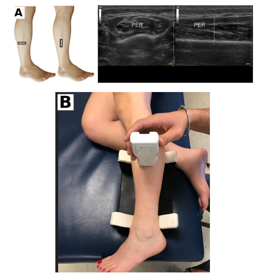
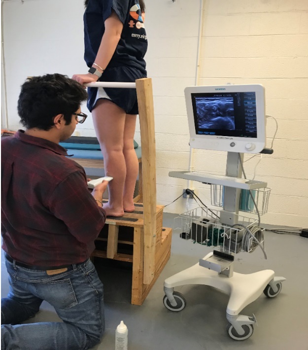
A customized step (Figure 2) was constructed to allow participants to stand in bipedal stance during imaging. US imaging was performed first in the sidelying position and then in bipedal stance. All imaging was performed at a depth of 3.5 cm, with a gain setting of 11 and a dynamic range of 70 dB.23–26 These parameters were selected to optimize visualization of superficial muscles like the peroneals.23–26 Ultrasound settings, including gain and depth, were standardized and consistently applied across all participants. The testing order for body positions was randomized using a computer-generated sequence to mitigate potential order effects. Probe placement followed previously validated methods,24,26 with the transducer positioned at 50% of the fibular length (Figure 1). This midpoint was determined by measuring the distance from the fibular head to the lateral malleolus using a tape measure and marking the halfway point.
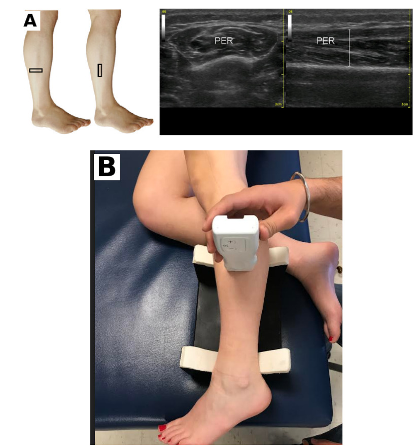
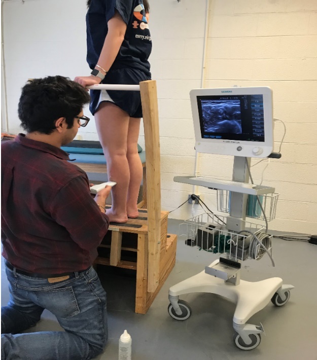
All images were collected at baseline and upon completion of the 4-week, 8-session rehabilitation protocol. Ultrasound gel was applied over the probe and on the imaging site before each image acquisition. Peroneus longus and brevis were imaged together in both sidelying and bipedal stance.
Impairment-Based Rehabilitation Protocol
Participants underwent a 4-week rehabilitation program targeting individual impairments through eight supervised sessions. Although all participants followed the same standardized framework, exercise selection, dosage, and progression were individualized to each participant’s presenting impairments and rate of progression. Rehabilitation was delivered by a physical therapist with five years of clinical experience and an athletic trainer with two years of experience. Comparable rehabilitation protocols have been implemented in previous studies.27,28 The supervised program addressed deficits in range of motion, balance, strength, and functional performance, and included targeted strengthening exercises for the peroneal muscle group including four-way ankle resistance exercises. Detailed information about the impairment-based rehabilitation protocol is available in the supplementary Appendix.
Range of motion was assessed through a comprehensive clinical evaluation conducted by the supervising clinician during the initial session. When restrictions were detected, joint mobilizations graded 2 to 3 on the Maitland scale were applied. Stretching exercises targeting the foot, ankle, and hip musculature were also incorporated.29 Ankle-focused exercises included heel and forefoot raises, four-directional manual resistance movements with progression, D1/D2 proprioceptive neuromuscular facilitation (PNF) patterns, and heel-to-toe walking drills. Hip strengthening included clamshell exercises using resistance bands and bird-dog movements. Balance training included reaching movements, single-leg stance, and hop-to-stabilization drills. Functional components involved lunges, step-ups and step-downs from a 30-cm platform, and jumping exercises.
Participants were also given a home exercise program (HEP) for non-supervised days (Appendix). The HEP incorporated single-leg balance training, calf stretches, and four-directional ankle exercises using a resistance band. Compliance was calculated as the percentage of completed HEP days relative to total days between supervised sessions. Rehabilitation was administered only to the participant-identified most-affected limb.
Data Processing
Muscle CSA of the peroneal muscle group was measured in square centimeters (cm2) within the fascial boundaries using ImageJ software (National Institutes of Health, Bethesda, MD). All ultrasound images were anonymized and analyzed by an investigator blinded to participant group assignment (pre vs. post) and testing position until all measurements were completed. A single examiner with five years of experience in ultrasound imaging conducted all analyses. Muscle echogenicity was quantified using grayscale analysis in ImageJ, applied to the same region of interest (ROI) used for CSA measurements. Echogenicity was assessed on a scale from 0 to 255 (black to white), with higher values indicating reduced muscle quality. For muscle quality measurements, only the bipedal stance position was utilized. Muscle size and quality were assessed using three images per position, and average values were calculated for each parameter.
Statistical Analysis
Statistical analysis was conducted using IBM SPSS Statistics (v26.0, SPSS Inc., Chicago, IL). Normality of dependent variables was assessed via skewness, kurtosis, and Levene’s test. Independent t-tests were used to examine differences in CSA and muscle quality between the gait training (n=13) and non-gait training groups (n=13). If no differences were found, groups were pooled (n=26) and pre-to-post rehabilitation changes were examined. Paired t-tests were used to assess differences in non-weight-bearing and weight-bearing positions for peroneal muscle group CSA (cm2) in both trained and untrained legs, and to examine post-rehabilitation changes in echogenicity. Mean differences with 95% confidence intervals were calculated, and Cohen’s d effect sizes were used to determine the magnitude of pre-to-post differences.
RESULTS
Dependent variables were normally distributed based on skewness, kurtosis, and Levene’s test (p > 0.05). Demographic information for the 26 participants with CAI (age: 21.88 ± 3.45 years; height: 171.11 ± 10.25 cm; mass: 71.16 ± 14.24 kg; 18 females, 8 males), including subjective outcomes following rehabilitation, is provided in Table 1. No significant differences (p > 0.05) in mean change scores of CSA or muscle quality were observed between the gait training and non-gait training groups; therefore, groups were pooled for further analysis.
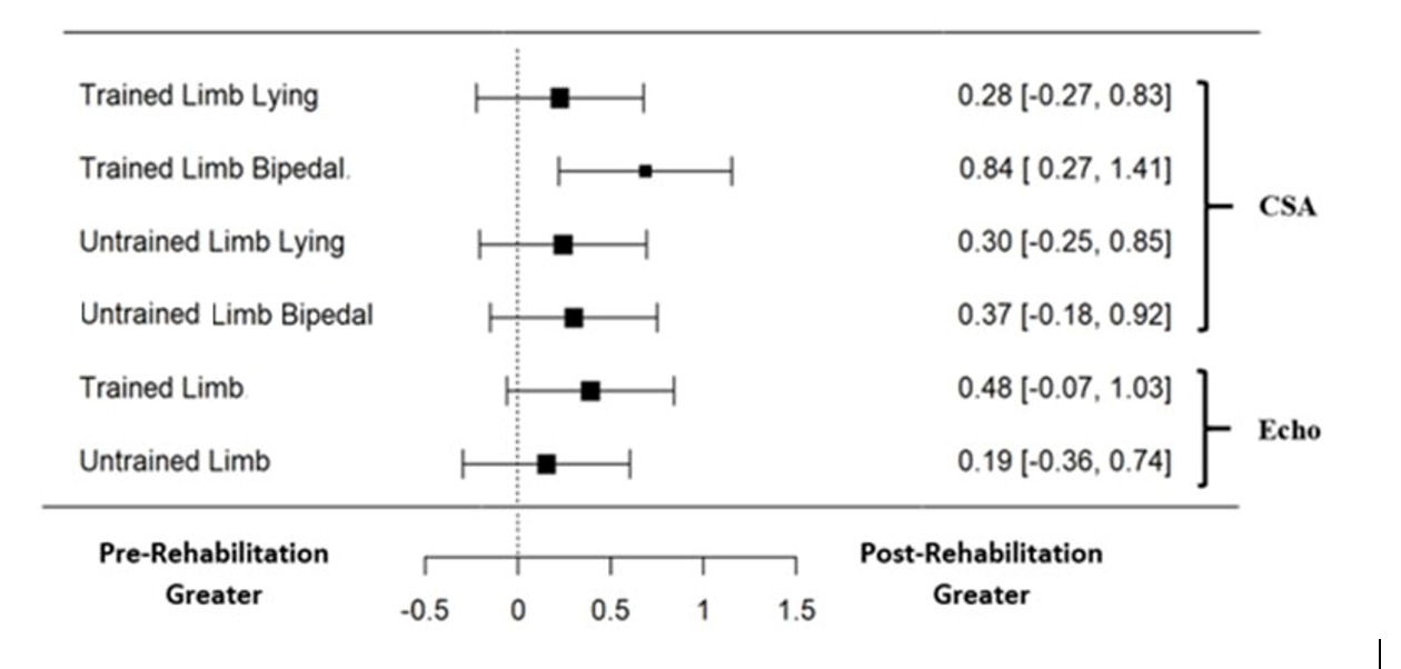
Significant gains (p < 0.01) in CSA were observed in both sidelying and bipedal stance positions in the trained leg after the 4-week rehabilitation program (Table 2). Significant improvements were also found in the untrained leg in both non-weight-bearing (p = 0.01) and weight-bearing (p < 0.01) positions. A significant decrease (p = 0.01) in echogenicity, indicating improved muscle quality, was found in the trained leg; however, no significant change (p = 0.15) was detected in the untrained leg. Moderate to large effect sizes (Cohen’s d = 0.37 to 0.84) were observed for the weight-bearing position compared to the non-weight-bearing position, which showed smaller effect sizes (Cohen’s d = 0.28 to 0.30) (Figure 3). Overall HEP compliance was 80.2%.
_with_95__confidence_intervals_for_pre-to-post_rehabilitation_chan.png)
DISCUSSION
Supporting the study hypothesis, significant increases in CSA of the peroneal muscle group were found after impairment-based rehabilitation in both non-weight-bearing (sidelying) and weight-bearing (bipedal stance) positions in the trained limb. Significant improvement in muscle quality was also demonstrated, reflected by a significant decrease in echogenicity measures in the trained limb. Significant improvements in the untrained leg in CSA were observed in both lying and bipedal stance positions; however, effect sizes were lower than those of the trained limbs (Figure 3). No significant differences in echogenicity were found in the untrained limb.
To the authors’ knowledge, this is the first study to use US imaging to characterize changes in size and muscle quality of the peroneal muscle group after 4 weeks of impairment-based rehabilitation in patients with CAI. Multiple reports document decreased strength, lower activation, and smaller CSA of peroneal muscles in patients with CAI.9,11,14,21 The present study adds valuable insight by characterizing the effects of rehabilitation on peroneal muscle CSA and quality. The mean ± SD CSA at baseline was 3.44 ± 0.99 cm2, similar to that reported by Angin et al.26 in participants with pes planus (3.26 ± 0.80 cm2). A 9.67% increase in peroneal CSA (3.72 ± 0.98 cm2) was observed after rehabilitation. Peroneal muscles were also examined in weight-bearing bipedal stance, which showed greater baseline CSA (3.46 ± 1.05 cm2) and a greater post-rehabilitation change of 29% compared to the lying position. Moderate to large effect sizes were found in the weight-bearing position (Figure 3), indicating that functional positions may be superior for detecting rehabilitation-related changes in peroneal muscles. Based on these findings, weight-bearing functional positions are recommended for testing peroneal muscles in patients with CAI.
Significant gains in peroneal CSA are consistent with known mechanisms of exercise-induced muscle adaptation.30 Molecular changes begin within hours of exercise, and fiber hypertrophy has been documented within 4 weeks of training.31 Seynnes et al.31 demonstrated considerable hypertrophy (3.2–5.2%) in quadriceps muscles after only 20 days of training. The present study found a 9.67% increase in CSA after four weeks of rehabilitation in the non-weight-bearing position, and a 29% increase in the weight-bearing position. The greater post-rehabilitation activation in the weight-bearing position reflects neuromuscular gains. Hodges et al.32 demonstrated that US measures can detect muscle activity through architectural changes at lower intensity levels compared to MVC testing. The greater improvements in the weight-bearing position may reflect improved capacity of the peroneal muscles to control the foot and ankle in functional positions—a critical requirement for patients with CAI, in whom the peroneal muscle group serves as the primary antagonist to sudden inversion movements associated with recurrent instability.
In addition to muscle CSA, muscle tissue quality was assessed using grayscale echogenicity analysis. Significantly lower echogenicity scores post-rehabilitation indicate improved muscle quality.20 Differences in echogenicity between injured and healthy groups have been reported previously,20 and changes in echogenicity after short-term rehabilitation programs have also been documented. A decrease in echogenicity of the vastus lateralis was reported after 6 weeks of resistance training in older adults.33 The present findings extend this evidence by demonstrating that peroneal muscle quality can improve in patients with CAI after 4 weeks of impairment-based rehabilitation.
Beyond improvements in the trained limb, significant improvement in CSA was observed on the untrained side, with a 9% gain in non-weight-bearing and 11.7% gain in bipedal stance. The gain in the lying position was similar to the trained side; however, the gain in the functional position was only 40% of the gains seen on the trained side. Similar contralateral gains of approximately 40–50% have been reported previously.34 These cross-education effects may reflect parallel adaptations mediated by synchronization of motor patterns and spillover from cortical, subcortical, or spinal levels.34
Limitations
Although peroneal muscles were captured in the weight-bearing functional position, measurement was not performed during dynamic activities. Assessment of CSA changes and activation during tasks such as walking would provide a more functionally representative picture of peroneal muscle performance. Additionally, while 4 weeks of impairment-based rehabilitation demonstrated significant improvements in size, quality, and neuromuscular activation post-treatment, the long-term benefits of the rehabilitation program remain unknown. The present study also used a convenience sample of participants as part of a larger investigation. Future studies should follow up patients with CAI at six months and one year post-rehabilitation to evaluate the sustainability of these improvements and should develop techniques to assess peroneal muscles using US imaging in dynamic contexts such as walking. Furthermore, a minimal detectable change for peroneal CSA has not been established, and the functional significance of the observed CSA changes remains to be determined; future work should define minimal detectable change values and relate these morphological changes to functional outcomes.
CONCLUSION
An 8-session, 4-week impairment-based rehabilitation program produced significant increases in peroneal muscle CSA and improved muscle quality in patients with CAI. Greater effect sizes and percentage changes were observed in the weight-bearing functional position than in the non-weight-bearing position. Cross-education effects were observed in the untrained limb, which showed significant improvement in muscle size; however, no changes in muscle quality were detected in the untrained limb.
Declarations of interest
None.