INTRODUCTION
Calcific tendinopathy (CT) is a relatively common, but complex pathology.1 CT can affect any tendon in the body but is most commonly reported in the rotator cuff.1 Women aged 30-50 appear to be affected greater than men, and other factors such as overuse, hypothyroidism, hyperlipidemia, and diabetes are commonly associated with CT.1–3 The pathogenesis of CT is controversial.1,3,4 Current theories revolve around a failed tendon healing response, leading to the inappropriate differentiation of progenitor cells into osteoblasts rather than tenocytes.3,4 Calcium hydroxyapatite crystals are focally deposited in areas of collagen degeneration or tearing.3 A coexisting tendon tear is not uncommon in CT.1 This physiological process may be a compensatory mechanism to increase tendon stiffness in the degenerated or damaged portion of the tendon.4
There are three distinct phases of calcific tendinopathy as described by Uhthoff et al.5 The pre-calcific stage, the calcific stage, and the post-calcific/repair stage.5 Patients are typically most symptomatic during the resorptive portion of the calcific stage; however, a large number of patients with CT are asymptomatic.1,3 During resorption, the calcific deposits undergo an inflammatory reaction with neovascularization at the calcification periphery.1 Macrophages are recruited to phagocytose the calcific deposits which alters the morphology into a toothpaste consistency, which may leak into peripheral tissues causing pain.1 Pain is also likely generated by the nerve fibers that grow and travel in proximity to the neovascularized tissue.3,6 Clinical findings during this phase can include pain, swelling, erythema, limited range of motion, and decreased strength.3 Symptoms tend to improve as they transition from the resorptive phase to the repair stage.3
The patellar tendon is a rare location for CT to manifest; therefore, the prevalence is unknown.4 This condition will typically present clinically with focal anterior knee pain at or slightly distal to the inferior pole of the patella.7 Patients will generally complain of pain with mechanical loading such as squatting, stairs, or jumping.7 Imaging is required to differentially diagnose traditional patellar tendinopathy versus calcific patellar tendinopathy (CPT). Diagnostic ultrasound is an ideal imaging modality for this condition due to its cost effectiveness, ability visualize the tendon echotexture dynamically at all stages, and to guide interventions.1,3 During diagnostic ultrasound imaging, calcium deposits appear as hyperechoic foci with or without posterior acoustic shadowing.1 Power Doppler can also be used to assess neovascularity which seems to correlate with pain.1
Treatment for CT can include non-invasive, minimally invasive, and/or operative interventions. Non-invasive interventions can include a combination of isometric and eccentric exercise, extracorporeal shockwave (ECSW), NSAIDs, therapeutic ultrasound, iontophoresis, osteopathic manipulative therapy, activity modifications, patient education, and/or friction massage.3 Minimally invasive procedures used include corticosteroid injections, platelet-rich plasma injections, barbotage, and dry needling.3,8 Operative debridement with lavage may be considered as a last resort in recalcitrant cases.3 The purpose of this case report was to describe the integration of diagnostic ultrasound in the successful evaluation and multi-modal treatment of a patient with CPT.
CASE PRESENTATION
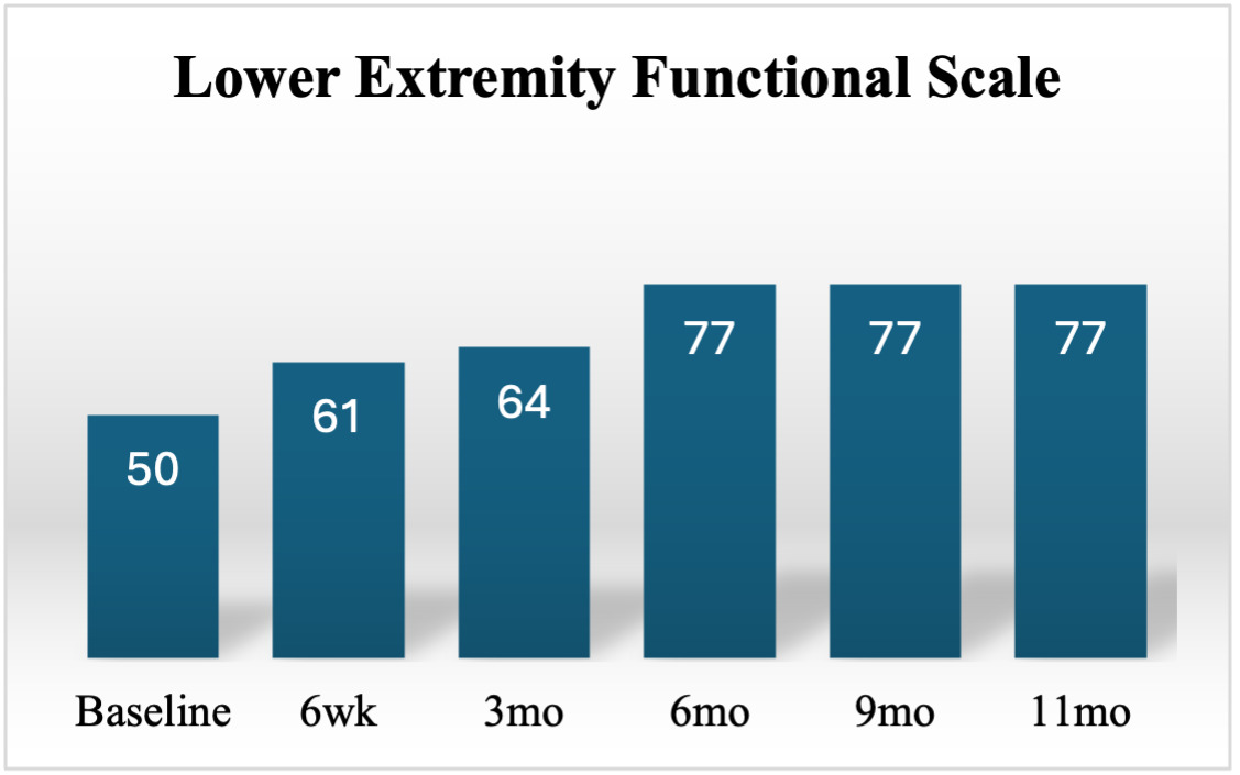
A 46-year-old active female with a history of high intensity strength training, was seen for a direct-access physical therapy evaluation. The chief complaint left anterior knee pain that developed gradually over the previous six weeks, with increased squatting volume in the gym. There was no reported traumatic mechanism of injury. A thorough evaluation of her medical history, surgical history, and medications were reviewed, and were unremarkable. Prior to examination, informed consent was obtained for clinical examination and management. The patient complained of pain and disability with mechanical loading such as squatting, lunging, stairs, running, and jumping. The patient reported 7/10 (worst pain) on the numeric pain rating scale (NPRS)9 and reported a 50/80 on the lower extremity functional scale (LEFS).10 Her goals included returning to her prior level of function which involved barbell squatting over 135 pounds, jogging, lunging, and using stairs pain-free.
Differential Diagnosis
The patient denied any dizziness, diplopia, dysphagia, dysarthria, drop attacks, numbness, nausea, or clumsiness during gait (ataxia). A neurological exam demonstrated normal (2+) deep tendon reflexes for the patella (L2-4) and the Achilles (S1). Clonus and Babinski upper motor neuron signs were negative bilaterally indicating no upper motor neuron involvement. Light touch sensation testing around anterior patellar region was equal bilaterally. Lower quarter myotome testing demonstrated normal strength for all nerve roots. Seated slump, straight leg-raise, femoral nerve tension test, and saphenous nerve tension test were performed and did not reproduce the patient’s symptoms. Lumbar AROM with overpressure was performed in standing and was within normal functional limits for flexion, extension, side-bending, and rotation. Secondary to the above findings, a local knee pathology was considered for the provisional diagnosis, and further examination of the region proceeded.
Examination
The treating therapist chose to observe a lateral step-down task11 and a bilateral bodyweight squat since these movements were reported to reproduce the patient’s symptoms. During the lateral step-down task, dynamic lower extremity valgus collapse was observed at the hip, knee, and ankle on the left leg with reproduction of comparable knee pain. Similarly, the bodyweight squat demonstrated a rightward weight shift on descent with reproduction of comparable knee pain. Observation of these movement compensations directed the treating therapist for further assessment at the hip, knee, and ankle joints.
AROM, PROM, and strength was assessed at the hip, knee, and ankle joints which can be found in Table 1. Hip AROM and PROM was impaired but did not reproduce the patient’s symptoms. Gross left sided weakness was found in the hip and knee with familiar symptom reproduction while testing resisted knee extension. Special testing including FABER,12,13 FADDIR,13,14 and Thomas test15 were positive with limited ROM compared to the contralateral side, but did not re-create the patient’s knee pain. Special testing of the anterior cruciate, posterior cruciate, collateral ligaments and menisci was also negative. Upon palpation, comparable pain was reproduced with direct pressure onto the inferior pole of the patella. There were no visible signs of effusion, with a negative sweep test.16 The contralateral right knee, hip and ankle exam was unremarkable. Additional baseline strength assessment included a 1-repitition max squat (unable to perform secondary to pain) and 10-repetition max unilateral knee extension test (Table 2).
Ultrasound Examination
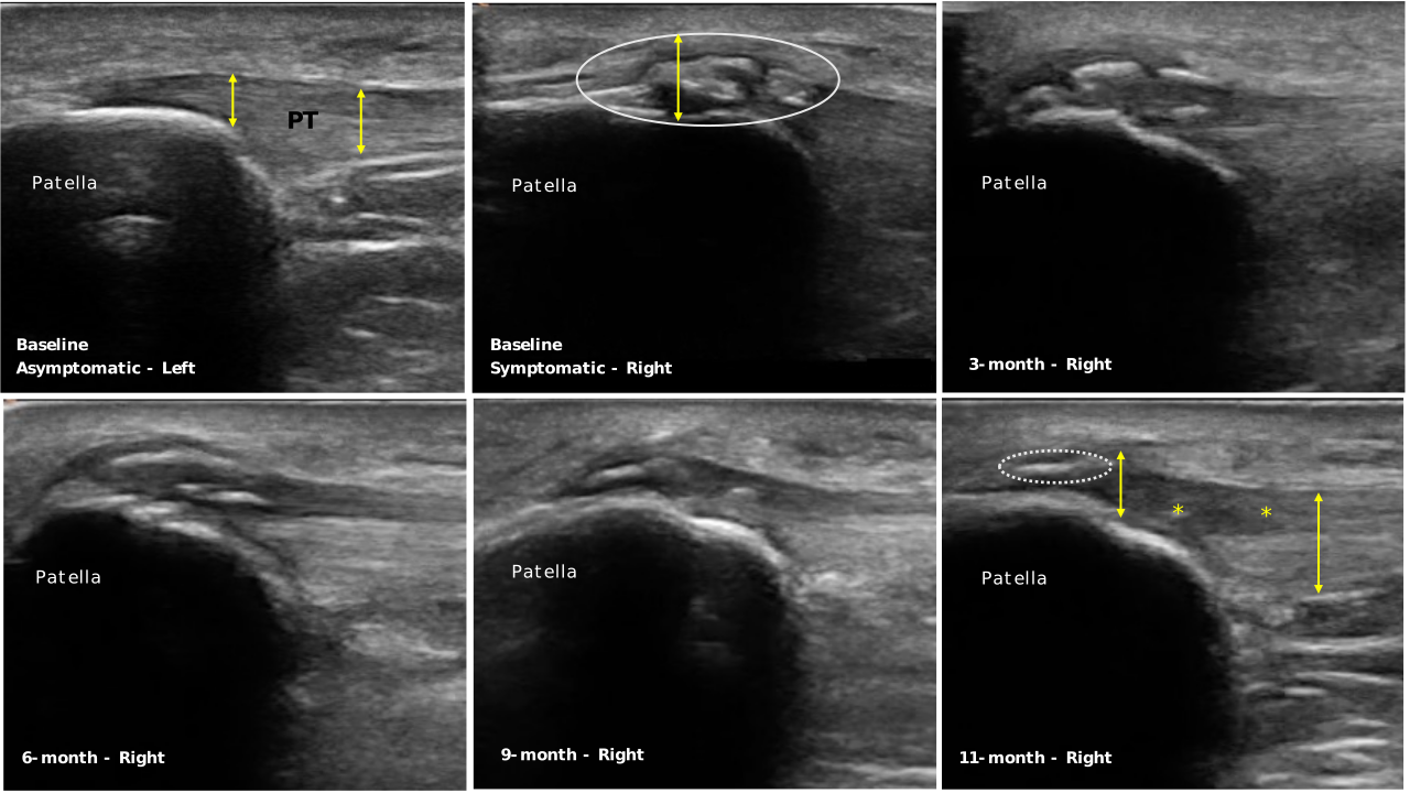
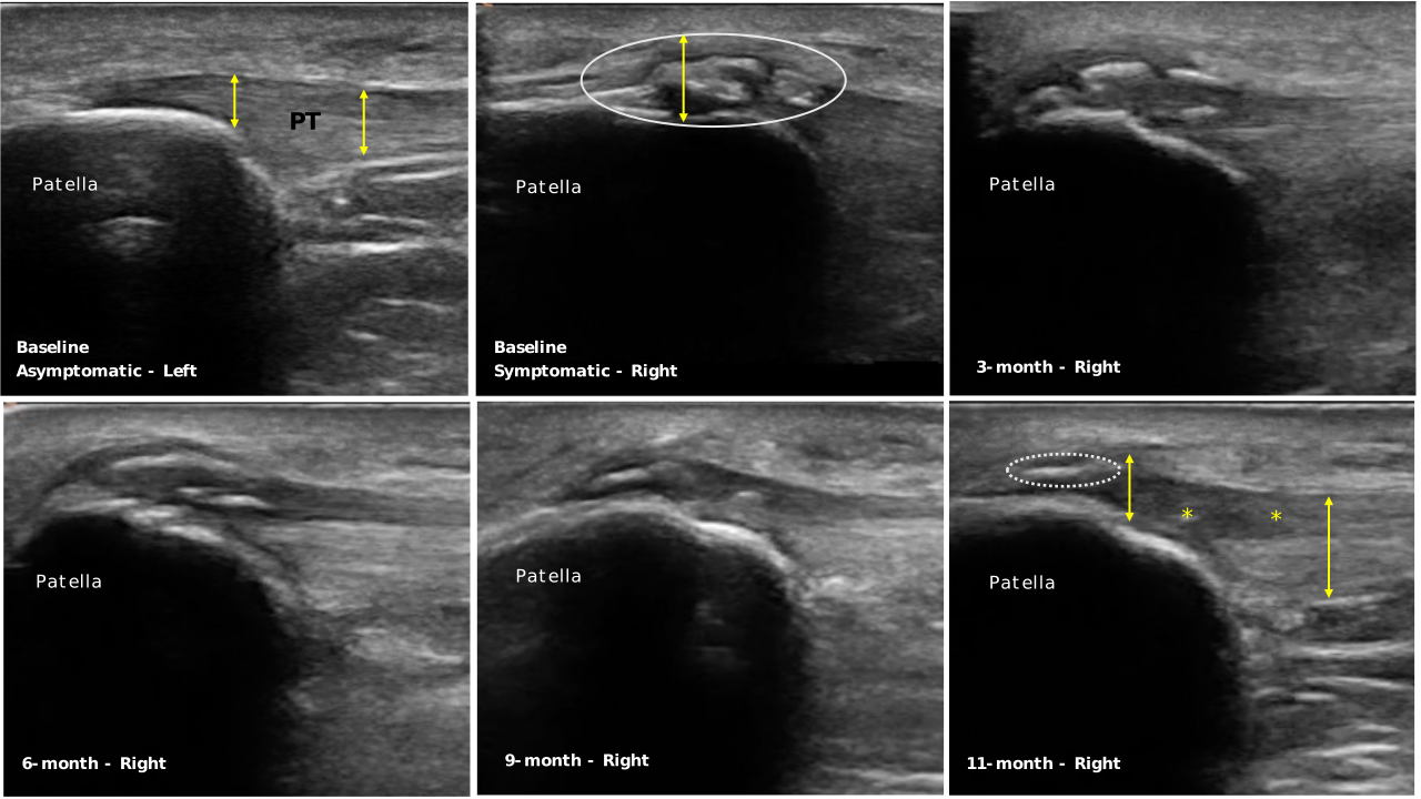
Based on the subjective and objective examination findings, the extensor mechanism of the knee was assessed using ultrasonography. A Clarius L7 HD3 linear scanner was used. The patient was positioned in supine with a bolster underneath her knee to create approximately 30 degrees of knee flexion. The image was captured in the center point in the sagittal plane of the distal patella during baseline and follow-up imaging to ensure consistency of the image over time. Hyperechoic intratendinous calcific foci, with acoustic shadowing deep to the deposits were appreciated at the inferior pole of the patellar tendon enthesis. Adjacent to the calcifications, hypertrophy and partial thickness tearing of the proximal patellar tendon was also appreciated, with disruption of the fibrillar echotexture (Figure 1).17 Comparable pain was reproduced with applied pressure using the ultrasound probe (sonographic palpation). The patient’s contralateral knee was also scanned for determining individualistic image findings, with no pathological findings and no pain with sonographic palpation. (Figure 1).
Treatment
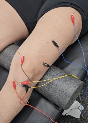
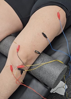
All sessions included a combination of cervical, thoracic, lumbar, long axis hip, and talocrural HVLA thrust manipulation at the start of each session for pain modulation, ROM, and motor output. Ultrasound guided dry needling (USGDN) with three sets of 30 seconds fenestration and winding targeting the calcific tendon region using two needles was performed at the end of each treatment session (Supplemental File 1). Additional needle points included the medial and lateral infrapatellar sulci, tibialis anterior, vastus lateralis, vastus medialis, and two needles in the muscle belly of the rectus femoris (Figure 2, Table 2). Needles were left in situ for 20 minutes with electrical stimulation at 2 Hertz/200 microseconds with a maximal tolerable intensity (ITO® ES-160 electrostimulator ITO co, Tokyo, Japan).18,19 Importantly, comparable pain was reported by the patient during the initial USGDN fenestration, aiding in confirmation of the local pain generator.
The patient was initally seen two times per week for 12 weeks and had at least 48 hours in between treatment sessions. USGDN was performed from weeks 2 to 12 at each session. After 12 weeks the patient was seen for “booster sessions” two times per month for the next six months. Booster sessions involved the standardized HVLA thrust manipulation protocol and progressive strength/plyometric training. USGDN was not performed after the three month follow-up.
During weeks 1-2, the treatment focus was on pain control, hip mobility and strength, and isometric tendon loading. Pain levels were kept ≤ 4/10 during exercise. USGDN was not incorporated until Week 2 due to the patient’s irritability; however, the supplemental protocol points for pain modulation were used during first two weeks. Weeks 3-6 utilized blood flow restriction exercise (BFR)20 and progressive hip and knee mobility and strengthening. During BFR (PUXHOE, Zhengzhou Trading Co., Zhengzhou, Henan Province, China), 80% limb occlusion pressure was used, as it has been found to be within the ideal range for maximal effect and patient tolerance.21 BFR was discontinued at Week 7 secondary to improved load tolerance (Table 3). Weeks 7-12 emphasized continued hip and knee strengthening. At 12 weeks, the unilateral 10-repitition max knee extension test was performed, and plyometrics were introduced secondary to greater than 90% quadriceps strength symmetry (Table 2). A detailed outline of the treatment plan can be found in Supplemental File 2.
OUTCOMES AND FOLLOW-UP
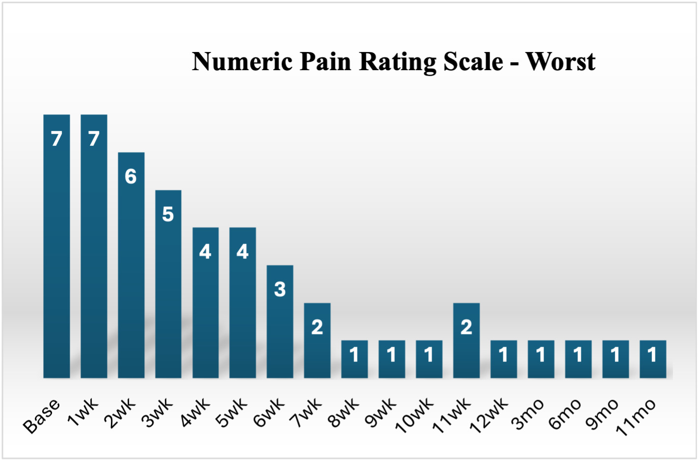
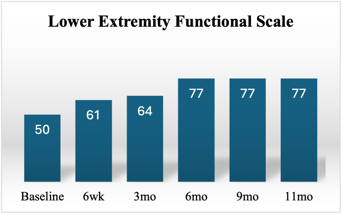
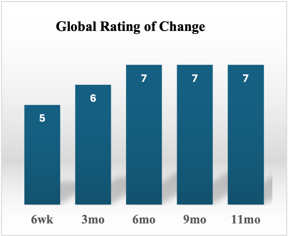
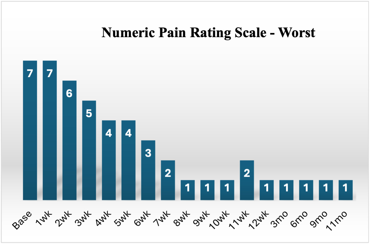
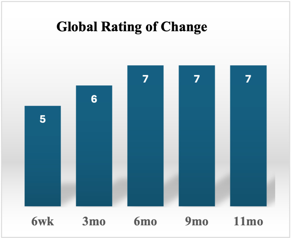
Patient reported outcome measures for pain (NPRS) and function (LEFS), and the global rating of change (GROC) were captured at baseline, 6-weeks, 3-months, 6-months, 9-months, and 11-months. Her NPRS scores showed improvements meeting the MCID (2-points) within the first three weeks of treatment and steadily improved throughout the plan of care (Figure 3).22 Her LEFS score exceeded the MCID of 9-points between baseline and all follow-up points (Figure 4).10 Perceived improvement scores using GROC23 are noted in Figure 5. Follow up measurements of ROM and strength can be found in Tables 1 and 3. The patient met all goals at the three-month follow-up including barbell squatting, jumping, and jogging and use of stairs without any pain or difficulty. Both pain and functional outcomes were maintained throughout the 11-month follow up (Figures 3-5). At the 11-month follow-up, the patient performed a 1-repetition max squat of 215 pounds without pain.
Ultrasound scans were performed at baseline and each follow-up time frame (Figure 1). As early as three months, changes in echotexture/morphology of the tendon as well as decrease in number and size of calcific foci were discernable. At six, nine, and 11-month assessments, the tendon appeared to have significantly smaller and reduced number of hyperechoic calcific foci over time as compared to the baseline image (Figure 1). At 11 months, the proximal tendon was still hypertrophied compared to the unaffected side, with hypoechoic echotexture noted superficially. However, a more fibrillar appearance of the tendon was appreciated compared to the baseline image (Figure 1). Importantly, there was no pain with sonographic palpation.
DISCUSSION
To the authors’ knowledge, this is the first case report to describe the use of USGDN, HVLA thrust manipulation, and exercise as a multi-modal treatment approach for CPT. Meaningful clinical improvements for this subject were noted in outcomes of pain, function, strength, range of motion, changes in tendon tissue morphology were noted in the short and long-term follow-up.
HVLA Thrust Manipulation
HVLA thrust manipulation is thought to influence a wide range of neurophysiological mechanisms to reduce pain.24–26 Although the patient’s neurological examination was unremarkable, tendon pain is complex and there is evidence for both peripheral nociceptive and centrally mediated mechanisms contributing to the patients pain state.27 Patients with tendinopathies often demonstrate sensory and motor deficits in their uninjured extremities suggesting a central influence.28 The patient’s objective examination revealed notable pain at the patellar tendon and quad strength deficits (Figure 3, Table 3). These strength deficits were likely to be a result of arthrogenic inhibition from pain/pathology at the patellar tendon,29 while the gross proximal and distal lower extremity weakness on the affected limb may have been a secondary maladaptive compensation. HVLA thrust manipulation has been shown to increase pain pressure threshold and decrease patient reported pain intensity.30 Fittingly, it has been suggested that HVLA may play a role the in management of tendinopathy, as a pre-conditioning process prior to formal exercise loading.30 Additionally, both lumbopelvic and hip manipulation have been shown to significantly increase quadriceps activation which is reduced in those with patellar tendinopathy.31–33 The analgesic and force production effects of HVLA thrust manipulation may have provided a window of opportunity to modulate this patients symptoms and progress more efficiently through the rehabilitation program.
Exercise
Isometric and eccentric exercise are considered the gold standard in exercise-based treatment approaches for tendinopathies; however, it is unclear whether isometrics and eccentrics are superior to other loading strategies.34–36 A recent Cochrane review concluded to be “very uncertain” whether strengthening exercise reduces pain or function in patellar tendinopathy compared to no treatment.37 However, other studies have shown isometrics and eccentrics to be effective in reducing pain and improving function, with or without the intension of strength changes.38,39 BFR was utilized in this case to assist in managing the patient’s pain response while increasing tendon loading.40 BFR allows low-load resistance to be useful in strengthening vs. conventional quadriceps strengthening, when symptoms are highly painful and irritable.41 It also has been suggested that BFR with low load resistance may be a viable option during rehabilitation aimed at inducing tendon adaptation,42 and has comparable results to high load resistance training on changes in tendon morphology.20,43 Finally, BFR has been found to cause exercise induced hypoalgesia through both opioid and endocannabinoid mechanisms.40 Limb occlusion pressure (50-80%) has been shown to be optimal for BFR,21 while repetition parameters range from 5-30, with some studies using muscular failure instead of predefined repetitions.40 In this case, 3 sets of 15 repetitions caused complete muscular failure and was reported as a high rate of perceived exertion (7-9) on a modified Borg scale. BFR was discontinued at Week 7, secondary to pain no longer being a limiting factor during rehabilitation (Figure 3).
Dry Needling
Dry needling (DN) uses thin monofilament needles, without the use of injectate, to treat a variety of neuromusculoskeletal conditions.44 DN is thought to promote tissue remodeling using mechanical stimulation to activate fibroblasts to produce type 1 collagen via winding and fenestration.36 The optimal number of insertions, treatment frequency, and duration of treatment is unclear in the literature.26,45 Animal models have shown increased collagen regeneration and tissue remodeling of the extracellular matrix in the Achilles tendon with three intra-tendon insertions and 30 seconds of rotation/winding at each insertion which guided the treating therapists dosage parameters.26 Electrical DN likely provides greater analgesia through both opioid and non-opioid mechanisms compared to dry needling without electrical stimulation.46 Treatment strategies with multiple needles left in situ for 20-30 minutes have been shown to produce superior biochemical, biomechanical, endocrinological, and neurovascular adaptations. compared to in and out/pistoning techniques.45
The patellar tendon is a rare place for CT so a “gold standard” treatment approach for this specific pathology is unknown. The shoulder and the hip are the most common anatomical locations for CT, with treatments often including physical therapy, (ESWT), ultrasound-guided percutaneous lavage (UGPL), and operative debridement.3 However, it has been speculated that the mineralogical structure of calcifications may vary in different tendon locations so it is unknown whether effective treatments in one region can be extrapolated to other areas.4
UGPL is considered the gold standard for medical treatment of CT due to its ability to reduce calcifications and pain while minimizing soft tissue damage.3 The procedure uses one or two needles to perform a lavage with or without aspiration of the calcifications using ultrasound guidance.8 However, a systematic review showed insufficient evidence to conclude UGPL is superior to other methods in management of CT and needle fenestration alone may be contributing to the therapeutic outcome.8,47 Tendon fenestration involves guiding a needle through an abnormal tendon multiple times to change a chronic degenerative process by stimulating an acute inflammatory cascade to re-initiate a healing response (Supplemental File 1).47 USGDN has been found to reduce pain, improve knee function, and improve ultrasonographic features/tendon morphology in patellar tendinopathy.48,49 There is minimal available evidence on USGDN for CT but DN appears as effective as other treatment methods for pain in tendinopathy in both the short and long term.50 Additionally, DN combined with exercise appears to provide greater reductions in pain and improved function than exercise alone.36
Limitations
Despite marked improvements in pain, function, and tendon tissue, several limitations are present. This is a single case report; therefore, the findings and conclusions are limited in generalizability and cannot establish direct causation between UGDN, HVLA thrust manipulation, and therapeutic exercise and the improved outcomes observed in this case. It is unknown whether this patient’s pathology would have resolved spontaneously due to natural progression without intervention. Furthermore, the absence of short axis/transverse images and Power Doppler flow at baseline and follow-up, limits the full diagnostic capabilities of ultrasound.
CONCLUSIONS
Incorporating diagnostic ultrasound in the evaluation and multimodal treatment of CPT may serve as a more comprehensive management strategy in the direct access physical therapy setting. Further study among multiple active/non-active individuals of varied age groups and populations are needed.
Appropriate diagnostic imaging of the pathology, combined with HVLA thrust manipulation, USGDN, and appropriate therapeutic exercise was successfully incorporated in management of this subject with CPT to improve pain, function and tendon morphology in both the short and long-term. Future research should investigate USGDN vs UGPL in treatment for CT.