INTRODUCTION
Anterior cruciate ligament (ACL) injuries affect a substantial number of youth athletes each year, leading to extended time away from sports with some athletes not returning to their previous competitive level for up to one year or more.1–3 As sports participation continues to rise, more young athletes are exposed to the risk of ACL tears, with female athletes four to six times more likely than their male counterparts to sustain a knee injury, potentially resulting in extensive recovery periods, financial burdens, and psychological distress.4–6 One strategy to mitigate these injuries is the implementation of validated movement screenings that identify biomechanical risk factors, such as excessive knee abduction.
Dynamic knee valgus (DKV), characterized by increased hip adduction, internal hip rotation, and knee abduction, has been established as a primary risk factor in non-contact ACL injuries.7,8 These altered lower-extremity mechanics place greater strain on the knee joint, thereby increasing the likelihood of ACL injury. Validated movement screens can detect undesirable movement patterns and risk factors, such as excessive knee abduction, to better guide training programs aimed at reducing injury risk. For instance, plyometric-based neuromuscular training interventions focus on improving landing mechanics and strength to correct excessive knee abduction and reduce injury risk.9 Although studies have examined knee biomechanics in adolescent athletes to identify injury risk factors, consensus on a standardized screening tool remains limited.10–12 Most existing screening approaches rely on two-dimensional (2D) video assessments, which are widely used due to their accessibility.13–16 However, the visually identified 2D knee abduction may not fully account for the multi-planar mechanics contributing to DKV and potential injury risk, as it does not distinguish between frontal-plane knee motion and accompanying hip and tibial movements. Previous research has explored how 2D measures such as frontal plane projection angle and knee separation distance relate to three-dimensional (3D) kinematics, illustrating that while 2D often demonstrates good reliability, its concurrent validity is still undetermined.17–23
Despite growing interest in using 2D video as a more accessible and cost-effective alternative to 3D motion capture for evaluating knee abduction and injury risk, it remains unclear how well 2D assessments reflect the multi-planar contributors to DKV. While 2D video can capture medial knee displacement, it primarily provides a heuristic for identifying knee collapse rather than a direct measure of potentially injurious biomechanics.7 Previous research has investigated the reliability of 2D assessments of tasks including a single-leg squat and step down, single-leg drop landing, and double-leg vertical jump; however, its reported validity varies with some studies reporting 2D techniques are unable to depict the multi-planar nature of DKV.11,19,24 Although previous research has established associations between 2D and 3D frontal-plane knee position, the multi-planar hip and tibial contributions that occur at peak medial knee position as well as the temporal agreement of these measures remains unknown. The temporal discrepancies between 2D and 3D medial knee position could lead to misidentification of knee loading phase, which may potentially overlook biomechanical components related to injury risk. Understanding timing differences is important in order to ensure practitioners utilizing 2D screening tools are accurately analyzing the periods of highest injury risk for screening and rehabilitation. Therefore, the purpose of this study was to evaluate how medial knee collapse corresponds to dynamic knee valgus multi-planar components by comparing timing and magnitude of knee abduction and medial knee position. It was hypothesized that the 2D analogous measure of most medial knee position (MMK) would demonstrate significant timing shifts and underestimate 3D peak knee abduction (MKA) angle due to the inability to account for out-of-plane motion. Similarly, the authors hypothesized that discrepancies across other planes of motion would also exist, suggesting that 2D video assessments may provide limited and potentially misaligned representation of multi-planar kinematic components of DKV.
METHODS
A convenience sample of 34 adolescent female volleyball players (10-18 years) were recruited from local club volleyball teams and participated in this study between September and December 2021. Participants were excluded from participation if they were diagnosed with an orthopedic condition that limited their ability to perform the required tasks or if they reported a recent musculoskeletal injury within three months prior to testing. A musculoskeletal injury was defined as any self-reported injury to the muscles, tendons, ligaments or bones that resulted in change to physical activity participation. The study was approved by a regional Institutional Review Board and all participants provided informed assent/consent before participating in the study.
Procedures: Twenty-one retroreflective markers were placed bilaterally on bony landmarks of each participant along with rigid clusters on both the thigh and shank segments according to a standardized maker set established by the Pediatric Research in Sports Medicine (PRiSM) Society’s Motion Analysis Research Interest Group.25,26 A standing static calibration was performed in which knee and ankle joint centers were created as determined by taking a bisection between the anatomical markers placed on the medial and lateral femoral condyles and malleoli. Additionally, the hip joint center was created according to Harrington’s regression equation and using anthropometric measurements such as leg length, pelvic depth, and ASIS distance.27 A 14-camera motion capture system (Vicon Motion System Ltd., Denver, Colorado, USA) was used to collect 3D kinematic data captured at 240 Hz while participants performed a three-task protocol.
The three-task protocol was administered by a biomechanist with 10 years of experience and included a single-leg squat (SL squat), a single-leg drop landing (SL drop landing), and a double-leg vertical jump (DL vertical jump), as established in prior work published by Ulman et al. which investigated the prevalence of volleyball injury risk factors.10 These tasks were selected to represent a range of task complexity and neuromuscular demand, and have been validated for identifying biomechanical risk factors in adolescent female athletes.10,11 For the SL squat, participants were instructed to begin by standing on one leg with the opposite leg bent behind them and their hands positioned on their hips. They were then instructed to squat as low as comfortably possible and return to the starting position. For the SL drop landing, participants were directed to jump from a 31-cm plyometric box, landing on one leg, holding the landing for at least two seconds. Lastly, for the DL vertical jump, participants were instructed to jump as high as possible from a standing position and were allowed to perform a countermovement and/or use their arms for momentum. The SL squat and SL drop landing were performed on each leg, while only the right leg was analyzed for the DL vertical jump. Given that the DL vertical jump is a bilateral task, only the right leg was used as a representative in order to avoid treating both limbs from the same participant as independent observations and to maintain consistency across participants. Participants were given three practice attempts for each task to ensure they were comfortable with the task protocol, and to verify their understanding of the instructions. After completing the practice trials, participants were given a single attempt for each task. The single attempt protocol was utilized in order to capture natural, unfatigued movement patterns and to minimize learning effects. If there was an observed error during the first attempt (e.g., loss of balance, failure to stabilize upon landing), participants were allowed a second attempt. Observed errors were defined based on previous literature and included falling upon landing, placing contralateral foot down during single-leg tasks, or lack of flight phase on jumping tasks.11,28
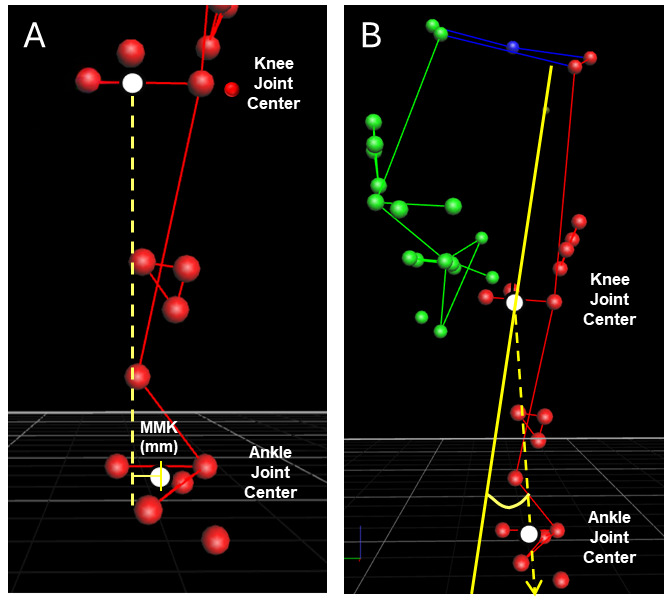
Data Analysis: Marker trajectories were filtered using a Woltring filter with a predicted mean square error of 10 mm.2,29 A custom MATLAB model (MATLAB 2022a; Natick, MA, USA) was used to compute joint angles for the trunk, pelvis, hip, knee, and ankle. The lower extremity model used three degrees of freedom for the hip and knee joints and six degrees of freedom for the ankle.26 Variables of interest included kinematic joint angles representing components of DKV, specifically pelvic obliquity, hip adduction, internal hip rotation, knee abduction, external tibial rotation, and ankle eversion. Additionally, the position and timing of when the knee joint center (midpoint between the medial and lateral epicondyle) was the most medial relative to the ankle joint center (midpoint between medial and lateral malleolus) were also extracted for each task (most medial knee: MMK; Figure 1A) to be analogous to 2D knee collapse. MMK position was computed using 3D motion capture as an analogous alternative to 2D assessment medial knee collapse. Lastly, maximum knee abduction (MKA) angle and associated timing were extracted from the motion capture data for the three tasks across the phases of interest (Figure 1B). Kinematic angles for the pelvis, hip, knee and ankle were calculated at the specific timepoints of MMK and MKA.
_most_medial_knee_position_(mmk)__distance_in_mm_between_knee_joint_center_and_ankle_jo.png)
The phases of interest were defined as the loading phase of the SL squat, the landing phase of the SL drop landing, and the loading and landing phases of the DL vertical jump. The loading phases for SL squat and DL vertical jump were defined as the period between initiation of knee flexion and cessation of knee flexion. The landing phases for both SL drop landing and DL vertical jump were defined as the period from initial foot contact until the cessation of knee flexion taking place after flight phase.12 Initial contact for the DL vertical jump landing was defined as the timepoint in which knee flexion exceeded 25% of the maximum knee flexion across landing phase. For the SL drop landing, initial contact was identified as the timepoint of minimum vertical velocity of the sacrum marker, representing the point of peak deceleration due to impact. Events were identified using a custom MATLAB (MATLAB 2022a, Natick, Massachusetts, USA) code and placed at the time points of interest, specifically including the initiation of knee flexion and the time point of maximum knee flexion. Timings were reported as the percent of loading/landing phase, with 0% defined as the initiation of knee flexion and 100% of the phase defined as the time point of maximum knee flexion.
Statistical Methods: Kinematic variables were extracted at two within-task events: maximum knee abduction (MKA) and most medial knee position (MMK) and compared using Wilcoxon signed-rank tests given significant tests of normality. Given these values were obtained from the same participant during the same task, comparisons between MKA and MMK were treated as paired comparisons. The timing and position of MMK were also compared to the timing and value of MKA using Wilcoxon signed-rank tests. Statistical significance was determined when p was less than 0.05. Effect sizes (rank-biserial coefficient, r) were also computed for each comparison test, and interpreted as small (r = 0.1), medium (r = 0.3), or large (r = 0.5).30
RESULTS
Of the 34 volleyball athletes tested, nine were excluded from the analysis due to errors made when completing the second attempt for the task. Athletes (n=26) ranged from 10.5 to 16.5 years of age (Mean age: 14.4 ± 1.2 years). Comparing the differences in the timings of when MKA and MMK occurred within the phases of interest, MMK occurred 6% earlier for SL drop landing (p=0.567), 23% earlier for DL vertical jump Load (p=0.005) and 39% earlier for DVLJ Land (p<0.001) while for SL squat, MMK occurred 17% later than MKA (p=0.022; Table1). Additionally, there were significant differences in the KJC-AJC distance at MKA compared to MMK for all tasks (p<0.001; Table 1).
Differences in kinematic variables at the two timepoints are shown in Tables 2-4. Specifically, during the SL squat, there was increased pelvic obliquity (MMK: 0.50° vs MKA: -1.52°) and slightly greater hip adduction (MMK: 13.86° vs MKA: 10.95°) at the timepoint of MMK while no differences were observed in pelvic obliquity and hip adduction with the remaining tasks. The hip was less externally rotated (or more internal) during DL vertical jump Load and Land at the timepoint of MMK position (Loading phase: MMK: 2.52° vs MKA: -0.93°; Landing phase: MMK: -1.11° vs MKA: -3.81°). No differences in hip rotation were measured at the two timepoints with SL squat or SL drop landing tasks. Across all tasks, there was greater external tibial rotation when the knee was at its most medial position (p<0.022). As expected, knee abduction was greater at MKA compared to the timepoint of MMK for all tasks, with the greatest difference occurring with DL vertical jump Land (3.6° difference). Ankle eversion only showed differences during the landing phase of DL vertical jump, with more ankle inversion measured at MKA versus MMK (p<0.001).
DISCUSSION
DKV predisposes athletes to ACL injury7,8,31 and has increased prevalence among females, particularly in the component of knee abduction.32,33 3D motion capture remains the gold standard as 2D screening metrics have shown both limited reliability and validity using risk factors such as poor frontal plane knee motion.17,22,34 While the three-task protocol was validated in a previous study for identifying the prevalence of biomechanical risk factors in adolescent volleyball players, the current study quantifies the validity gap between 2D and 3D methods.10,11 Notably, the current study compared simultaneous 2D-equivalent and 3D data, providing a reference to directly quantify the validity of 2D medial knee position by identifying kinematic differences between methods. Results of the current study indicate that 2D analysis represented by the MMK demonstrated significant differences in timing compared to 3D analysis represented by MKA during most tasks. In the DL vertical jump loading and DL vertical jump landing phases, MMK occurred earlier than MKA, 28.5% and 18.4% of the loading phase, respectively, compared to 51.4% and 57.7% for MKA.
One possible explanation for MMK occurring prior to MKA for the more dynamic tasks, is that medial knee movement occurs prior to peak knee abduction due to contributions at the hip, knee, and other aspects of DKV during high stress tasks. In the SL drop landing and DL vertical jump landing phases, specifically, a larger ground reaction force is absorbed upon impact, which could cause the medial collapse of the knee as a mechanism for balance while also corresponding to the greatest load on the ACL.35,36 Medial knee movement preceding peak knee abduction is supported by previous findings by Myer et al., which identified that 2D medial knee collapse commonly occurs prior to peak 3D joint angles in high-impact landing tasks.37 The lack of a significant difference in timing for the SL drop landing could correspond to the shorter landing phase of the task, which limits the opportunity for medialization to occur. In contrast, MMK occurred after the MKA timepoint in the SL squat (59.0% vs 42.4% of the loading phase). Given that only a loading phase was assessed, more neuromuscular control might be employed during this period along with later integration of hip, trunk, and other compensatory mechanisms which required participants to achieve a greater depth compared to the SL drop landing.38 Despite the DL vertical jump including a loading phase like the SL squat, integration of a preparatory requirement for takeoff may reduce similarities. The apparent discrepancy between timing of the MMK and MKA also indicates that 2D analysis, while appropriate for screening, is not directly indicative of DKV which involves a more complex and multiplanar movement. This timing shift suggests that 2D analysis of most medial knee position may be assessing movement that occurs before the actual 3D peak abduction load, therefore 2D analysis may overlook the timepoint of maximum joint strain. This is crucial in evaluating risk for ACL injury, as non-contact ACL ruptures typically occur within the first 40 milliseconds after initial contact suggesting that 2D measures may underestimate the mechanical demand on the ACL.16 While 2D measure of medial knee position is a practical screening tool for movement strategies, 2D knee medialization and 3D peak knee abduction are distinct biomechanical events that may occur at different timepoints.
Comparison of other DKV measures at the pelvis, hip, tibia, and ankle at MMK and MKA generally revealed greater DKV components at the point of MMK, which supports the utility of MMK in identifying peak risk components that might be overlooked at the time of greatest knee abduction. For example, at the time of MMK compared to MKA, hip adduction was 2.9° greater in the SL squat. Additionally, hip rotation was 3.5° and 2.7° more internal (or less external) during both DL vertical jump loading and landing phases respectively, external tibial rotation was greater in for all tasks (1.2° - 3.0°), and there was 4.1° less ankle inversion in the DL vertical jump landing phase. The clinical relevance of these kinematic differences remains unknown, and while significant, further research is needed to determine if these differences would influence lower extremity injury risk. Though time of peak knee abduction was largely incongruent with peak knee medialization, coronal plane hip and pelvic kinematics were more often similar between the two timepoints for the higher impact tasks. Of note, the single DKV component which was greater during MKA was pelvic obliquity during the SL squat - corresponding to increased pelvic drop and potentially less neuromuscular activation of the hip abductor muscles on the stance leg. Still, the similar pelvic obliquity during the two landing tasks (SL drop landing and DL vertical jump) further supports that the hip abductors and other stabilizers are important and may aid in preparation for higher ground reaction forces while loading tasks involve more gradual, controlled movement. The three tasks employed in this established protocol represent a range of neuromuscular demands.10,11 Researchers should consider that loading tasks, particularly the SL squat, present altered neuromuscular and DKV patterns in comparison to landing tasks. By utilizing both loading and landing tasks, the current study offers a comprehensive evaluation of DKV and how movement patterns are altered across different task demands. These alterations do not consistently transfer between the 2D and 3D systems for all DKV components.
Given the timing discrepancy between MMK and MKA and overall greater DKV components observed at MMK, consideration should be made whenever 2D techniques are used for additional DKV contributors other than frontal plane knee collapse. While 2D frontal plane assessment is a clinically accessible screening tool which may be integrated into routine athletic screening for early detection of DKV, its value is in identifying broader movement patterns and poor neuromuscular control throughout the loading phase rather than determining peak knee loading. Specifically, the current findings help identify which specific kinematic differences are consistent across 2D and 3D timepoints, offering clinicians reliable measures for when 3D analysis is inaccessible. By identifying those athletes at risk early, targeted rehabilitation programs can successfully reduce DKV by targeting the knee joint muscles and the posterior chain.9,36,39,40 However, these results underscore the importance of synthesizing multiple components of DKV rather than a singular component during in-depth laboratory analysis.
Limitations
Limitations of the current study include participants of a single sport and of the same gender, which reduces generalizability of the study. Additionally, the age range of adolescent participants spans a period of various stages of physical development. While this age range represents a youth club volleyball population, movement patterns are likely to change during adolescence which may result in variability in biomechanical comparisons in the cohort. Future work should investigate whether similar findings exist in male athletes, those who play other sports, as well as across varying stages of development. Second, most medial knee position was determined using motion capture as an alternative to 2D assessment. While this measure was obtained from a single plane and thus similar to 2D video analysis, it is likely that 2D analysis may introduce additional out of plane error depending on the camera view. Lastly, participants completed a single representative trial for analysis. While this method is susceptible to fluctuations in technique, it was chosen to ensure natural movement patterns without fatigue-induced changes commonly observed with repeated maximal effort attempts. While practice attempts were completed to ensure familiarity with the task, future research could include multiple trials in order to investigate reliability within subjects. Future studies may investigate larger, more diverse cohorts, examine athletes with pathological knee valgus, and compare components of DKV across the entire functional task.
CONCLUSION
Analysis of peak knee medialization position and timing captures components of DKV with equal or potentially greater injury risk than at the timepoint of peak 3D knee abduction. Therefore, assessing medial knee collapse as is commonly employed in 2D video analysis may be useful for screening given its greater accessibility, but it cannot be considered a valid indicator of dynamic knee valgus and might overlook peak knee abduction loading depending on the performed task. In future studies of 2D screening, researchers should investigate multiple timepoints when investigating DKV rather than only peak kinematics. Future work may also examine the individual components of DKV to determine which 2D measures are most predictive of ACL injury risk.
Conflict of Interest Disclosure
None