INTRODUCTION
Hamstring injuries involving the so-called “T-junction” or distal musculotendinous junction (DMTJ) remain a true enigma for clinicians in sports medicine, as demonstrated by recurrence rates reported to be as high as 54%.1 The challenge in managing these injuries arises from the complex anatomy of this structure, the high inter-individual anatomical variability, and the different innervations of the biceps femoris long head (sciatic nerve) and short head (peroneal nerve), which can lead to asynchronous movement of the two heads and place significant mechanical stress on the DMTJ.2,3 Magnetic Resonance Imaging (MRI) is the gold standard for muscle injuries assessment but may sometimes underestimate DMTJ injuries. For instance, Kayani et al. reported a 35% discrepancy rate between MRI and intraoperative findings.4 The static nature of MRI may limit its ability to assess structures such as the DMTJ, which demonstrates its true functional relevance under dynamic conditions, where it must manage the opposite force vectors generated by the two heads of the biceps femoris.1,3 Dynamic ultrasound has been proposed as a complementary tool to detect dissociative movement between the two heads of biceps femoris, although assessments in previous work were performed only during isometric knee flexion and not under functional multi-joint loading conditions.5 The aim of the present case report was to describe a novel method for dynamic ultrasound evaluation of the DMTJ performed during functional loading exercises and to explore its potential role in return-to-play (RTP) decision-making.
CASE DESCRIPTION
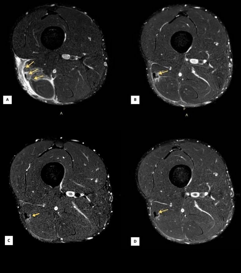
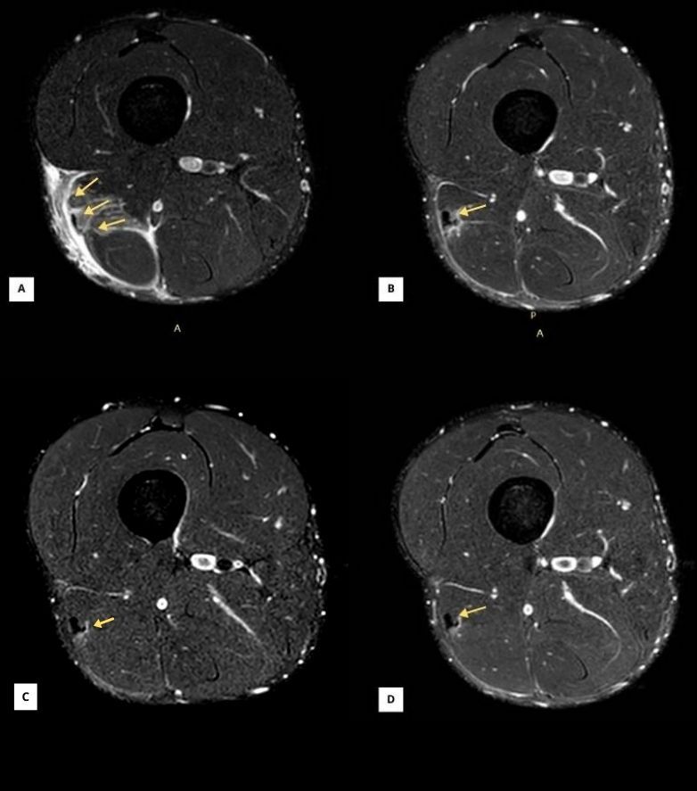
A 23-year-old male elite football (soccer) player sustained a DMTJ injury. The athlete’s pre-injury characteristics were as follows: height 190 cm; body mass 100 kg; sum of six skinfolds 40 mm; 30–15 Intermittent Fitness Test (IFT) velocity 21 km/h; and maximal sprinting speed 34.5 km/h. MRI revealed a complete rupture of the superficial portion (myotendinous) and the deep portion (myoaponeurotic) of the T-junction. When interpreted within existing classification systems, the present injury could be considered consistent with a grade 3c lesion according to the British Athletic Muscle Injury Classification (BAMIC) classification.6 This classification should be regarded as an academic categorization rather than a comprehensive radiological description, as such classification may obscure some of the lesion-specific imaging details observed in this case.7
The injury occurred initially during a high-speed nonlinear running action. The player continued to play and felt another acute pain at the same point after a pass leading to removal from competitive match participation for 10 weeks. An expert musculoskeletal radiologist made the initial diagnosis using a 3T MRI scan. Subsequently, three dynamic ultrasound evaluations were performed at weeks 3, 6 and 9 to monitor the RTP process. Dynamic ultrasound assessment of the healthy side was also performed to detect any abnormalities and identify potential asynchronous movements between the two heads of the biceps femoris, serving as a comparative reference (at week 6). A follow-up 3T MRI was conducted in the middle of the rehabilitation process (week 5) and another one before the reintroduction of high-speed running (HSR) & sprinting (week 8). A final MRI was performed 4 weeks after RTP (week 14) to check the evolution of the scar. (Table 1)
Procedures
Ultrasound assessments were performed using a Versana Active ultrasound system (GE HealthCare, Italy), with the transducer positioned over the injury site and stabilized using a ProbeFix Dynamic device (PFD) (Usono, Netherlands).
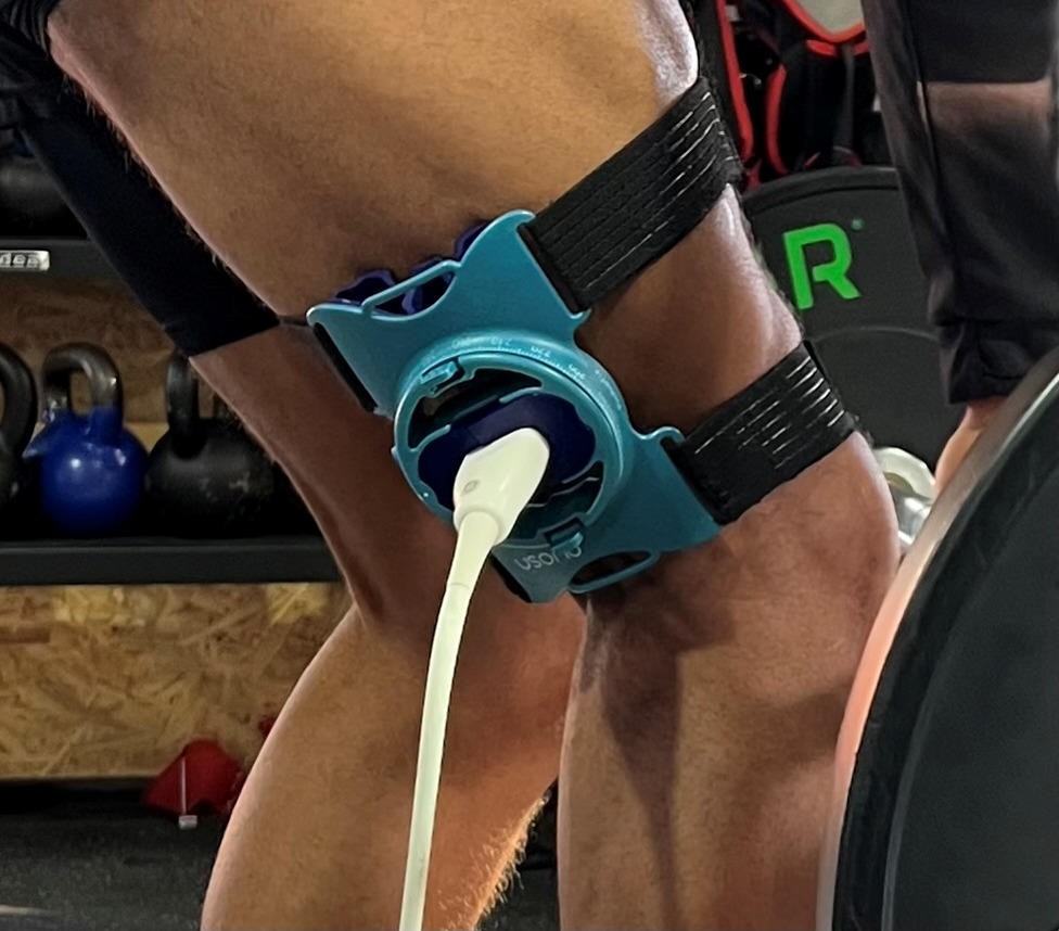
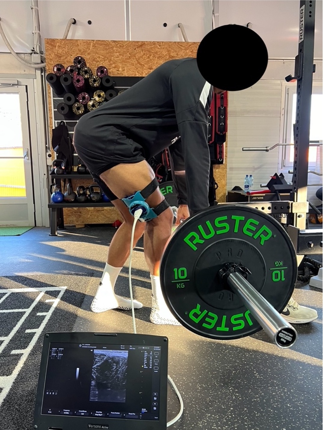
For the dynamic assessment of both exercises, a 12L-RS linear transducer (4–13 MHz) was used. The probe was placed transversely over the distal third of the biceps femoris muscle until the myoaponeurotic tissue of the T-Junction between long and short heads was visualized at the center of the image, with the myotendinous portion visualized as horizontal echogenic striation in the upper part.9 At this point, the probe was fixed to the player’s thigh using PFD (Figure 1).
.png)
Each evaluation included dynamic imaging performed during two gym-based exercises:
-
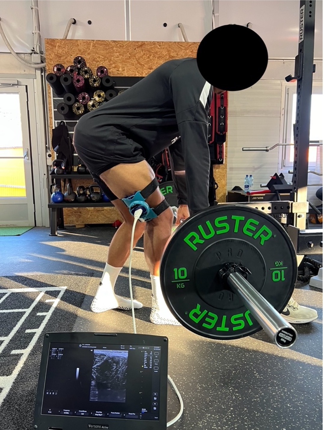
Single-leg barbell Romanian deadlift (SL-RDL): The athlete was instructed to grasp the barbell with both hands and perform a SL-RDL on the affected limb. The stance knee was kept slightly flexed while the barbell was lowered along the anterior aspect of the knee toward the tibial tuberosity, with the contralateral foot lightly in contact with the ground for balance purposes. The athlete then performed a 1-second isometric pause at the bottom position. This was followed by ascent through a controlled hip-hinge movement (Figure 2). The external load was set at 30 kg during the first assessment and increased to 40 kg for both the second and third evaluations. The progression during rehabilitation from 30 to 40 kg was based on symptom response, movement quality, and an estimated repetitions-in-reserve approach, with progression allowed when the player completed the prescribed sets without symptom exacerbation and with approximately two repetitions in reserve. The load was not increased beyond 40 kg to maintain a submaximal rehabilitation stimulus while preserving movement quality and symptom control.
-
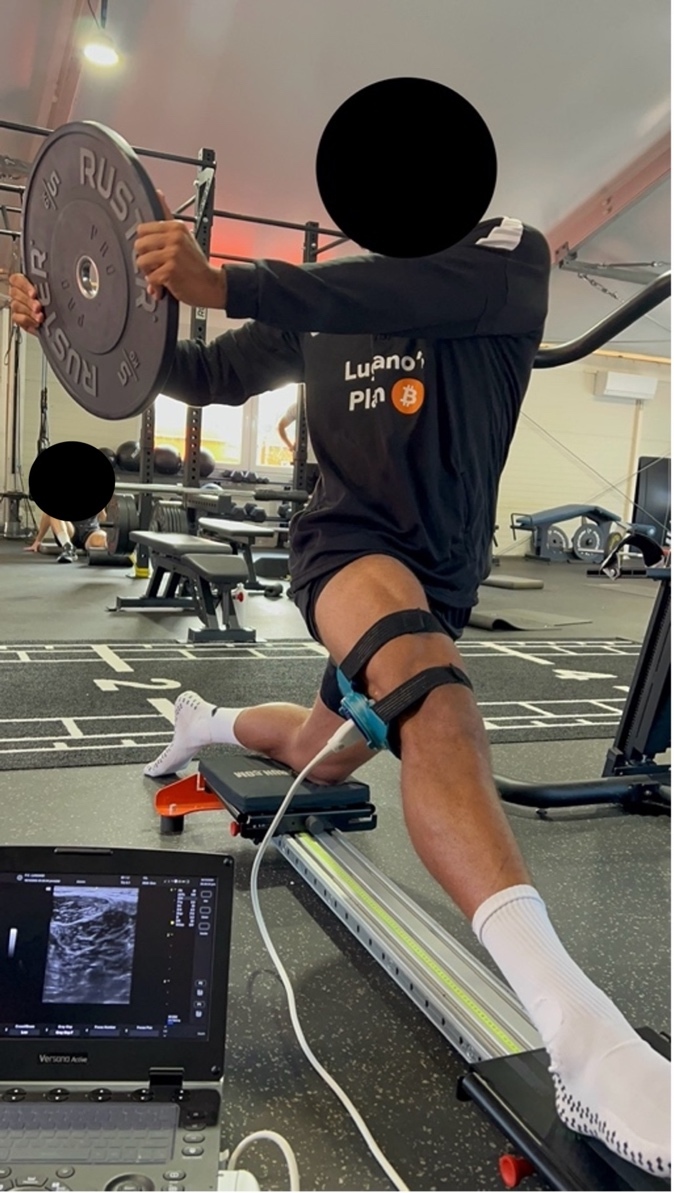
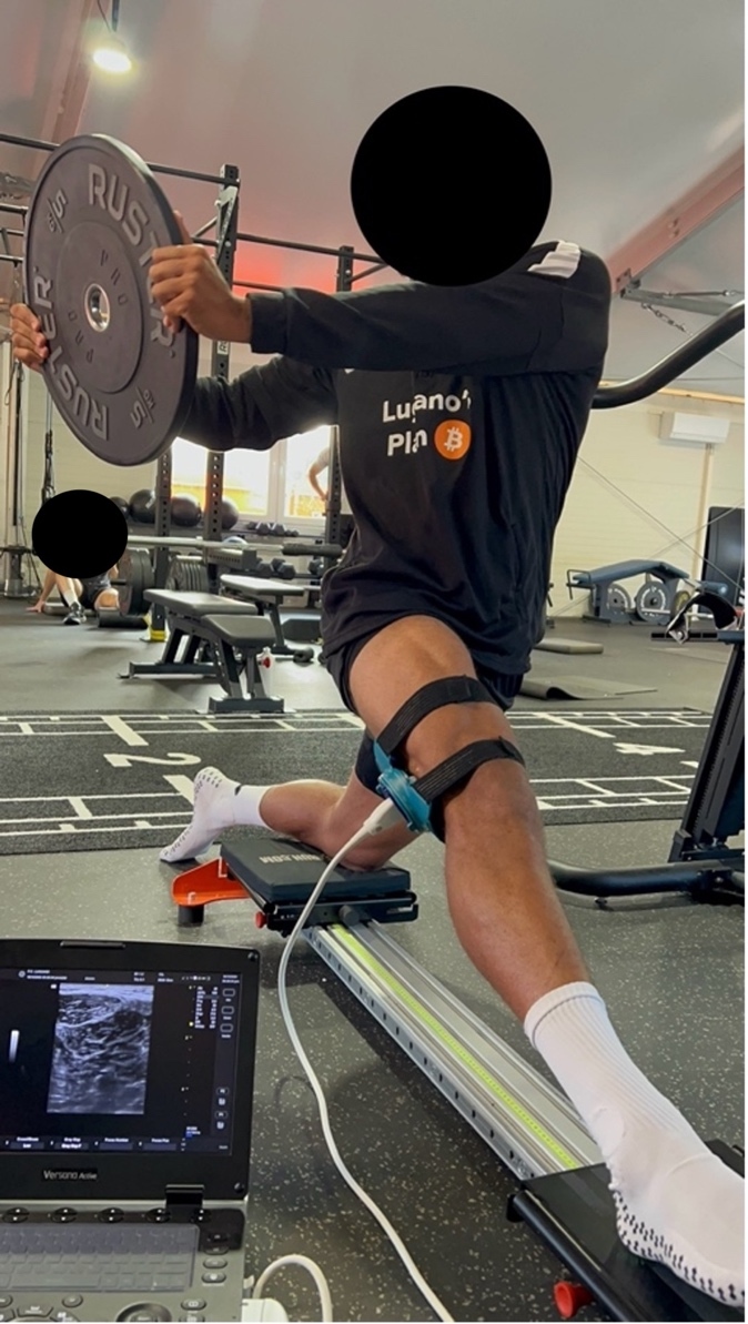
Single-leg hamstring slider with ipsilateral trunk rotation: the exercise was performed using a commercial slider device (Battle4run, Spain). The contralateral knee was positioned on a static plate, whereas the limb under evaluation was placed on a sliding plate and moved progressively into knee extension with simultaneous hip flexion. Simultaneously, the athlete performed ipsilateral trunk rotation while holding a 5-kg weight plate (Figure 3).
The athlete was familiar with and had previously trained using both exercises. Each exercise was performed for three repetitions with 3-second eccentric and concentric phases. Dynamic ultrasound videos were recorded for each trial (Supplemental File 1) and also for the uninjured limb evaluation (Supplemental File 2).
OUTCOMES
The player returned to running and soccer-specific activities at intensities below 70% of maximal sprint speed (MSS) after 6 weeks. At week 8, he progressed to HSR and sprint exposure, and at week 9 he was cleared for full team training. The player successfully returned to play, according to the adopted return-to-play definition,10,11 10 weeks post-injury with no re-injury at 4 months follow-up. Across serial evaluations, progressive changes were observed in both tissue appearance and movement behavior at the distal myotendinous T-junction. Dynamic ultrasound demonstrated a gradual reduction in visible interhead asymmetry and progressive restoration of synchronous movement between the BFlh and BFsh during functional tasks. In parallel, ultrasound imaging showed progressive maturation of the scar tissue and improvement in tissue continuity over time.
DISCUSSION
Previous studies have shown that MRI may have limitations in detecting DMTJ injuries and that ultrasound evaluation can assist clinicians in completing the diagnosis and monitoring the healing process during RTP phases.4,5,12 Rapid symptom resolution despite persisting structural damage is characteristic of T-junction injuries, making it unreliable to rely solely on patient-reported feedback to guide rehabilitation decisions.1,12 Cronin et al. demonstrated that dynamic ultrasound evaluation during isometric knee flexion can detect gap widening between the short and long heads of the biceps femoris, monitor hematoma evolution, and identify asynchronous movement of the two heads.12 Normal dynamic movement of the T-shaped hamstring region is characterized by synchronous motion, in which mechanical and temporal coordination between the long and short heads ensures uniform distal force transmission without intratendinous shear during contraction. The key indicator of normal function is the absence of “dissociative movement” (sliding or separation) between the long and short heads during contraction.1,3
In the described approach, dynamic ultrasound evaluation was performed during gym-based exercises, which impose greater demands on muscle tissue than a simple isometric contraction. The SL-RDL and slider exercises with ipsilateral trunk rotation were selected primarily because they provide mechanical stress on the biceps femoris, allow progressive loading, and offer sufficient space to secure the device without interfering with movement. This setup helped enhance the precision and reliability of dynamic ultrasound evaluation. Ipsilateral rotation was added to the slider exercise because this movement pattern is commonly involved in the mechanism of DMTJ injuries.13 As suggested by Kerin, a multiplanar evaluation approach may represent a clinically relevant advancement, offering a more functionally meaningful assessment compared with traditional static or linear imaging assessments.12
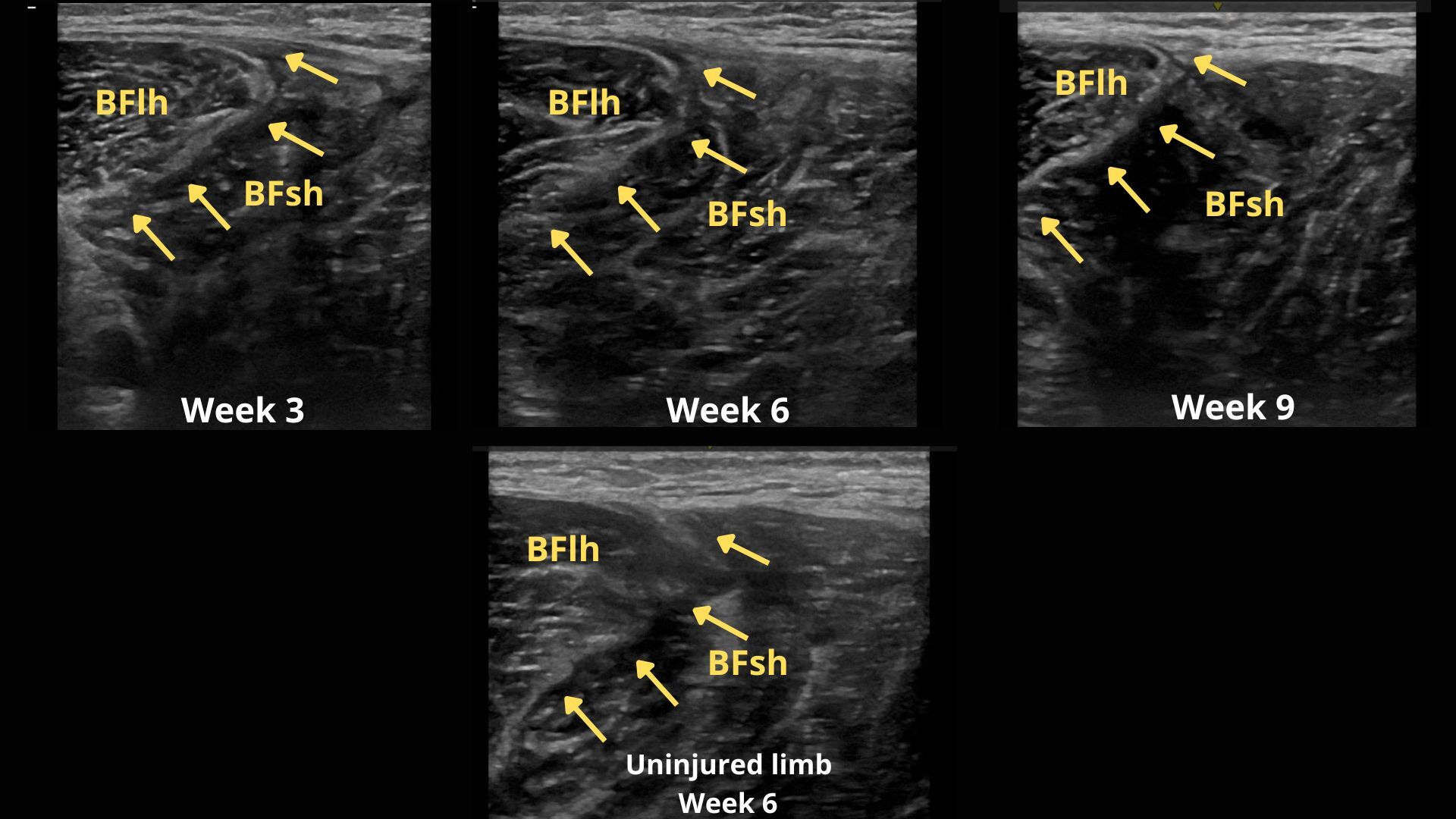
In the present study, tissue healing was monitored using dynamic ultrasound evaluations at three different time points. The first evaluation was conducted at week 3 (Figure 4), when the athlete had already begun performing the assessed exercises with low loads during rehabilitation sessions and was able to execute them without symptoms. During the first assessment in addition to superficial edema and intermuscular hematoma, interhead movement was not smooth and contraction appeared predominantly localized around the scar tissue. At week 6 (Figure 4), a second evaluation was conducted while the athlete was engaged in an intensive gym-based strength and conditioning phase and running at <70% of maximal sprinting speed (MSS).8 Resolution of the intermuscular hematoma was observed, with residual edema around the scar. Interhead movement appeared smoother and more synchronous. The final evaluation (week 9; Figure 4) was performed during the last week before RTP, when the athlete had progressed to high-speed running (>70% MSS) and partial team training. Dynamic ultrasound assessment showed reduced edema and near-normal movement between the two heads of the biceps femoris (Supplemental File 1). These findings were consistent with the week 8 MRI results (Figure 5). Following these evaluations, the player was cleared for full team training (week 9) and subsequently returned to play at week 10. At the 4-month follow-up, no reinjury was reported.
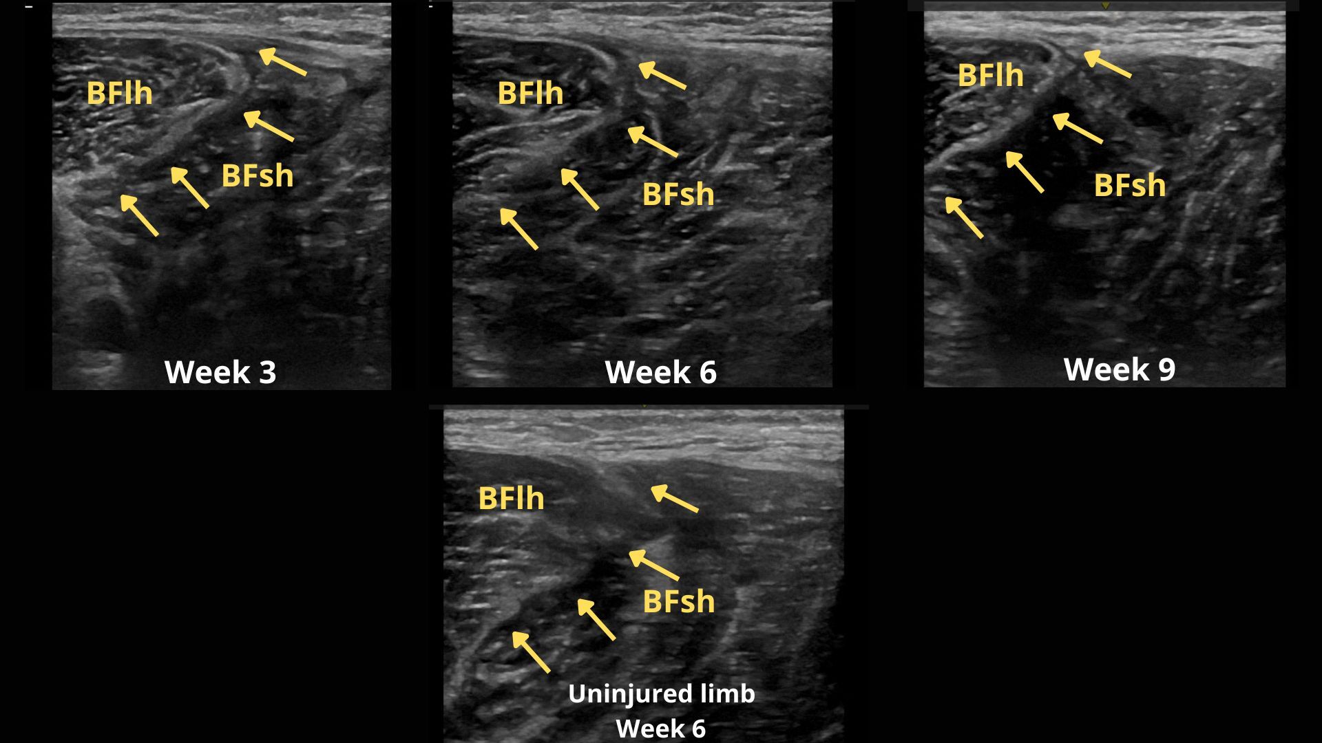
Healing progression, particularly the normalization of the synchronous movement between the two heads of the biceps femoris, was observed primarily during the SL-RDL exercise, whereas it was less easily seen during the slider exercise. This difference may reflect the higher contraction demands of the deadlift exercise, which allows greater loading and more targeted stress on the affected tissue.
CONCLUSIONS
This case report highlights the potential value of dynamic assessment of the DMTJ in hamstring injuries. Dynamic ultrasound performed under progressive functional loading may provide clinically relevant information during the return-to-play process, particularly in evaluating the movement synchrony between the long and short heads of the biceps femoris. Among the exercises tested, the SL-RDL appeared to allow clearer visualization of the restoration of coordinated interhead movement, likely due to its higher loading demands. Further research is needed to establish the reliability, validity, and prognostic utility of this novel assessment modality.
Declaration of competing interest
The authors declare that they have no known competing financial interests or personal relationships that could have appeared to influence the work reported in this paper.
Funding statement
The authors received no financial support for the research, authorship, and/or publication of this article.
The subject included in this case report was informed that the data concerning the case would be submitted for publication in IJSPT