INTRODUCTION
Running and jumping performance depend on efficient stretch–shortening cycle mechanics and optimal regulation of lower-limb stiffness.1,2 Appropriate plantar stiffness contributes to energy storage and reutilization during explosive movements.3 Intrinsic foot muscles (IFMs) play a key role in modulating plantar stiffness and force transmission during late stance.4 IFM morphology has been associated with contact time and jump performance, suggesting a potential contribution to reactive strength.5 Because reactive strength index (RSI), calculated as the ratio of flight time to contact time, reflects stretch–shortening cycle efficiency, alterations in plantar stiffness mediated by IFMs may influence contact time and force transmission during rapid ground interactions.
The short-foot exercise (SF-ex) is commonly used to target IFMs; however, its transfer to dynamic and sport-specific performance remains uncertain.2,6–8 SF-ex requires precise motor control and may not reflect the dynamic mechanical demands of running and jumping.9 Elastic resistance training provides continuous load throughout joint motion and may enhance neuromuscular coordination.10–12 Combining toe flexion with ankle motion under elastic resistance may provide a more functionally relevant stimulus by simultaneously engaging intrinsic muscles under dynamic loading conditions.13 This approach may better mimic late-stance mechanics during explosive tasks.14
Ultrasound imaging allows the detection of acute exercise-induced changes in muscle cross-sectional area (CSA), reflecting transient muscle swelling.15 Acute increases in CSA are interpreted as reflecting muscle fiber recruitment and local hemodynamic responses. However, the relationship between acute IFM morphological changes and reactive performance remains unclear. Clarifying this relationship is clinically relevant because acute neuromuscular responses may inform warm-up strategies and early-stage rehabilitation protocols. Therefore, this study aimed to examine the acute effects of an elastic toe-tube exercise (Tube-ex) on IFM CSA and reactive strength compared with SF-ex and a control condition. It was hypothesized that the elastic toe-tube exercise would produce greater increases in IFM CSA and reactive strength than SF-ex and the control condition.
METHODS
Participants
The sample size was calculated using G*Power (version 3.1.9.7, Heinrich-Heine-University Düsseldorf, Germany) based on a one-factor, three-level repeated-measures design. Although the main analysis included a time × condition interaction, sample size estimation was based on condition effects as a pragmatic proxy because effect size estimates for interaction effects were not available in previous studies. Previous studies investigating immediate changes in IFM activity during short-foot or toe-flexion tasks have reported clear within-subject differences16; however, effect sizes were not explicitly provided in these studies. Because acute within-subject designs typically yield large standardized effects, a conservative large effect estimate (Cohen’s d = 1.0) was selected. Based on this assumption, a priori power analysis (α = 0.05, power = 0.80, correlation among repeated measures r = 0.50, sphericity correction ε = 0.90) indicated that a minimum of 10 participants would be required to detect a significant condition effect. To enhance statistical robustness and account for inter-individual variability and potential dropouts, 22 participants were recruited, of whom 20 completed all conditions and were included in the final analysis.
Participants were healthy recreationally active young men who engaged in regular recreational physical activity. “Recreationally active” was defined, based on the guidelines of the American College of Sports Medicine (ACSM), as individuals who had continuously performed at least 150 minutes of moderate-intensity exercise per week for the preceding six months.17 Exclusion criteria included any lower limb injury or surgery within the previous three months that required cessation of sports activity, the presence of current lower limb pain or instability preventing safe performance of repeated jump tasks, and a history of neuromuscular disorders. All participants were fully informed of the study’s purpose, procedures, and potential risks, and provided written and verbal informed consent. The study was conducted in accordance with the Declaration of Helsinki and approved by the Ethics Committee of Daiichi Institute of Technology (approval number: 21-003). Of the 22 participants initially recruited, two were excluded prior to analysis due to meeting the predefined exclusion criteria, resulting in a final sample of 20 participants included in the analyses.
Procedures
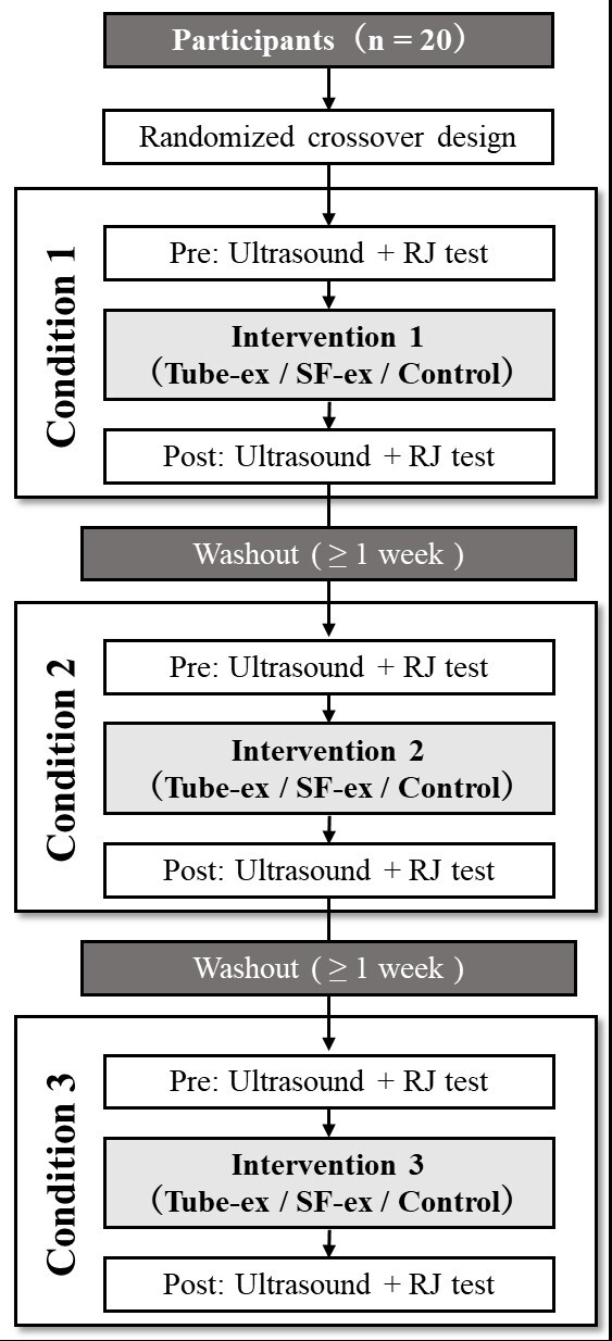
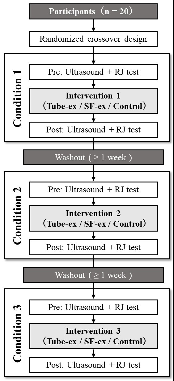
The experimental procedure for each participant is illustrated in Figure 1. This study employed a randomized crossover design comparing three conditions: the Tube-ex, the SF-ex, and a control condition with no intervention. Each participant completed all three conditions in a randomized order, with a washout period of at least one week between sessions. The order of interventions was randomly assigned using a random number table generated in Microsoft Excel. In each session, ultrasound images of three IFMs—the abductor hallucis (AbH), flexor hallucis brevis (FHB), and flexor digitorum brevis (FDB)—were obtained three times each using a standardized imaging protocol as pre-intervention measurements. Participants then performed the rebound jump test, followed by the assigned intervention (SF-ex, Tube-ex, or Control). Immediately after the intervention, the same ultrasound measurements were repeated (post-intervention), and the rebound jump test was performed again to complete the session. All measurements were conducted by the same examiner under consistent environmental and procedural conditions to minimize measurement variability.
Outcome measures
Morphological characteristics
Foot length was measured under non–weight-bearing conditions using a digital caliper (Shinwa Rules Co., Ltd., Japan). Participants were seated with the knee flexed to approximately 90°, and the plantar surface was placed lightly in contact with the floor in a natural resting position. Foot length was defined as the linear distance from the most posterior aspect of the calcaneus to the most anterior projection of the toes (typically the tip of the hallux). The non-dominant foot (the side not used for ball-kicking) was selected because the dominant supporting limb is more influenced by postural stabilization demands, making the non-dominant side more suitable for obtaining stable strength measurements.18
Toe grip strength
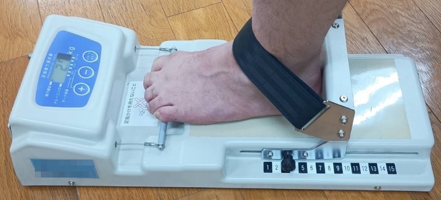
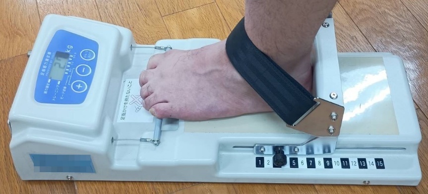
Toe grip strength was measured on the non-dominant limb using a toe-grip dynamometer (Figure 2; T.K.K. 3364; Takei Scientific Instruments Co., Ltd., Japan). This procedure has demonstrated high reliability in previous studies and was performed according to an established protocol.19 Participants sat on a 40-cm-high chair with their arms crossed over the chest and the pelvis maintained in a neutral position. The hip, knee, and ankle joints were positioned at 90°of flexion, and the foot was positioned in line with the body’s midline. The plantar surface was placed on the dynamometer, with the heel secured against the stopper and the dorsum of the foot lightly fixed using a non-elastic strap. The proximal phalanx of the hallux was aligned with the grip bar. Participants were instructed to gradually increase force over three seconds and maintain maximal voluntary contraction for approximately two seconds. Two trials were performed with sufficient rest between attempts, and the greater value was used for analysis. Measurements were obtained from the non-dominant foot to maintain consistency with the foot length assessment.
Ultrasound measurements of intrinsic foot muscles
Changes in the CSA of the IFMs due to acute muscle swelling were assessed using an ultrasound imaging system (SONIMAGE MX1α, KONICA MINOLTA, Japan) equipped with a high-sensitivity linear probe (L11-3). All measurements were performed on the non-dominant foot. Participants were positioned in the prone position with the knee flexed to 90°, and the ankle was maintained in a neutral position (0° dorsiflexion) using a cushion to ensure relaxation. The measurement procedures followed previously validated and reliable protocols described in earlier studies.20 All ultrasound examinations were performed by an experienced examiner. To ensure consistent probe positioning and minimize tissue deformation during scanning, a custom-made probe fixation device was used. The examiner carefully monitored probe contact to avoid excessive pressure on the underlying tissue. The AbH, FHB, and FDB muscles were selected as target muscles because they play essential roles in supporting the medial longitudinal arch.21,22 Measurement landmarks were identified on the skin using an oil-based marker to ensure consistency across sessions. For the AbH, the probe was placed on the anterior aspect of the medial malleolus, perpendicular to the long axis of the foot. For the FHB, the probe was positioned perpendicular to the muscle fiber orientation at the level of the first metatarsal base. For the FDB, the probe was applied perpendicular to the line connecting the medial calcaneal tubercle and the base of the third toe, and the CSA image was obtained at the midpoint of this line. For each muscle, three transverse images were acquired. Image analysis was performed using ImageJ software (version 1.53t; National Institutes of Health, Bethesda, MD, USA). The region of interest was manually traced along the inner boundary of the muscle fascia, and the CSA was calculated from the delineated area. The mean value of the three images was used as the representative CSA for each muscle. This analytical approach has demonstrated excellent intra- and inter-rater reliability in previous studies,23,24 and the same methodological considerations were applied in the present study. To evaluate measurement reliability, intra-session consistency was assessed using the intraclass correlation coefficient [ICC(3,1)], based on a two-way mixed-effects model with absolute agreement among the three trials conducted by the same examiner. To estimate practical measurement error, the standard error of measurement (SEM) and minimal detectable change at the 95% confidence level (MDC95) were calculated. The MDC95 was derived from the first and second trials of each condition using the formula: MDC95 = 1.96 × √2 × SEM.
Repeated rebound jump test and reactive strength index
The repeated rebound jump test was performed using the non-dominant limb. To eliminate the influence of arm swing, participants performed all jumps with their hands placed on their hips. From an initial upright standing position with the knee fully extended, participants were instructed to slightly flex the knee and jump as high as possible. Upon landing, they were instructed to minimize ground contact time and immediately transition to the subsequent jump. Seven consecutive single-leg jumps were performed using the non-dominant limb, and two sets were completed. A rest period of one minute was provided between sets. During the trials, participants were verbally instructed to keep their gaze directed forward and to land at the same location for each jump. Jump performance was measured using an optical timing system (Optojump™ system; Microgate, Bolzano, Italy), which automatically recorded contact time and flight time for each jump. Of the seven consecutive jumps, the maximum and minimum values were excluded, and the mean of the remaining five jumps was calculated to obtain a representative value, thereby reducing the influence of outliers. The higher value obtained from the two sets was used for subsequent analyses. The RSI was calculated as the ratio of flight time to contact time (RSI = flight time / contact time). The repeated rebound jump test and the calculation of RSI have demonstrated high day-to-day reliability in previous studies,25,26 and the same testing procedures were applied in the present study.
Subjective evaluation
After completing each exercise, participants rated their subjective impressions regarding the exercise in three separate questions, using a 5-point Likert scale (5 = strongly agree, 4 = agree, 3 = neutral, 2 = disagree, 1 = strongly disagree). This measure was included to assess perceived feasibility and acceptability of each exercise. They were asked to evaluate whether the exercise was easy to perform, whether it was perceived as effective, and whether they felt that it strengthened the toes and plantar muscles. Higher scores indicated greater perceived ease of performance, effectiveness, or sense of muscle engagement.
Interventions
Toe-tube exercise
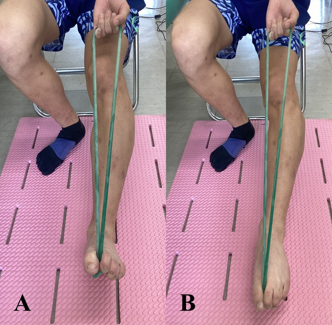
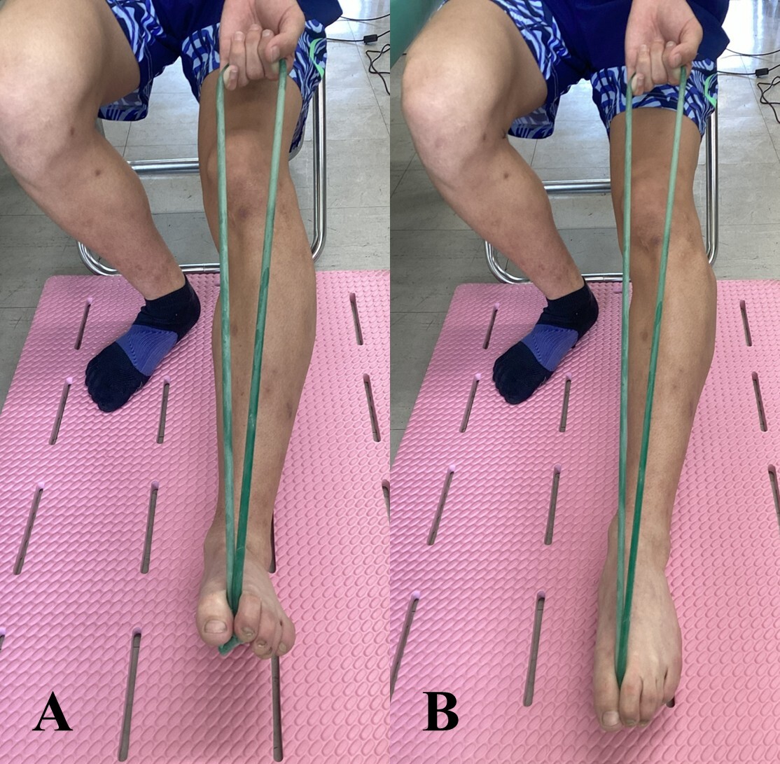
The Tube-ex was performed in a seated position with the knee of the exercising limb fully extended and the heel in contact with the floor (Figure 2). An elastic tube (TheraBand®, The Hygenic Corp., Akron, OH, USA; green, heavy resistance; length 1 m; natural rubber) was used. A knot was tied at each end of the tube, and the two knots were positioned between the hallux and second toe at the midfoot level to stabilize the tube around the toes. The opposite end of the tube was held by the participant’s hand and positioned to provide sufficient resistance for maximal voluntary contraction of the IFMs. Participants performed ankle plantarflexion and dorsiflexion against the elastic resistance. At end-range plantarflexion, they were instructed to maintain flexion of the metatarsophalangeal (MTP) and interphalangeal (IP) joints of all toes, including the hallux, and to produce a maximal voluntary contraction of the IFMs for 5 seconds. Verbal cues were provided to prevent unwanted IP joint extension and to ensure consistent execution of the task. Each set consisted of 10 maximal-effort repetitions, and three sets were performed with 45 seconds of rest between sets. Throughout the exercise, the direction of resistance was kept parallel to the longitudinal axis of the lower limb, and participants were instructed to coordinate toe flexion with ankle motion.
Short-foot exercise
The SF-ex was performed in a seated position with the knees flexed to 90°.27 Participants were instructed to shorten the foot in the anteroposterior direction by drawing the first metatarsal head toward the heel without curling the toes. Care was taken to avoid toe flexion or lifting of the foot, and participants maintained the contraction for 5 seconds at a position where plantar muscle activation could be perceived. Visual feedback was provided to monitor arch elevation and minimize excessive ankle or toe motion. Each set consisted of 10 maximal voluntary contractions, and three sets were performed with 45 seconds of rest between sets. All participants received direct instruction from a strength and conditioning specialist with 16 years of professional experience. Both exercise conditions were performed with identical volume and rest intervals to allow direct comparison of acute effects between interventions.
Control condition
In the control condition, participants remained seated in the same posture for five minutes without performing any voluntary movements of the toes or ankle. They were instructed to stay relaxed and minimize muscle activity throughout the rest period. The duration of the control condition was matched to the exercise conditions.
STATISTICAL METHODS
All statistical analyses were performed using Modified R Commander (version 4.5.0), based on the R statistical software package (R Foundation for Statistical Computing, Vienna, Austria; https://www.r-project.org/). The normality of each variable was verified using the Shapiro–Wilk test. For normally distributed data, a two-way repeated-measures analysis of variance (ANOVA) with factors of condition (three levels: SF-ex, Tube-ex, and Control) and time (two levels: pre- and post-intervention) was used to examine main effects and interaction effects on muscle CSA and the RSI. When a significant interaction or main effect was observed, post hoc pairwise comparisons were performed using Bonferroni correction. For variables that did not meet the assumption of normality, within-subject comparisons between conditions or time points were conducted using the Wilcoxon signed-rank test. The same nonparametric approach was applied to analyze subjective evaluation scores (ease of performance, perceived effectiveness, and sense of muscle engagement) across exercise conditions. Effect sizes were calculated as Cohen’s d for pairwise comparisons and partial eta squared (partial η²) for ANOVA results and were interpreted according to conventional thresholds (small: d = 0.2, η² = 0.01; medium: d = 0.5, η² = 0.06; large: d = 0.8, η² = 0.14). The level of statistical significance was set at p < 0.05 (two-tailed).
RESULTS
Twenty healthy male university students participated in this study. The mean age, height, and body mass were 20.8 ± 1.2 years, 173.8 ± 5.9 cm, and 73.3 ± 10.9 kg, respectively, with a mean body mass index of 24.2 ± 3.3 kg/m². Mean foot length was 25.4 ± 1.4 cm, and maximal toe-grip strength of the non-dominant foot averaged 23.0 ± 8.3 kgf.
The within-session reliability of ultrasound measurements was good to excellent across all IFMs and conditions. Intraclass correlation coefficients [ICC(3,1)] ranged from 0.969 to 0.997, indicating high consistency among repeated measurements obtained by the same examiner (Table 1). Measurement error was small, with the SEM ranging from 2.1 to 3.8 mm² and the MDC95 ranging from 5.9 to 10.5 mm², corresponding to 1.1–1.9% and 3.1–5.3% of the grand mean, respectively. The magnitude of CSA change observed after the Tube-ex exceeded the MDC95 values for all IFMs, indicating changes beyond measurement error. In contrast, CSA changes following the SF-ex exceeded the MDC95 threshold only for selected muscles, whereas no CSA changes in the control condition exceeded the MDC95 values.
Significant time × condition interactions were observed for all IFMs (Table 2). For the AbH, a significant interaction was found (F(1.46, 27.73)=9.65, p=0.002, partial η²=0.34), representing a large effect. Post hoc analyses showed that CSA increased after SF-ex and Tube-ex compared with Control, with the largest increase following Tube-ex (approximately +20 mm²). In the FHB, a significant time × condition interaction was also observed (F(1.52, 28.84)=5.14, p=0.019, partial η²=0.21), representing a large effect. CSA increased after Tube-ex (approximately +11 mm²) and to a lesser extent after SF-ex (approximately +5 mm²), whereas no meaningful change was observed in the Control condition. For the FDB, a highly significant interaction was detected (F(2, 38)=29.80, p<0.001, partial η²=0.61), representing a large effect. CSA increased markedly after Tube-ex (approximately +25 mm²), exceeding the changes observed after SF-ex (approximately +17 mm²) and Control. Overall, Tube-ex induced the greatest immediate increases in CSA across all IFMs.
Regarding the RSI, a significant time × condition interaction was observed (F(2, 38) = 3.68, p = 0.035, partial η² = 0.16, la arge effect; Table 3). Post hoc analyses indicated that only the Tube-ex resulted in a significant increase in RSI from pre- to post-intervention (simple main effect of time: F(1, 19) = 14.92, p = 0.001), whereas no significant changes were observed following the SF-ex or the control condition. Furthermore, Shaffer-corrected post hoc comparisons confirmed that RSI values after Tube-ex were significantly higher than those observed after both SF-ex (d = 0.80, large effect) and Control (d = 0.66, moderate effect).
Subjective evaluations (Table 4) further supported these findings. Participants rated Tube-ex as significantly easier to perform (p=0.005, r=0.61) and providing a stronger training sensation (p=0.042, r=0.45) than SF-ex.
Taken together, these morphological, functional, and perceptual results consistently demonstrate that the newly developed Tube-ex produced greater acute morphological and functional responses than SF-ex, providing greater acute activation of IFMs and enhanced reactive performance.
DISCUSSION
This study aimed to compare the immediate effects of a newly developed Tube-ex with those of the conventional SF-ex and a control condition. Significant time × condition interactions were observed in the CSA of the AbH, FHB, and FDB muscles, with the greatest increases following Tube-ex. In addition, only Tube-ex significantly improved the RSI, suggesting an acute enhancement of reactive jump performance. Participants also rated Tube-ex as easier to perform and more effective than SF-ex, indicating its practical feasibility. These findings indicate that Tube-ex acutely enhanced ultrasound-derived IFM morphology and RSI. However, because electromyographic activity was not assessed, the present study could not determine the relative contribution of intrinsic versus extrinsic toe flexor muscles to these responses.
Tube-ex involves coordinated toe flexion with ankle motion while maintaining MTP joint flexion against elastic resistance. This dynamic configuration likely increases intrinsic muscle demand under external load, consistent with previous reports demonstrating enhanced neuromuscular activation during elastic resistance exercise.12 In contrast, SF-ex primarily involves static arch shortening with limited resistance, potentially explaining the smaller morphological and performance responses observed. Because Tube-ex incorporates both ankle plantar- and dorsiflexion, it may better reflect late-stance mechanics during rapid ground contact, thereby providing a more functionally relevant stimulus.
The present increase in muscle CSA aligns with prior findings reporting a 7–10% enlargement in the AbH, FHB, and FDB immediately after toe flexion tasks, which has been interpreted as transient muscle swelling associated with increased intramuscular pressure and perfusion.28 In the current study, the 6–13% increases in CSA after Tube-ex exceeded MDC thresholds, indicating that the response was unlikely attributable to measurement error. These transient morphological changes may reflect increased muscle recruitment and local hemodynamic responses under dynamic loading. Furthermore, findings in adolescent athletes have shown that RSI values around 0.35 are significantly associated with the muscle thickness and CSA of the AbH and FDB.20 Because RSI reflects the ratio of flight time to contact time and is commonly used as an indicator of stretch–shortening cycle function in the lower limbs, the observed improvement suggests enhanced efficiency of force transmission during rapid ground contact, potentially mediated by improved plantar stiffness regulation.29 Coordinated activation of intrinsic and extrinsic plantar flexors may contribute to improved dynamic stiffness regulation during explosive tasks.13,30 Both intrinsic and extrinsic muscles collectively generate MTP flexion moments, emphasizing the importance of their coordinated function. Thus, Tube-ex may promote this coordinated activity, optimizing dynamic arch support and plantar stiffness and contributing to reactive performance during explosive tasks.
From a performance perspective, the rapid improvement in reactive strength suggests that Tube-ex may serve as a neuromuscular priming strategy prior to explosive activities. These findings may have clinical relevance for early-stage rehabilitation, where low-load exercises capable of eliciting neuromuscular activation are desirable. Because the exercise requires only elastic resistance and can be easily implemented within training routines, it may represent a practical preparation modality for sports involving rapid push-off and landing tasks. However, whether the acute improvements in RSI observed in the present study translate to more complex athletic movements such as sprinting, cutting, or change-of-direction tasks remains unclear. Future studies should investigate this transfer by assessing sprint acceleration, cutting and change-of-direction performance, ground contact time, lower-limb stiffness, plantar pressure distribution, and lower-extremity kinematics and kinetics during sport-specific tasks.
This study examined only the acute effects in healthy young male participants. Future research should include diverse populations to assess sex and age differences, investigate longer-term adaptations (e.g., muscle morphology, arch-related measures, and functional performance), and evaluate the effectiveness of Tube-ex in injured or clinical populations. Optimal resistance magnitude and direction should also be determined. Finally, electromyographic assessment was not performed in the present study. Therefore, although ultrasound measurements demonstrated acute increases in the CSA of muscles anatomically classified as IFM, it was not possible to directly confirm selective activation of IFM or distinguish their contribution from that of extrinsic toe flexors. Because toe flexion tasks generally involve coordinated activation of both intrinsic and extrinsic musculature, the observed improvements in RSI may reflect integrated neuromuscular responses rather than isolated IFM activation. Combining electromyography with plantar pressure measures may help clarify the underlying neuromechanical mechanisms in future studies. By integrating toe flexion with ankle motion under elastic resistance, this exercise may nevertheless provide a functionally relevant stimulus for optimizing plantar stiffness during explosive tasks.
CONCLUSIONS
Tube-ex produced acute increases in IFM CSA and RSI in healthy young adults. The observed changes were generally greater than those following SF-ex or a control condition. Further research is warranted to investigate long-term adaptations and potential applications in clinical populations.
Conflict of Interest
The authors declare no conflicts of interest.
ACKNOWLEDGMENTS
The authors would like to thank all participants for their time and commitment to this study. We also acknowledge the assistance of laboratory staff in data collection and the constructive discussions with colleagues during the development of the study protocol. This work was supported by the Japan Society for the Promotion of Science (JSPS KAKENHI) [grant number JP24K14419].