INTRODUCTION
Anterior cruciate ligament (ACL) injury is one of the most common injuries in sports activities that cause functional problems, such as knee joint instability and muscle weakness, as well as increased risk of reinjury.1 Traditionally, functional impairments after ACL injury have been understood mainly as issues within the peripheral musculoskeletal system. However, recent neurological and neurophysiological studies have indicated that these dysfunctions may stem from alterations in the central nervous system (CNS).1–3 Although evidence mainly comes from animal studies, mechanoreceptors have been identified in the ACL, which are thought to transmit afferent information on joint position and kinesthesia to the CNS.4 Therefore, ACL injury may affect not only peripheral structures but also sensorimotor integration mechanisms through afferent input loss. Indeed, individuals with ACL injury demonstrate abnormal control of quadriceps muscle activity and muscle output imbalance during static and dynamic tasks.5 These findings are accompanied by adaptations in motor-related brain regions and corticospinal excitability alterations.6,7 Studies using functional magnetic resonance imaging and transcranial magnetic stimulation have reported reduced activation in the primary motor and somatosensory cortices, alongside increased activation in subcortical regions, such as the cerebellum and basal ganglia, after ACL injury.2,8 These neural alterations may extend beyond the injured limb to the contralateral intact side,9,10 suggesting that neural adaptations following injury occur systemically and bilaterally. Furthermore, previous studies have reported persistent increases in cortical inhibition and deficits in quadriceps force control even after ACL reconstruction,11,12 indicating the importance of neural function evaluation during the rehabilitation process. Based on these findings, increasing attention has been paid to viewing ACL injury not merely as a peripheral joint injury but as a neurophysiological dysfunction.13,14 CNS reorganization after peripheral ligament injuries, such as ACL injury, is thought to involve sensory input alteration, cortical inhibition and facilitation imbalance, as well as motor planning and execution network reconfiguration. Incomplete neural adaptation in these systems may contribute to residual muscle weakness and increase reinjury risk.3,15 In addition, increased cognitive-motor interference during simultaneous cognitive and motor tasks16 and impaired proprioceptive reweighting17 has been reported, indicating progressive disruption of sensorimotor integration at the central level. Recently, neuron-targeted rehabilitation , motor imagery, and brain–computer interface-based interventions have been proposed as novel approaches to the restoration of optimal neuromuscular control via CNS plasticity modulation.15,18 Collectively, these findings suggest that comprehensive understanding of peripheral and central neurophysiological changes is essential for addressing persistent muscle weakness and functional impairments after ACL injury.
After injury, despite continued effort, the action potentials of a voluntarily contracting muscle undergo transient suppression after electric stimulation of the mixed-nerve innervating that muscle.19 This period or electrical inactivity, referred to as the mixed-nerve silent period (SP), results from several physiologic mechanisms.19 Voluntary muscle activity is regulated by complex neural control mechanisms at the peripheral and central levels. In particular, the electromyographic SP observed after electrical stimulation appears as transient muscle activity suppression following supramaximal stimulation of the peripheral nerve. SP termination has been reported to be determined by spinal inhibitory reflexes dependent on afferent impulses.20 Comparative studies using cutaneous and mixed-nerve stimulation have also been conducted to elucidate the mechanisms underlying SP expression. In one study, patients with paroxysmal kinesigenic dyskinesia showed discrepancies between the cutaneous SP and mixed-nerve SP, particularly characterized by the disappearance of the third phase of the mixed-nerve SP and loss of the overlapping phase.21 These findings indicate the presence of abnormal integration of the spinal interneurons and support the idea that SP reflects both peripheral and central integration processes. Further studies have investigated the temporal characteristics and reexcitation phenomena associated with SP during motor control. When a load is suddenly released during voluntary contraction, a transient SP is followed by the emergence of a high-amplitude motor potential known as the terminal volley.22 This reexcitation is believed to be mediated by proprioceptive feedback through gamma motor neurons, indicating an association between spinal reflex loops and fusimotor activity. At the peripheral level, tendon vibration and stretching have been investigated for their effects on electromyographic responses; however, Achilles tendon vibration did not affect the H-reflex, T-reflex, or SP-related parameters, suggesting limited influence of peripheral interventions on SP modulation.23 During sustained muscle contraction, SP is prolonged at 30% MVC and shortened during recovery, suggesting that fatigue-related changes in central drive influence the interaction between peripheral inhibitory and central excitatory mechanisms.24 Collectively, these findings suggest that SP is not merely an inhibitory response but rather a multilevel neural integration phenomenon that involves the central, spinal, and peripheral systems. SP has been considered to represent a comprehensive index reflecting the degree of excitability or disinhibition of the CNS, as it encompasses the total circuit time from the point of peripheral nerve stimulation to CNS, including the M wave, F wave, and long-latency reflex (LLR).25 Moreover, SP alterations following ACL reconstruction have been previously reported,26 suggesting that SP can provide a foundation for novel approaches in clinical assessment for neurophysiology and rehabilitation. By integrating current knowledge on central plasticity following ACL injury, with findings from SP studies on inhibitory neural mechanisms, it may be possible to achieve a more comprehensive understanding of the neurophysiological basis of motor control impairment. Specifically, this framework posits that sensory input loss and motor experience modifications induce reorganization of spinal and cortical inhibitory neural networks, resulting in deficits in force production and coordinated movement. From this perspective, in addition to conventional musculoskeletal focused rehabilitation, neuron-targeted rehabilitation aimed at modulating neuroplasticity is required.20 Clinically, tissue healing, muscle strength, range of motion, and performance tests are often employed to assess return-to-play following sports injuries. However, muscle strength itself can sometimes cause injury, and a smaller range of motion may be beneficial in sprinting. Therefore, these may be insufficient for recovery and return-to-play. In addition, standard performance tests may fail to evaluate the complex motor skills required in sports.
This study aimed to elucidate central nervous CNS excitability during muscle output regulation and explore neurophysiological characteristics in healthy athletes and contrast with findings from an athlete with delayed return to play. Moreover, this study aimed to describe the neurophysiological characteristics of a postoperative athlete with delayed return to sport by comparing her SP findings with those of healthy athletes, as a preliminary exploration of potential neurophysiological indicators for rehabilitation and return-to-sport assessment.
METHODS
This study included 13 healthy female university basketball players and one postoperative athlete who was evaluated three months after right lateral meniscal repair and had not yet returned to running. Healthy participants had no current musculoskeletal symptoms, neurological disorders, or recent lower-extremity injuries affecting sports participation. One participant (age, 18 years; height, 174 cm), was three months post right lateral meniscal repair, was unable to run.
An a priori power analysis was conducted to determine the sample size required to detect a difference in SP between the dominant and nondominant limbs among healthy athletes. Based on an expected mean SP of approximately 105 ms, a standard deviation of 8 ms, α = 0.05, and power = 0.80, the required sample size was estimated to be 11 participants. Therefore, this study included 13 participants, comprising at least 11 healthy individuals. This study was approved by the Hokuriku University Research Ethics Review Committee for Human Subjects (approval no. 2023-16, 27/June/2023). All study tasks were conducted in accordance with the Declaration of Helsinki. Before the experiment, the participants were informed about the study details, potential risks, and publication plans with the document, according to the decision of the Hokuriku University Research Ethics Review Committee for Human Subjects. Subsequently, they provided written informed consent.
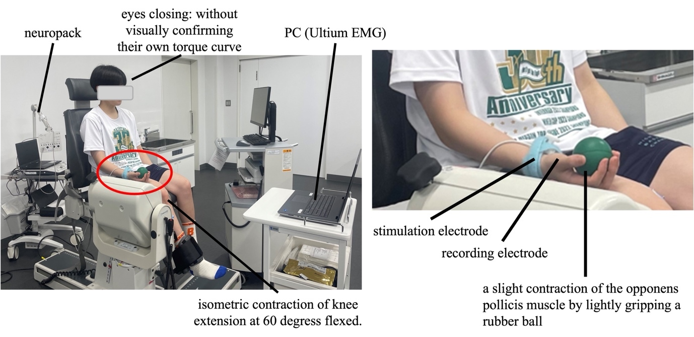
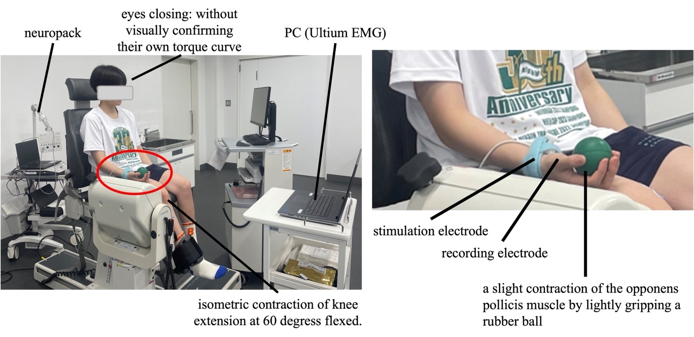
The muscle output adjustment task was maintaining a constant knee extension torque, which was performed one side at a time. The participants’ lower limb laterality was determined as the side they found easier to kick a ball, designated as the dominant leg. The order of task execution was randomized. The task was performed using the BIODEX System 4 BDX-4X (Biodex Medical System Inc., USA). The participants were seated with their knee flexed at 60°. They were instructed to maintain a knee extension torque corresponding to 25% of their measured maximal voluntary isometric contraction for 60 s. The BIODEX system was synchronized with the electromyography measurement system Ultium EMG EM-U880 (Noraxon, USA). Torque data from BIODEX were imported into Ultium EMG and displayed on its connected computer (Figure 1). The participants maintained their muscle output without visual feedback of their torque curve, indicating that the torque was self-generated. Muscle output was adjusted based on verbal feedback provided by the same examiner for all participants throughout testing to minimize inter-examiner variability. Standardized verbal instructions were used throughout testing.
During the muscle output adjustment task, a mixed-nerve SP was derived from the ipsilateral opponens pollicis muscle using NeuroPack (Nihon Kohden, Japan) (Figure 1). The recording conditions for SP measurement were as follows: muscle, opponens pollicis; side, ipsilateral to knee extension; contraction intensity, light contraction achieved by gently gripping a rubber ball; active electrode, placed over the belly of the opponens pollicis muscle; and reference electrode, placed at the distal end of the proximal phalanx of the thumb. Electrode placement and recording procedures were standardized across participants. The stimulation conditions for SP measurement were as follows: median nerve- center of the wrist; intensity- supramaximal stimulation at 120% of the intensity that produced the maximal M-wave amplitude; frequency- 0.5 Hz; duration- 0.2 ms. To improve measurement stability, SP recordings were obtained 16 times under each condition. This is because standard neurophysiological evoked electromyography tests typically involve 16 repetitive stimuli. Stimulation intensity was adjusted individually to ensure supramaximal stimulation throughout the test. The obtained 16 recorded SPs were measured, and their average was used as the representative value for each condition. Based on the obtained results, the SP values of the dominant and nondominant legs were compared among healthy participants (n = 13). In addition, the SP values of the single case subject were compared with those of the healthy participants. Specifically, the comparison was made between the dominant (injured/surgical) and nondominant legs and between the nondominant (uninjured) and nondominant ones.
Statistical methods
Statistical analyses were conducted using SPSS (version 31.0.0.0, IBM, Armonk, NY, USA). To compare the SP between the dominant and nondominant legs of the healthy participants, normality was first evaluated using the Shapiro–Wilk test. Normality was confirmed (n = 13; dominant leg, p = 0.164, and nondominant leg, p = 0.838); therefore, a paired t-test was conducted for further analysis with a significance level set at 5%. Furthermore, as the data of healthy participants were normally distributed, the case’s data were compared by evaluating the extent of deviation from the healthy participants’ mean values. In addition, because the study included a single postoperative case comparison with the small healthy reference sample, a Crawford and Howell modified t-test was additionally performed to evaluate whether the case’s SP value significantly deviated from the normative distribution. The analysis was conducted using Singlims_ES software developed by Crawford and colleagues.27,28 Statistical significance was set at p < 0.05.
RESULTS
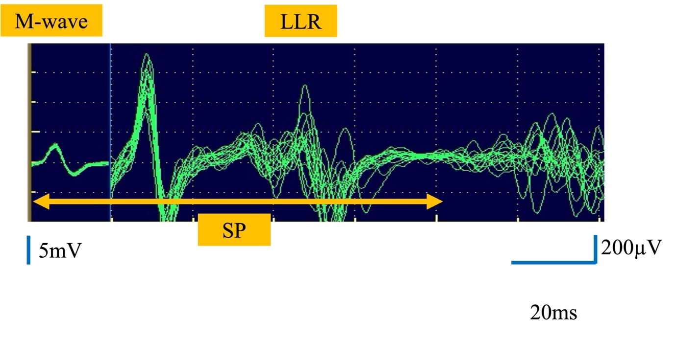
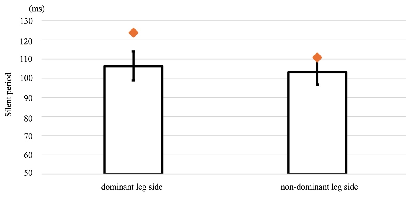
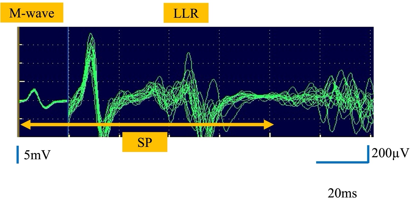
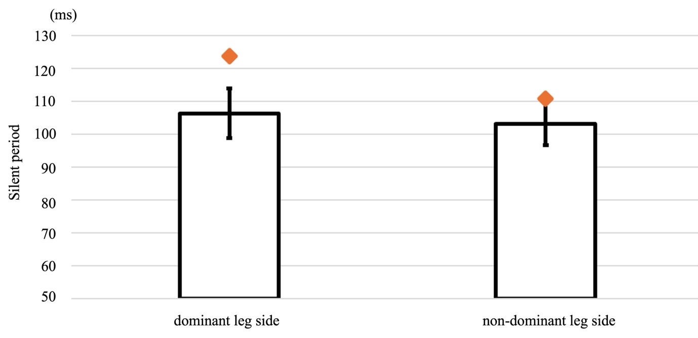
The healthy reference group consisted of 13 female university basketball players (age, 19.6 ± 1.1 years [range, 18–22 years]; height, 164.2 ± 5.5 cm [range, 156–173 cm]). One additional athlete was evaluated three months after meniscal repair who had not yet returned to running (age, 18 years; height, 174 cm). Figure 2 presents an example of typical electromyography waveforms obtained in this study. The waveforms show that the SP was recorded with good reproducibility. The SP for the healthy participants’ dominant leg was 106.3 ± 7.5 ms, whereas that for the nondominant leg was 103.2 ± 6.5 ms. On the other hand, in the participant who was three months post right meniscal repair (Case subject), the injured leg, which was the dominant leg had an SP of 123.7 and the noninjured leg side, (nondominant leg) showed SP of 110.8 ms, 110.8 ms. No significant difference was observed in the SP between the dominant and nondominant legs of the healthy participants (Table 1). For the single subject case, no differences were observed in the noninjured (nondominant) leg compared with the nondominant leg of the healthy participants. However, for the injured (dominant) leg, the results deviated by >2 SD from the data of the healthy participants’ dominant leg (Figure 3). And the SP for injured (dominant) leg demonstrated significantly prolonged SP compared with the healthy athlete reference sample (t = 2.236, two-tailed p = 0.045, effect size Z-CC = 2.320).
DISCUSSION
As noted previously, a transient suppression of muscle activity, referred to as the mixed-nerve SP, occurs after electrical stimulation of the innervating nerve during sustained voluntary contraction and is attributed to several physiological mechanisms.19 The SP analyzed in this study encompasses the M wave, F wave, and LLR. The M wave reflects the conductive properties of the peripheral nerve and the physiological state of the muscle.29–31 The F wave represents the excitability of spinal motor neurons,32 whereas LLR indicates the excitability of the brainstem or motor cortex.33–35 Accordingly, SP variations are considered to reflect the level of facilitation within the brainstem or motor cortex, influenced by muscle spindle activity, Golgi tendon organ inhibition, recurrent spinal inhibition, and cortical inhibitory mechanisms.34,36–38 In particular, SP recorded from the upper limbs includes long-latency components involving supraspinal pathways, the measurement was intended to reflect generalized CNS excitability modulation during motor output regulation rather than local lower-limb peripheral function alone. Consequently, SP variations are considered potential indices of changes in central excitability, reflecting both facilitatory and inhibitory processes within the brainstem or motor cortex. Based on the concept that SP is used to evaluate brainstem and cortical facilitation in physical therapy and to examine neuromuscular function in healthy individuals under various conditions, baseline data from healthy subjects were collected and compared with data from the patient who underwent lower limb surgery. The authors plan to continue additional basic research to use SP as a tool for assessing recovery status and return-to-play indicators in athletes recovering from injury.25,26,38–44
The outcomes of this study demonstrated that the SP of the ipsilateral opponens pollicis muscle measured during isometric knee extension at a constant intensity showed values comparable to those observed in previous studies involving healthy individuals.39,44 No significant difference in SP was observed between the dominant and nondominant legs among athletes belonging to a single competitive team. These findings suggest that CNS excitability during the motor task was comparable between these healthy athletes and the general healthy population. This consistency may be considered one of the strengths of this study, as it indicates methodological reproducibility across populations and supports the validity of SP parameters as stable neurophysiological indicators.
Contrarily, in the single case of an athlete who had undergone meniscal repair and was unable to resume running despite passing the expected return-to-sport period, the SP on the noninjured leg showed values nearly identical to those of healthy athletes (within 2 SD), whereas the SP value on the injured side deviated by >2 SD from the mean of the healthy group, a moderate deviation from the distribution observed in healthy athletes. As the results for the healthy participants were assumed to follow a normal distribution, approximately 95% of the data would fall within the range of the mean ± 2 SD. In clinical neurophysiology, values exceeding approximately 2 standard deviations from normative means are commonly interpreted as being outside the expected reference range, assuming normal distribution of the data.45 Thus, an SP exceeding the mean + 2 SD indicates a high likelihood of being an outlier, implying that this single athlete may demonstrate properties different from those of healthy athletes. This interpretation is supported by the results of the Crawford and Howell modified t-test. Furthermore, because SP reflects the excitability and inhibitory mechanisms of the CNS at supraspinal levels, a prolonged SP of >2 SD suggests the possible occurrence of CNS excitability alterations, potentially involving polysynaptic reflex pathways within suprabrainstem regions. However, since fluctuations in SP may be influenced by changes in central nervous system excitability resulting from variations in effort level (% maximum effort) during muscle contraction, this study must take into account the effort level of the knee extensor muscles during task performance, particularly in the healthy subjects. In this experiment, the authors adopted a methodology consistent with their previous studies.26,38–40,43,44 Given that the output level was light (25%) and the SP measurement time (duration of knee extensor output) was 32 seconds, the influence of fatigue, which would be reflected in changes in effort level, is believed to be minimal.
This observation highlights a potentially clinically significant outcome—namely, that SP assessment may help detect subtle post-injury neuromuscular dysfunction not captured by conventional return-to-sport criteria, representing another strength of this study. Conventional return-to-sport evaluations often emphasize peripheral factors such as muscle strength, joint range of motion, and performance tests. However, these assessments may not fully capture residual alterations in central motor control and inhibitory processing. The present findings suggest that SP assessment may provide supplementary neurophysiological information regarding motor output regulation during rehabilitation after sports injury.
However, some weaknesses and limitations must also be acknowledged. Notably, although the number of participants in this study was determined based on power analysis, the final sample size was close to the minimum value required. This may limit the sensitivity of the study to detect subtle differences in SP between conditions or subgroups. And also, although the SP has long been a widely used measure in the field of clinical neurology, the fact that its test-retest reliability has not been assessed remains a challenge for future research. Additionally, the use of a single athletic team limits generalizability to broader athlete populations or different sports, and the absence of longitudinal assessment in the postoperative athlete limits interpretation of how SP may change throughout recovery.
Although this study focused on basketball athletes and a single postoperative meniscal injury case, altered CNS excitability and sensorimotor dysfunction have also been reported in various musculoskeletal and sports-related conditions. Therefore, SP assessment may have broader applicability as an exploratory neurophysiological measure across different sports and injury categories. Nevertheless, further studies involving larger and more diverse cohorts (especially including greater numbers of injured athletes) are required before clinical generalization can be established.
Conclusion
The results of this study indicate that SP values were symmetrical between the dominant and nondominant limbs in healthy athletes, whereas prolonged SP was observed in the postoperative athlete with delayed return to sport. These findings may reflect altered CNS excitability associated with incomplete recovery during muscle output regulation.
While the outcomes of this study provide supportive evidence for the usefulness of SP as a neurophysiological measure of CNS excitability in athletes, future research with larger heterogeneous samples, longitudinal assessment designs, and clinical outcome correlations will be necessary to further validate its applicability in rehabilitation and performance evaluation contexts. Finally, because the present interpretation is based on a single postoperative case, the findings should be considered hypothesis-generating rather than confirmatory.
Conflicts of interest
The authors report no conflicts of interest.
ACKNOWLEDGMENT
The authors report that this study received no external funding and that no individuals or organizations contributed to the work in a manner that requires acknowledgment. The authors also extend their sincere appreciation to all participants for their time and cooperation.