INTRODUCTION
In recent years, the participation of masters athletes in competitive sports has increased significantly. While this population serves as a model for active aging, they inevitably face age-related neuromuscular alterations, such as dynapenia—the loss of muscle strength and power exceeding the loss of muscle mass.1–3 Crucially, for sprinters, the selective loss of neural drive to the posterior chain not only impairs performance but also increases the risk of musculoskeletal injuries, particularly hamstring strains.4 From a sports physical therapy perspective, identifying and addressing these specific activation deficits is essential for injury prevention and performance optimization.
A phenomenon frequently observed in asymptomatic masters sprinters is functional inhibition or specific muscle activation failure during closed kinetic chain movements.5,6 Clinically, profound muscle inhibition following joint injury is well-documented as Arthrogenic Muscle Inhibition (AMI).7,8 However, asymptomatic athletes without acute trauma or pain may exhibit a functional analogue of AMI. This condition, often driven by chronic maladaptive movement patterns or sedentary behavior, is best described as learned non-use, where altered body schema in the central nervous system results in a selective downregulation of neural drive to specific muscles.9
To overcome such centrally-mediated inhibition, biofeedback using surface electromyography (sEMG) is a promising neuro-cognitive intervention. Electromyographic biofeedback converts invisible muscle activity into real-time visual signals, allowing athletes to objectively perceive their motor output.10 By providing an external focus of attention, VFB may facilitate the bypass of inhibitory neural pathways that traditional internal focus coaching cues (e.g., instructing the athlete to squeeze the muscle) are often insufficient to overcome.11
While the efficacy of biofeedback is established in post-injury rehabilitation,12 its application for re-activating dormant muscles in asymptomatic masters athletes remains under-explored. The purpose of this case report was to describe the acute neuromuscular effects of real-time visual biofeedback (VFB) in a masters sprinter with a 20-year history of hamstring activation failure. We aim to highlight the potential of VFB as a practical strategy for optimizing motor control and restoring neural drive in the aging athletic population.
CASE DESCRIPTION
Participant The participant was a 41-year-old male masters sprinter (height: 183 cm, body mass: 62 kg). While maintaining a continuous routine of running and resistance training throughout adulthood, the athlete formally resumed dedicated sprint training in 2019 and began competing in masters athletics in 2023. The primary athletic goal for this individual is to break the national age-group record in the 400-m sprint. Outside of track, the sprinter maintains an active lifestyle, which includes weekly two-hour round trip cycling sessions. The habitual weekly training regimen consists of two to three days of short-distance sprint sessions and two to three days of resistance training. The athlete’s daily occupation is sedentary, which likely contributed to his chronic maladaptive movement patterns.
At the time of testing, the participant was asymptomatic, reporting no current musculoskeletal pain or history of neurological disorders. Despite an extensive training background, the athlete reported a specific, subjective difficulty in activating the hamstring muscles during Romanian deadlift exercises, persisting for approximately two decades. This activation failure occurred despite the absence of any structural injury. This clinical presentation is highly suggestive of a functional neuromuscular deficit, often described as learned non-use.
At the time of the laboratory visit in July 2025, the athlete was in a preparatory phase for an upcoming August competition. Conditioning was at approximately 80-90% of peak condition following a minor injury sustained two months prior. Some accumulated training fatigue was reported during the week of the intervention, with a light hill-sprint session completed the day before testing.
Ethical Considerations The study was conducted in accordance with the Declaration of Helsinki and was approved by the Ethical Review Committee at Kanagawa University (Approval No. 202222-2). Written informed consent was obtained from the participant for the publication of this case report and any accompanying images.
Intervention and Procedures A crossover design was employed to assess the acute effects of different interventions on hamstring activation. Three conditions were performed in a randomized order on the same day, separated by 5-minute rest intervals to minimize fatigue:
-
Control Condition: Conventional Romanian deadlifts performed with the participant’s habitual form without any specific feedback or warm-up intervention.
-
Exercise Condition (Supplemental File): Romanian Deadlifts performed immediately after a specific mobility and stability routine aimed at disinhibiting the posterior chain. The routine consisted of four exercises: Swiss Ball Rollout, Single-Leg Step-Up with Overhead Reach, Side Plank with Dumbbell Row, and Single-Leg Romanian Deadlift with Overhead Press. Each exercise was performed for 60 seconds.13
-
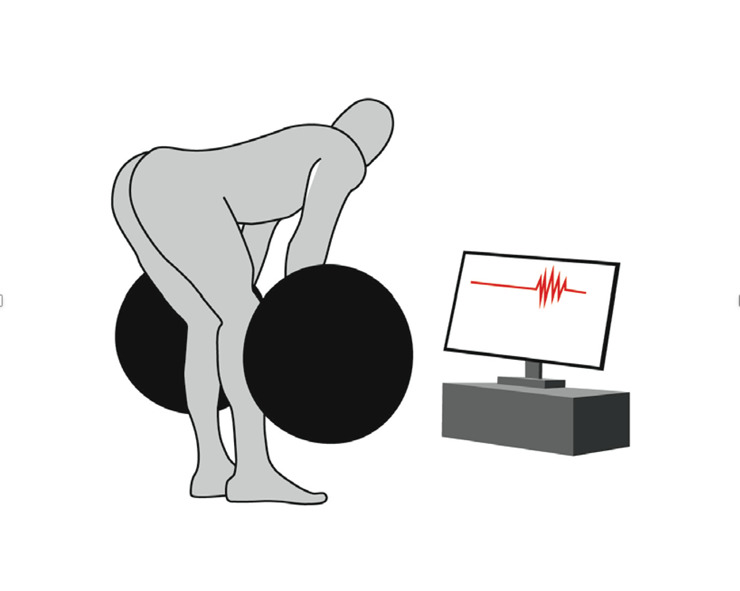
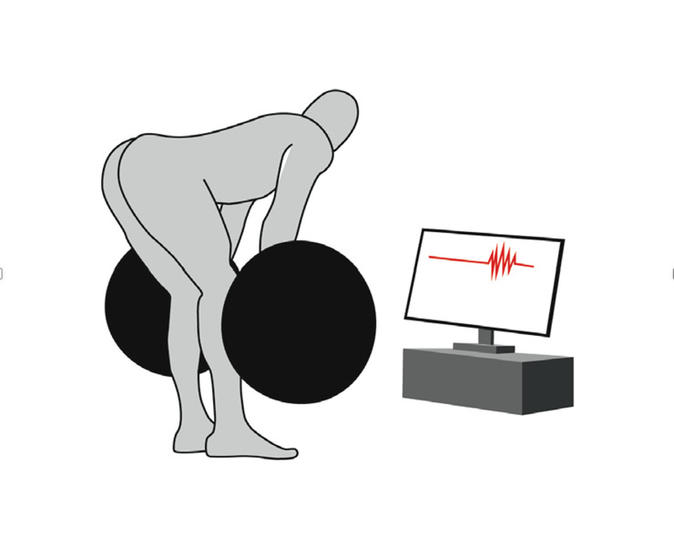
Visual Feedback (VFB) Condition: Romanian Deadlifts performed while the participant viewed real-time EMG waveforms of his hamstring muscles. A laptop monitor displaying the EMG signals was positioned directly in the participant’s field of view. The participant was instructed to maximize the amplitude of the waveform during the concentric phase of the lift. Figure 1 depicts an athlete performing deadlifts while receiving real-time visual feedback on hamstring activation via an EMG waveform display. The setup included a computer monitor displaying EMG signals and providing immediate visual feedback to optimize muscle activation during exercise. For each condition, the participant performed five repetitions of the deadlift at loads of 50 kg and 70 kg, corresponding to his typical training intensity. The cadence was controlled at approximately 35 repetitions per minute.
EMG Data Acquisition and Analysis Surface EMG (sEMG) signals were acquired using a wireless EMG system (Trigno, Delsys, Boston, MA, USA). Following skin preparation, Trigno Avanti sensors were placed on the dominant leg (right side) according to SENIAM guidelines.14 Electromyographic signals were recorded via the analog output of the Trigno Base Station into a PowerLab 16/35 data acquisition system (ADInstruments, Sydney, Australia). Crucially, the analog output of the Delsys Trigno system applies a hardware gain of 909 V/V to scale low-voltage biological signals to the system’s full dynamic range (±5 V).15,16 Therefore, to reconstruct the physiological EMG amplitude (mV) at the skin surface, all recorded voltage data were post-processed by dividing by a factor of 909. The average peak amplitude of the five repetitions was calculated for analysis.
Subjective Assessment To assess the subjective perception of muscle engagement, the participant rated his hamstring activation immediately after each trial using a 10-cm Visual Analog Scale (VAS), where 0 cm represented no activation and 10 cm represented maximal activation.
OUTCOMES
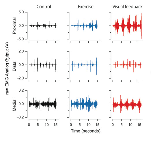
Electromyographic Assessment The raw EMG waveforms (Figure 2) visually corroborated these findings, showing a transition from near-silence in the control condition to distinct, high-amplitude burst patterns during the VFB condition.
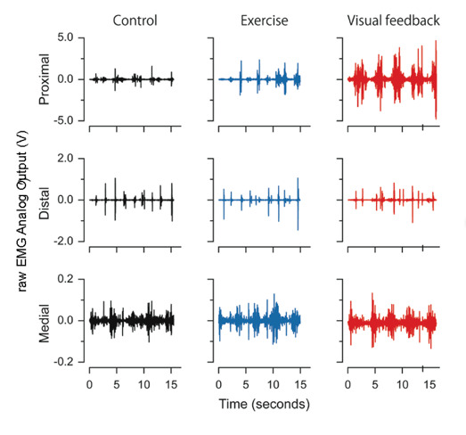
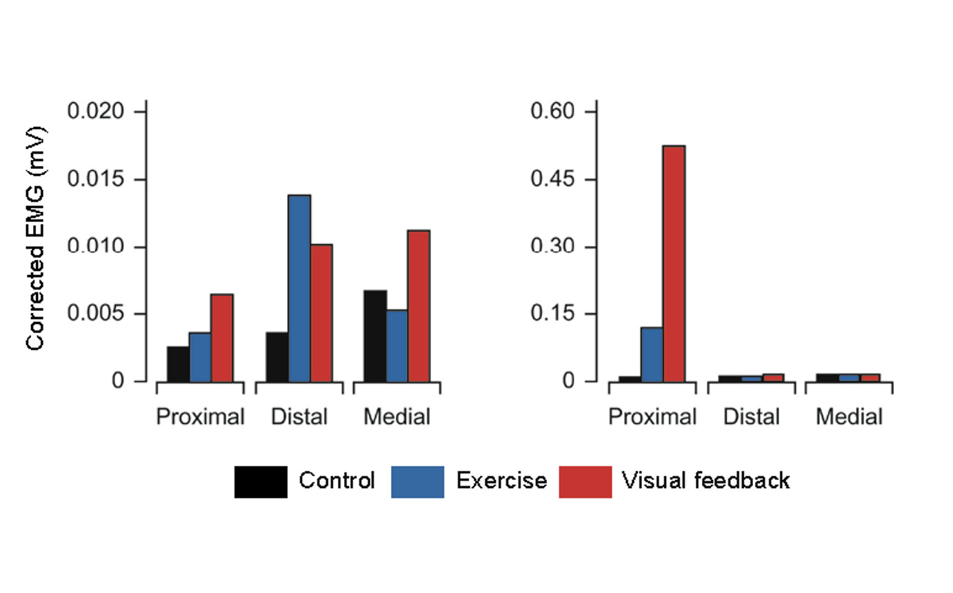
Gain-corrected EMG analysis revealed a marked restoration of hamstring activity (Figure 3).
-
Control Condition: Under the 70 kg Romanian deadlift load, the proximal hamstring amplitude was negligible (0.014 mV), confirming the presence of severe functional inhibition despite the athlete’s effort.
-
Exercise Condition: The mobility and stability routine elicited a moderate increase in amplitude (0.130 mV), representing a ~9-fold improvement over the control.
-
Visual Feedback (VFB) Condition: The VFB intervention resulted in a dramatic ~40-fold increase in average peak amplitude compared to the control, reaching 0.574 mV. This value is consistent with functional levels of muscle activation expected during vigorous voluntary contraction.
_and_70_kg_(right)_romanian_d.png)
Subjective Assessment Subjective ratings of muscle engagement were consistent with the objective EMG findings. The participant’s rating on the Visual Analog Scale (VAS, 0–10 cm) improved from 3.8 cm in the Control condition to 5.8 cm following the exercise routine. The highest rating was recorded during the VFB condition (7.8 cm), confirming that the athlete could consciously perceive the enhanced motor recruitment facilitated by the visual feedback.
DISCUSSION
Disinhibition of Neural Drive
The most striking finding of this case report is the immediate, approximately 40-fold increase in proximal hamstring EMG amplitude during the VFB condition compared to the control. Such a magnitude of change within a single session cannot be attributed to morphological adaptations, such as muscle hypertrophy. Instead, it strongly supports the hypothesis that the athlete’s functional deficit was due to functional neuromuscular inhibition or a learned non-use phenomenon, rather than true physiological weakness.8,9
The baseline EMG amplitude (~0.014 mV) in the control condition indicates that despite the structural integrity of the hamstrings, the central nervous system had downregulated the neural drive to this muscle group. This state closely resembles Arthrogenic Muscle Inhibition (AMI), typically observed after joint injury.7 In the present case, in the absence of acute injury, we posit that chronic maladaptive movement patterns led to a similar functional inhibition. The VFB intervention effectively acted as a disinhibition stimulus, releasing the neural brake and restoring the drive to a functional level consistent with vigorous voluntary contraction.
Mechanisms of Efficacy: The Visual Bypass and Attentional Focus
The efficacy of VFB can be explained through the lens of motor learning and attentional focus theories. Traditional coaching cues (e.g., instructing the athlete to squeeze the hamstring) induce an internal focus of attention. Research by Wulf and colleagues has consistently shown that an internal focus can constrain the motor system and degrade performance.11 In contrast, the real-time EMG waveform provided a clear external focus target (i.e., directing them to make the wave bigger), which likely facilitated more automatic motor control.
Furthermore, the authors propose a visual bypass mechanism (conceptually distinct from the thalamo-amygdala pathway often termed visual bypass in fear conditioning literature17) to explain the rapid restoration of activation. Neuroplasticity research on ACL injuries suggests that when proprioceptive feedback is compromised or inhibited, the brain undergoes cross-modal reorganization to rely more heavily on visual information for motor control.10,18 In this study, the high-fidelity visual feedback likely engaged the visual cortex and premotor areas, bypassing the compromised or inhibited proprioceptive feedback loops at the spinal level.18 By converting the internal physiological state (muscle contraction) into an external visual goal, the athlete was able to access high-threshold motor units that were previously dormant.19
Clinical Implications
These findings have significant implications for the management of masters athletes. Aging is associated with dynapenia, often driven by age-related remodeling of motor units and a decline in neural drive.20 The participant’s subjective report of a 20-year history of activation failure suggests a chronic sensory-motor deficit that conventional training failed to address. The dramatic response to VFB highlights that perceived weakness in masters athletes may often be a reversible neural deficit rather than purely structural atrophy. Visual feedback serves as a potent neurocognitive intervention to reactivate dormant motor units, compensating for age-related sensory decline by substituting proprioception with visual information.
Limitations This study is a single-case report (N=1), limiting the generalizability of the findings. Additionally, while the hardware gain was corrected for to estimate physiological EMG amplitudes, surface EMG is inherently subject to crosstalk and skin impedance variations. Future research should investigate the long-term retention of this reacquired potential neural pathway for muscle activation.
CONCLUSION
This case report demonstrates that real-time EMG visual biofeedback can acutely reverse long-standing functional hamstring inhibition in an asymptomatic masters sprinter. The findings suggest that VFB may act as a powerful neuro-cognitive tool to improve muscle activation. For clinicians working with aging athletes, VFB represents a viable strategy to address learned non-use and optimize neuromuscular function beyond traditional strength training methods.
Conflict of Interest
The authors declare that this study was conducted in the absence of any commercial or financial relationships that could be construed as potential conflicts of interest.
Funding
This research was funded by the Tokyo Metropolitan Industrial Technology Research Institute (no grant number).
Data Availability Statement
The original contributions presented in the study are included in the article/Supplementary Material. Further inquiries can be directed to the corresponding author.
ACKNOWLEDGMENTS
The authors gratefully acknowledge the participant. The authors are also grateful to Mr. Keita Ishida for teaching them the exercises.