Introduction
Carpal tunnel syndrome is the most frequent compressive neuropathy that is treated by healthcare professionals.1 This condition accounts for 90% of the peripheral entrapments yet affects only 5% of the population.2,3 Paresthesia, motor loss, and atrophy of the thenar muscles, and sensory loss in the median nerve distribution are the clinical signs and symptoms. Without a timely diagnosis, morbidity from this condition can have serious consequences. Chronic compression of the median nerve can cause irreversible damage, resulting in muscle atrophy, weakness, or total loss of sensation. Numerous causes of carpal tunnel syndrome exist and include malunion following fractures, rheumatic synovitis, amyloidosis, diabetes, pregnancy, and or soft tissue tumors.4 The diagnosis is made via a comprehensive subjective medical history, description of symptoms, and clinical examination. Symptoms include numbness and impaired sensation in the sensory distribution of the median nerve; nocturnal numbness, tingling, and pain; thenar muscle atrophy if chronic; and occasional swelling on the volar side of the wrist. Electrophysiological testing may be useful to determine the severity of nerve injury.
Anatomy of the Carpal Tunnel
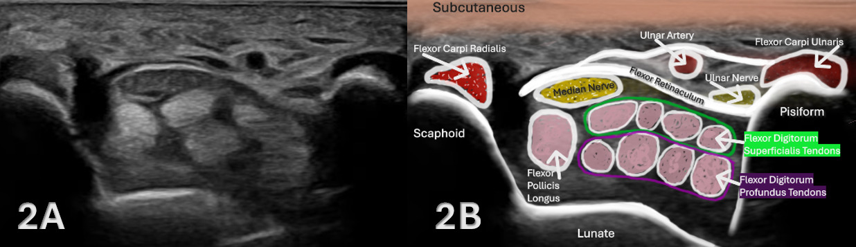
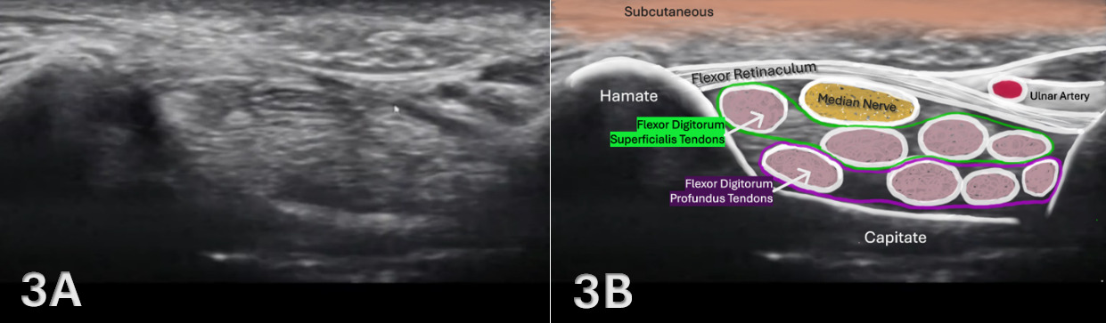
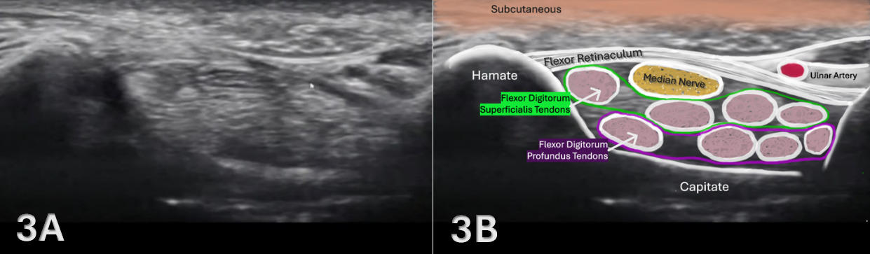
The carpal tunnel is a narrow fibro-osseous canal at the wrist that is formed by the retinaculum and bony structures. The superficial border is composed of the flexor retinaculum, the floor of the tunnel is formed by the carpal bones, the medial (ulnar) border by the hamate, and the lateral (radial) border by the trapezium. Contents that originate in the forearm and run distally through the carpal tunnel include the nine finger and thumb flexors and the median nerve. Carpal tunnel syndrome occurs when the median nerve is compressed or entrapped within the carpal tunnel, causing symptoms.
The Role of MSK Ultrasound in the Radiocarpal Joint
Advantages
-
Non-Invasive: MSKUS is a non-invasive imaging technique that can be used on a variety of soft tissue pathologies, including those at the carpal tunnel.
-
Real-Time Imaging: MSKUS allows evaluation of the carpal tunnel and its contents, and can be performed statically or during dynamic motion of wrist and forearm movement.
-
High-Resolution Visualization: Sonographic assessment provides detailed images of the size and shape of the carpal tunnel, the diameter of the median nerve, and the thickness of the flexor tendons that run through the carpal tunnel.
-
Accessibility and Cost-Effectiveness: MSKUS is portable, widely available, and less expensive than magnetic resonance imaging (MRI).
-
Accuracy: MSKUS has a combined sensitivity of 86% to 88% and specificity of 84% for cross-sectional area measurements of the median nerve at the carpal tunnel inlet.5,6
-
Reliability: Sonographic assessment of median nerve volume demonstrates great reliability with ICC values of 0.90.7
Limitations
-
Operator Dependency: MSKUS requires skill and experience for accurate interpretation of findings. The ability to sonograph the carpal tunnel is largely influenced by the operator and by the availability and technical considerations of state-of-the-art equipment.
-
Depth Limitations: Visualization is usually not a problem for the carpal tunnel due to its superficial location on the volar wrist.
-
Artifacts and Shadows: Bone and calcifications may create image artifacts, requiring adjustments in probe positioning and frequency.
Sonographic Technique for Evaluating the Carpal Tunnel
Equipment Setup
-
Transducer Type: Because of the superficial nature of the carpal tunnel, a standard high-frequency, linear array transducer is normally used.
-
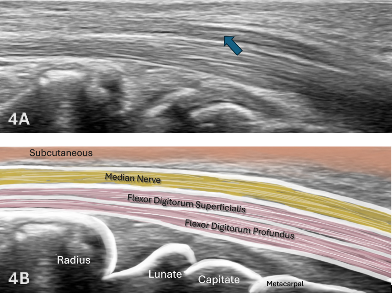
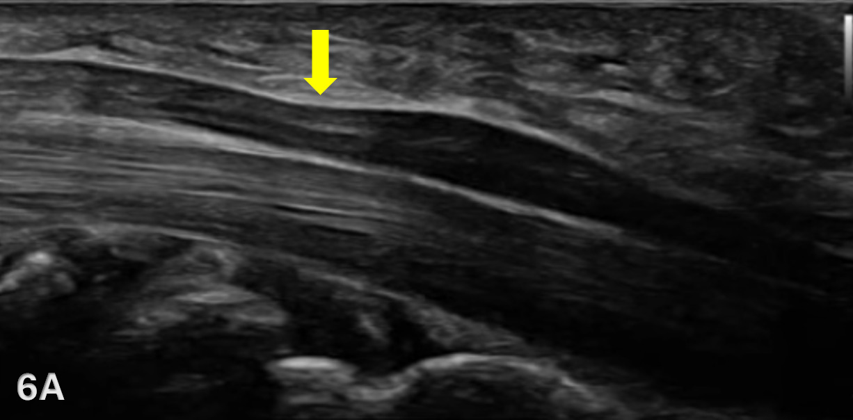
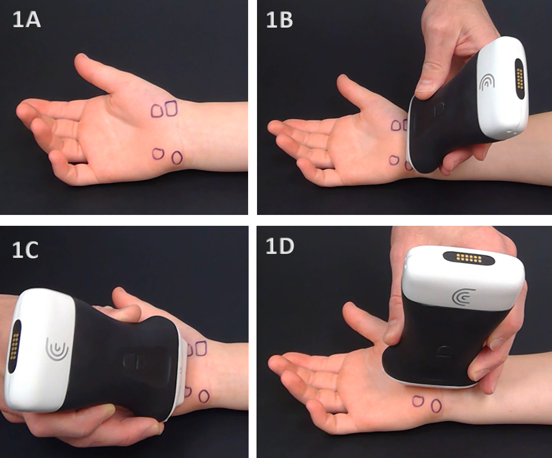
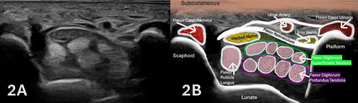
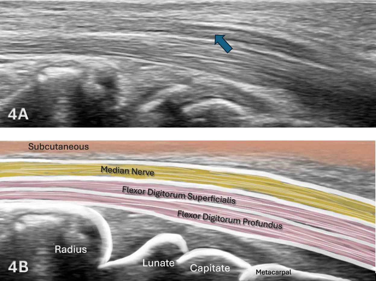
Patient Positioning: The patient’s forearm and wrist are placed in a supinated position with the dorsal forearm on the table. The transducer can be easily placed in either the long axis (LAX) or the short axis (SAX) to visualize the contents of the carpal tunnel. In the SAX view, the pisiform can be used as a bony landmark for the starting position of the medial side of the wrist. The transducer is placed in SAX across the wrist. Once the median nerve is found in the SAX view, the transducer can be rotated 90 degrees to the LAX view of the median nerve.
-
Dynamic Assessment: Dynamic movements of wrist flexion and extension can be used to visualize tendon and nerve excursion in the carpal tunnel.
Examination Protocol
Normal Sonographic Appearance
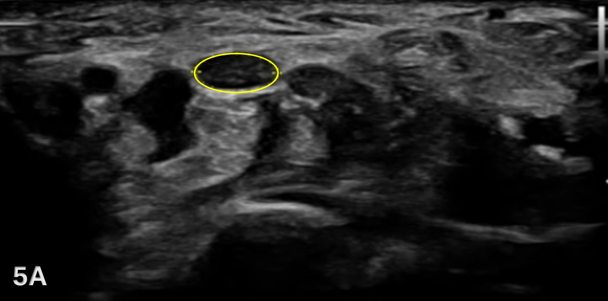
- When viewing images in SAX, the median nerve is easily seen as a honeycomb structure surrounded by a hyperechoic epineurium.
Pathologic Findings in Injured Carpal Tunnel
-
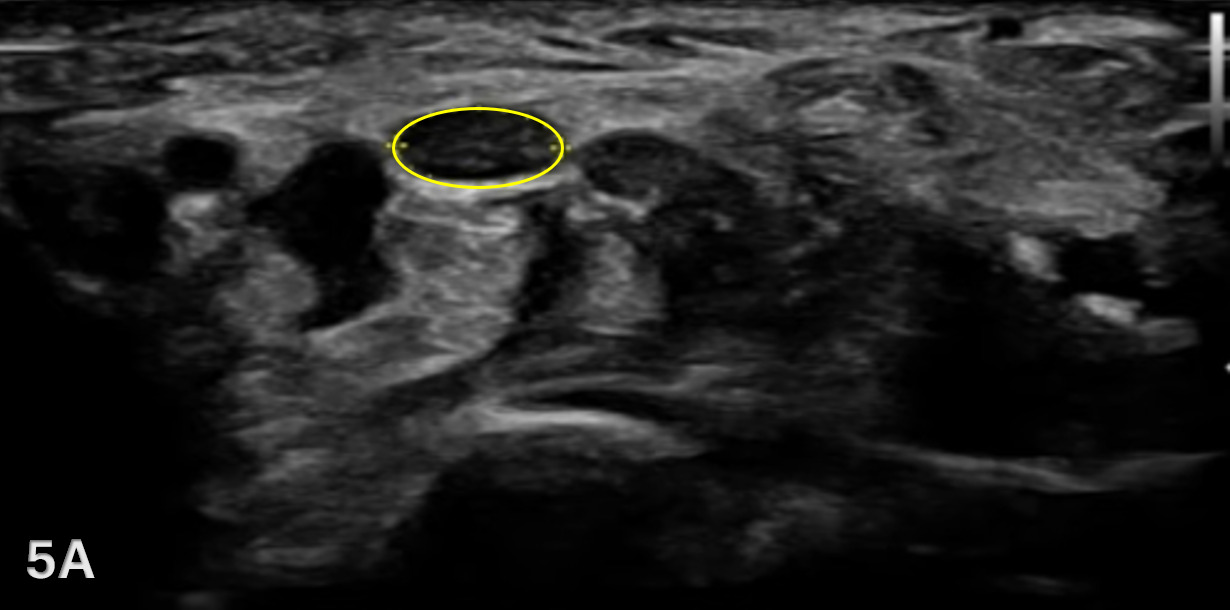
Swelling or thickening of the median nerve. The cross-sectional area of the median nerve at the level of the pisiform is normally 10 mm2.8 In a separate analysis of various locations from 74 studies, it was shown that a mean pooled cross-sectional area of the median nerve is 8.3 mm2 at the wrist, 6.4 mm2 at the forearm, and 8.3 mm2 at the upper arm.9 In those with carpel tunnel syndrome, the cross-sectional area jumps to 16.8 mm2.10
-
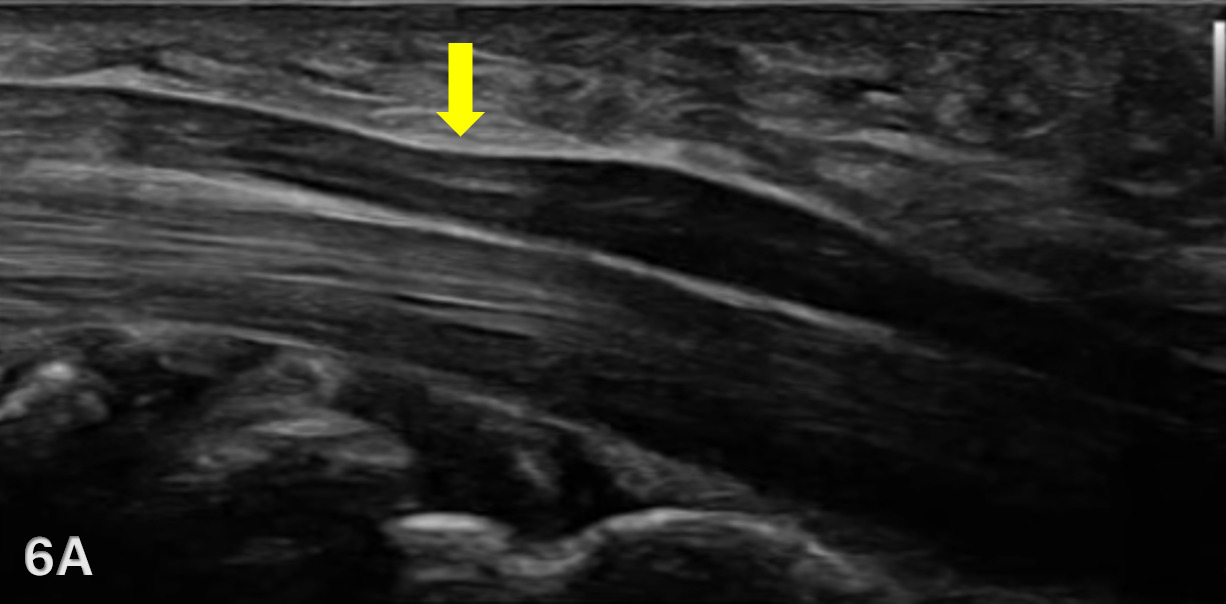
Flattening of the median nerve can be seen at the compression site at the flexor retinaculum. One can find the median nerve stenosis rate, which is the ratio of the minimum to the maximum anteroposterior diameter. The median nerve stenosis rate is 41.8% of those with carpal tunnel syndrome compared to 17.4% of those without.11
-
It is not uncommon for the nerve to have an hourglass appearance due to swelling both proximal and distal to the area of compression. This is known as the “notch” sign.12,13
-
Other structural findings can be viewed including “bowing” of the flexor retinaculum, swelling from tenosynovitis of the flexor tendons, ganglion cysts and other space-occupying lesions.14
Clinical Implications for Rehabilitation Providers
MSKUS provides real-time feedback for rehabilitation professionals, facilitating early diagnosis of median nerve compression and determining the need for intervention. Key applications include:
-
Early Detection of Injury / Accurate Injury Grading: MSKUS can quickly examine swelling, thickening, or compression of the median nerve, and this assessment can help guide treatment planning.
-
Dynamic Functional Testing: Rehabilitation professionals can use MSKUS during active movements of the wrist and forearm to assess functional movement on nerve compression and nerve mobility or assess limited nerve mobility.
-
Guided Interventions: MSKUS imaging assists in dry needling and precision-guided injections, such as corticosteroids for inflammation.
-
Patient Education: Real-time imaging with MSKUS serves as a visual aid to explain the nature of the injury and set realistic expectations for recovery.
Limitations and Challenges
Due to its superficial nature, the median nerve is easily visualized within the carpal tunnel. Evaluation of the median nerve can be done very expediently with MSKUS. This can be done actively in the office, at much lower cost than other imaging modalities, and pathologic findings can often be seen.
Conclusion
The integration of MSKUS into physical therapy practice gives therapists an advantage in evaluating carpal tunnel syndrome. By providing real-time, non-invasive visualization of the carpal tunnel, the median nerve, and surrounding tissues, MSKUS can help enhance clinical decision-making beyond what a traditional examination alone can offer. It allows physical therapists to more accurately identify swelling, observe actual structural changes, monitor progression, and tailor interventions with greater overall precision. These enhancements to practice ultimately improve the outcomes for individuals experiencing carpal tunnel syndrome.
_patient_positioning_for_viewing_median_nerve_in_the_carpal_tunnel._(b--c)_transducer_p.png)