INTRODUCTION
The Jefferson curl, a loaded spinal-flexion exercise, has grown in popularity in rehabilitation practice and strength training but remains uncharacterized in the scientific literature.1 Practitioners prescribe it without empirical data on which muscles it actually recruits. Loading the spine in flexion has long been considered hazardous. Early biomechanical work linked spinal flexion under load to disc herniation, annular tearing, disc prolapse, and elevated intradiscal pressure, framing flexion under load as a risk factor for injury.2–7 But those studies relied on in-vitro porcine and cadaveric preparations and radiographic imaging. More recent in-vivo work using motion tracking and electromyography finds no clear link between disc pressure, muscle activity, peak moments, lifting technique, and low back pain (LBP).8–11 A recent review reframes deadlift injury risk around training-load management, movement variability, and dynamic trunk control rather than postural rigidity.12
Spinal flexion is a normal movement, not a fault to be trained away.12 Progressive loading can build lumbar extensor strength in healthy adults,13 and people with back pain can tolerate lifting well.14 Yet the spinal extensors are rarely loaded through the range in which they are longest, and the demand that loaded flexion places on them has not been measured. Rehabilitation professionals, however, still commonly view lifting with a rounded back as dangerous. Those beliefs may drive nocebo-like fear-avoidance, disability, and movement deficits.15–19
Two exercises exemplify those contrasting approaches. The Romanian deadlift (RDL) is a hip-hinge movement that minimizes spinal motion: the lifter begins upright, hinges at the hip to lower a barbell toward mid-shin while keeping a neutral spine, then returns to standing. It emphasizes hamstring and gluteal recruitment while minimizing lumbar flexion, and its activation patterns are well documented.20–23 The Jefferson curl, by contrast, uses controlled segmental spinal flexion: standing on an elevated platform, the lifter curls down vertebra by vertebra to lower a weighted barbell, then reverses the movement. Practitioners use it to build posterior-chain strength and flexibility. The two exercises use distinct recruitment patterns. The Jefferson curl lengthens the erector spinae under load and increases their moment arm relative to the spine. The RDL, by contrast, loads the glutes and hamstrings at the hip while the spine acts largely isometrically.
Using surface electromyography (sEMG) to quantify muscle activation during the RDL and the Jefferson curl can provide new insights into human movement principles and the development of targeted rehabilitation plans.24,25 Specifically, understanding muscle activation patterns during spinal flexion and hip-hinge movements is essential for designing exercise protocols that optimize strength, flexibility, and injury prevention in individuals with LBP and athletes.26 Controlled spinal flexion lengthens the erector spinae and increases their moment arm, which provides the rationale for a directional hypothesis for these muscles. Therefore, the aims of this study were to characterize erector spinae activation during the Jefferson curl at a submaximal load and test the hypothesis that, at the same absolute load, the Jefferson curl elicits greater erector spinae activation than the RDL. A secondary aim was to describe gluteus maximus and biceps femoris activation without a directional hypothesis.
METHODS
Participants and ethical approval
Ethical approval was obtained from the institutional review board of the Technion - Israel Institute of Technology (Number: 171-2023). The study was conducted between July and October 2024 at the Fischer Biomotion lab. Reporting follows the STROBE statement for cross-sectional observational studies.27
This was a cross-sectional, within-participant repeated-measures study conducted in a single session. Participants were recruited through advertisements on social media platforms (sports-related groups) and physical posters in the university gym. Inclusion criteria were healthy males and females aged 18-45 years who performed resistance training at least three times per week in most weeks over the past year, with the deadlift regularly included in their training during that period. A researcher assessed eligibility, including training history, in a structured screening interview. Individuals were excluded if they reported having back or lower limb pain in the prior month, had undergone orthopedic surgery, or had cardiovascular, neurological, or respiratory conditions. Participants were instructed to avoid high-intensity activity for 48 hours before testing.
Exercise protocol
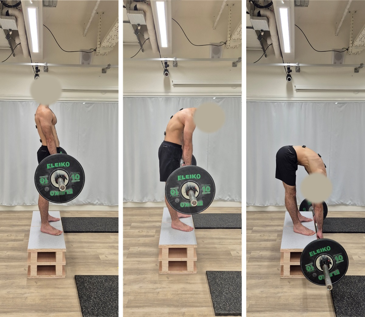
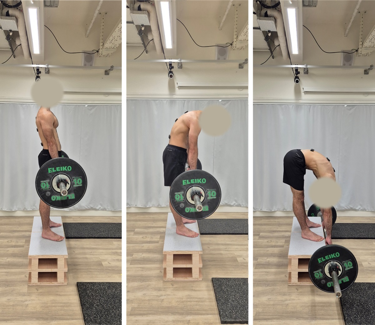
The Jefferson curl was performed on a 30-cm wooden platform with the participant standing upright, feet hip-width apart and barefoot, holding a standard Olympic barbell in a double-overhand, shoulder-width grip in front of the thighs (Figure 1). Participants were instructed to flex the spine in a controlled, sequential manner from the cervical region downward, lowering the barbell toward the floor until reaching their maximum comfortable spinal flexion, then to reverse the movement to the upright starting position while maintaining a neutral neck. Cadence was participant-driven. A brief familiarization with ten unloaded repetitions preceded testing.
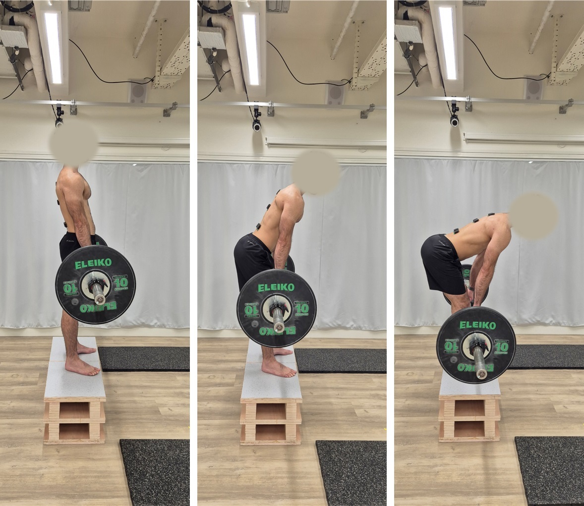
The RDL was performed from the same 30-cm platform, feet hip-width apart and barefoot, holding the barbell in a double-overhand, shoulder-width grip (Figure 2). With knees slightly bent, participants pushed their hips backward in a hip hinge, lowered the barbell to approximately mid-shin while maintaining a neutral spine, and reversed the movement to an upright position. Cadence was participant-driven. A trained gym instructor visually inspected all lifts for gross technique conformance.
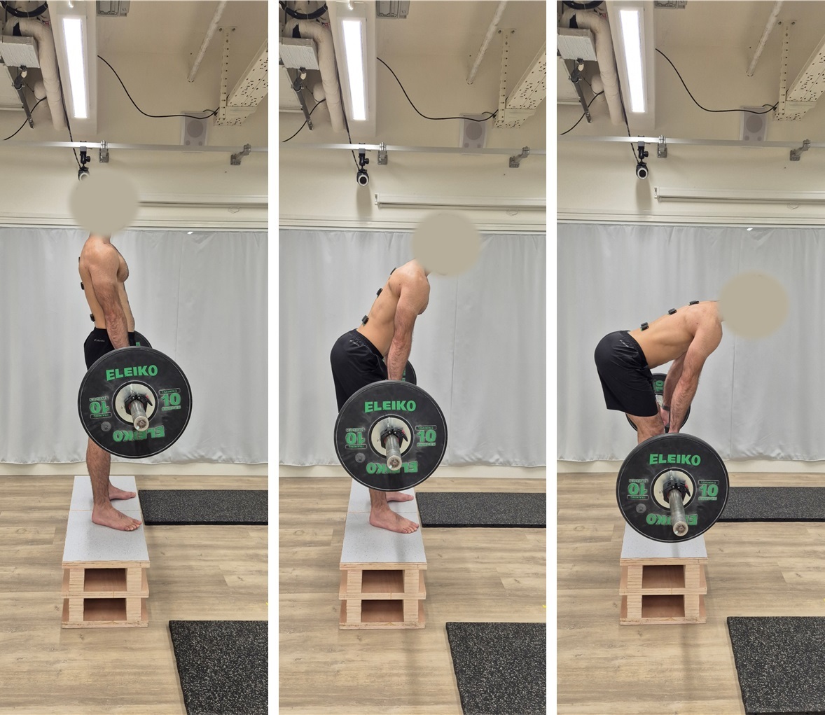
The Borg Rating of Perceived Exertion (RPE) scale, which ranges from 6 (very light exertion) to 20 (maximal exertion), was used to assess physical activity intensity based on self-reported perceived exertion. The RPE has demonstrated a moderate correlation with load intensity in resistance training.28,29 RPE was also recorded at the final tested load of each exercise.
After providing informed consent, each participant completed an approximately 10-minute standardized warm-up organized into four blocks of 2–3 minutes: (i) self-paced walking and light jogging within the laboratory, (ii) dynamic lower-limb and trunk mobility drills, (iii) a self-selected movement-specific preparation, and (iv) ten unloaded repetitions of the Jefferson curl followed by ten unloaded repetitions of the RDL performed with a lightweight wooden dowel, with a trained gym instructor (B.K) coaching the technique. Maximal voluntary isometric contraction (MVIC) testing followed the warm-up and preceded the loaded trials. Loaded trials began with an 8 kg barbell; participants performed five repetitions, rested for 5 minutes, then repeated with progressively heavier loads in 5-10 kg increments until the subject reached a Borg RPE of 16 or chose to stop earlier. The average EMG across the five repetitions at the final tested load was used for analysis. Following a 5-minute rest period, participants then performed five repetitions of the RDL using the same absolute load previously achieved during the Jefferson curl. Throughout the exercise protocol, participants were continuously monitored for any signs of discomfort or pain.
The loading protocol was anchored to the Jefferson curl rather than to a percentage of one-repetition maximum (1RM). The Jefferson curl has no established 1RM protocol or normative data, and a maximal-effort trial on an unfamiliar lift involving loaded end-range spinal flexion would reflect task novelty and motor caution more than muscular capacity. A validated perceived-effort threshold (Borg RPE 16) provided a submaximal anchor. Applying the resulting absolute load to the RDL enabled a matched-load comparison aligned with the study question: how trunk and hip musculature respond to the same external resistance under two different movement patterns. The exercise order was fixed, because the RDL load was defined only after the Jefferson-curl RPE-16 load was determined within the same session.
EMG data acquisition and signal processing
The erector spinae, gluteus maximus, and biceps femoris were selected due to their critical roles in spinal stability, hip extension, and posterior chain activation during lifting movements. The erector spinae are key spinal stabilizers, while the gluteus maximus and biceps femoris are primary movers in hip extension, making them essential for understanding the demands of both exercises. According to the SENIAM (surface EMG for non-invasive assessment of muscles) recommendations, each participant’s skin was scrubbed and cleaned with alcohol before electrode placement. EMG activity was recorded bilaterally using Delsys Trigno Avanti wireless surface electrodes (Delsys Inc., Boston, MA; sensor 27 x 37 x 13 mm, 10-mm fixed inter-electrode spacing, 99.9% silver contacts). Sensors were secured using manufacturer-supplied adhesive interfaces, with additional tape over the back sensors to maintain contact during spinal flexion. Back sensors were placed bilaterally 2 cm lateral to the spinous processes of T3, T8, and L4 to sample muscle activity in the upper thoracic, mid-thoracic, and lumbar regions, respectively. Sensors were also placed on the muscle bellies of the gluteus maximus and biceps femoris. All were aligned with the direction of the underlying muscle fibers.
Maximal voluntary isometric contraction (MVIC) trials were performed before the loaded trials. For the gluteus maximus, participants lay prone with the knee extended while the examiner provided manual resistance to hip extension. For the biceps femoris, participants lay prone with the knee flexed to approximately 45° against manual resistance. For the erector spinae (upper, middle, and lower segments), MVICs were obtained in two prone positions reported by Vera-Garcia et al. (2010): (i) upper trunk extension in the Biering-Sørensen position, with the pelvis and legs strapped to the bench and the torso extending horizontally beyond the end of the bench, while the participant extended the upper trunk against manual resistance applied at the shoulders; and (ii) lower trunk extension, with the torso on the bench and the legs extending horizontally beyond the end of the bench, while the participant extended the hips and lower trunk against manual resistance applied to the legs.30 The higher of the two peak values per segment was retained as the reference MVIC. Two MVIC trials per muscle (per position, where applicable) were separated by 5 minutes of rest to minimize fatigue. Standardized verbal encouragement was given. The highest peak EMG amplitude across trials was designated as the MVIC reference for each muscle. A multi-joint task such as the isometric mid-thigh pull was not used, as normalization required a separate maximal reference for each of the five muscles.
For each exercise, repetitions were performed consecutively at a self-selected cadence, without a prescribed rest interval between repetitions. One assessor monitored movement technique while another monitored the live EMG signals. Trials were repeated in case of a signal drop. EMG signals were band-pass filtered with a 4th-order Butterworth filter (20-500 Hz), full-wave rectified, and smoothed with a 200-ms moving-average window. The %MVIC signal was computed per rep as the mean of the smoothed, MVIC-normalized amplitude across the full repetition cycle, and then averaged across the five repetitions at the final tested load per exercise.
Statistical methods
Continuous demographic variables are reported as median [minimum, maximum], categorical variables as count (%), and the lifted load as mean ± standard deviation. For each participant and muscle, left and right sEMG amplitudes (normalized to %MVIC) were averaged to obtain a single activation value per exercise. For 11 of 210 participant × muscle × exercise observations, the left-side channel was unusable because of signal dropout (middle erector spinae: 5 on the RDL, 3 on the Jefferson curl; lower erector spinae: 2 on the RDL, 1 on the Jefferson curl). In these cells, the activation value was taken from the right side alone. No cell had both sides missing, so all 21 participants contributed to every muscle comparison.
A linear mixed-effects model was fitted by restricted maximum likelihood, with %MVIC as the outcome, exercise (Jefferson curl, RDL), muscle (upper, middle, and lower erector spinae, gluteus maximus, biceps femoris), and their interaction as fixed effects, and participant as a random intercept. Per-muscle Jefferson curl and RDL comparisons were then estimated from the model. Estimated marginal means are reported with standard errors and 95% confidence intervals, together with mean differences, and Benjamini-Hochberg-adjusted p-values (5% false discovery rate across the five muscles).31 Effect sizes are reported as Hedges’ g with 95% bias-corrected and accelerated bootstrap confidence intervals (5,000 paired-participant resamples). Thresholds of 0.20, 0.50, and 0.80 denote small, medium, and large effects.32,33
As a non-parametric robustness check, a Wilcoxon signed-rank test was applied to each muscle’s paired within-participant difference with the same Benjamini-Hochberg adjustment; median differences are reported. A sensitivity power analysis indicated that n = 21 provided approximately 80% power (α = .05, two-tailed) to detect a paired effect with Cohen’s d = 0.64. Smaller true effects are likely to have been missed. All analyses were performed in Python 3.13 (statsmodels 0.14.6, scipy 1.17.1) at α = .05 (two-tailed). The analysis notebook and the de-identified tabular data are provided in the Appendix.
RESULTS
Twenty-one resistance-trained adults (12 men, 9 women) completed the protocol (Table 1). The mean final tested load was 41.9 ± 19.3 kg. The RPE at the final tested load was 17 [14-19] for the Jefferson curl and 16 [11-18] for the RDL. Eighteen of 21 participants rated the Jefferson curl equal to or harder than the RDL.
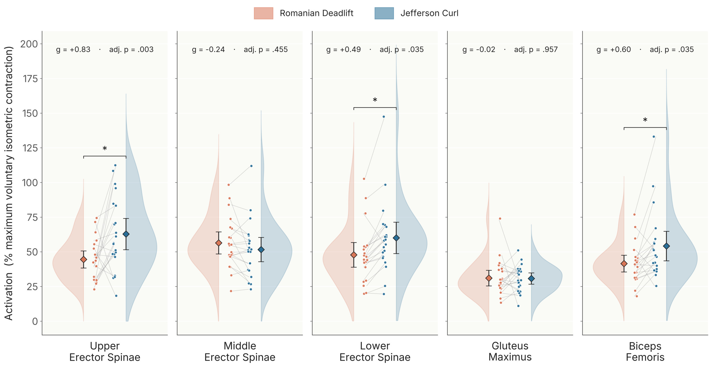
Per-muscle Jefferson-curl and RDL comparisons from the mixed-effects model, Benjamini-Hochberg adjusted across the five muscles (Table 2, Figure 3), showed that the Jefferson curl elicited significantly greater activation than the RDL in the upper erector spinae, lower erector spinae, and biceps femoris, and no difference in the middle erector spinae or gluteus maximus. Activation of the upper erector spinae was significantly greater during the Jefferson curl (62.84 ± 4.30 %MVIC, 95 % CI [54.41, 71.27]) than during the RDL (44.52 ± 4.30 %MVIC, 95 % CI [36.09, 52.95]; mean difference = 18.32 ± 5.30 %MVIC, adjusted p = 0.003; Hedges’ g = 0.83, 95 % CI [0.19, 1.39]). The lower erector spinae were also significantly more active during the Jefferson curl (60.07 ± 4.30 %MVIC, 95 % CI [51.64, 68.50]) than during the RDL (47.85 ± 4.30 %MVIC, 95 % CI [39.42, 56.28]; mean difference = 12.22 ± 5.30 %MVIC, adjusted p = 0.035; Hedges’ g = 0.49, 95 % CI [0.22, 0.78]). The biceps femoris were also more active during the Jefferson curl (54.18 ± 4.30 %MVIC, 95 % CI [45.75, 62.61]) than during the RDL (41.54 ± 4.30 %MVIC, 95 % CI [33.11, 49.97]; mean difference = 12.64 ± 5.30 %MVIC, adjusted p = 0.035; Hedges’ g = 0.60, 95 % CI [0.21, 0.92]).
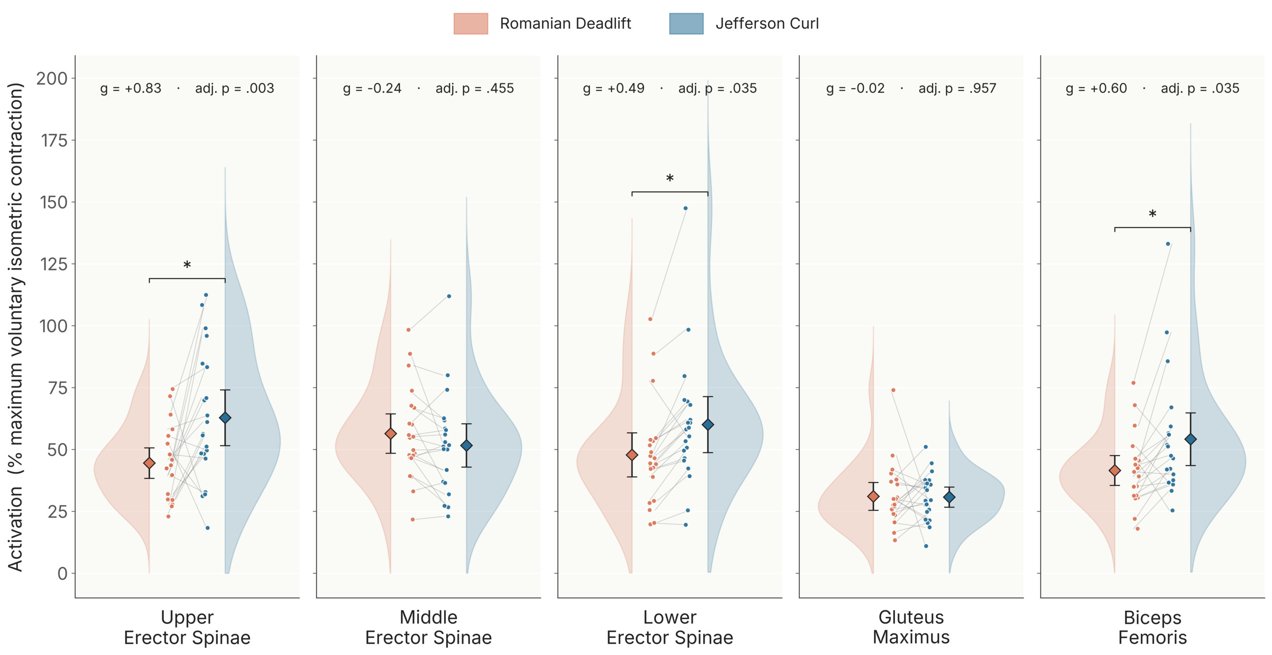
The middle erector spinae, in contrast, did not differ in activation significantly between exercises (Jefferson curl 51.66 ± 4.30 %MVIC vs RDL 56.46 ± 4.30 %MVIC; mean difference = -4.80 ± 5.30 %MVIC, adjusted p = 0.455; Hedges’ g = -0.24, 95 % CI [-0.55, 0.03]). For the gluteus maximus, no significant difference was detected (Jefferson curl 30.79 ± 4.30 %MVIC vs RDL 31.07 ± 4.30 %MVIC; mean difference = -0.29 ± 5.30 %MVIC, adjusted p = 0.957; Hedges’ g = -0.02, 95 % CI [-0.65, 0.60]). The Wilcoxon signed-rank sensitivity check reproduced the conclusions of the primary analysis: upper erector spinae (median difference = 18.4 %MVIC, adjusted p = 0.026), lower erector spinae (median difference = 10.9 %MVIC, adjusted p = 0.002), and biceps femoris (median difference = 11.4 %MVIC, adjusted p = 0.016) all showed significantly greater activation during the Jefferson curl, whereas the middle erector spinae (adjusted p = 0.129) and the gluteus maximus (adjusted p = 0.452) did not.
DISCUSSION
At the same absolute submaximal load, the Jefferson curl recruited the upper erector spinae, lower erector spinae, and biceps femoris more than the RDL. In contrast, activation of the middle erector spinae and gluteus maximus was not statistically different. This is the first surface EMG characterization of the Jefferson curl and its activation profile relative to the RDL.
Earlier sEMG studies involving spinal flexion lifting used low external loads and lifts starting from the floor. Pan et al. and Van der Have et al. reported lower erector spinae activation during stoop lifting than during squat lifting, with the difference being non-significant in Pan et al. In both studies, the load was 5-10 kg, and the lift started at floor level, limiting the range of motion at the hip and the spine.9,34 The larger load and greater spinal range of motion in the present study may account for the greater erector spinae activation observed during the Jefferson curl.
Coratella et al. extended the RDL’s range of motion by raising the lifter on a 15-cm platform while retaining a hip-hinge technique with a straight spine.35 In the Jefferson curl, the additional range comes from segmental spinal flexion, which lengthens the moment arm relative to the spine and raises demand on the erector spinae. In the step-RDL, the added range also increases recruitment of the hip extensors. Upper and lower erector-spinae activation was greater during the Jefferson curl than during the RDL in the present study. Coratella et al. also found greater longissimus excitation during the ascending phase of the step-RDL than during the standard RDL, with no significant difference during descent or for iliocostalis during either phase. The present findings are therefore consistent with those of Coratella et al., in that a larger range of motion under load was accompanied by greater erector spinae activation, although the two variants have not been compared directly at a matched load.
Recent in-vivo evidence challenges the long-held view that stoop lifting places greater loads on the spine than squat lifting.8,36,37 Upper-lumbar spinal forces measured from instrumented vertebral implants and motion capture differ by roughly 4% between the two techniques, and ergonomic assessments suggest that squat lifting may carry a higher musculoskeletal burden despite comparable spinal loads.8,36,37 In healthy populations, the evidence does not clearly link spinal flexion to elevated LBP risk, and fear-avoidance beliefs about spinal flexion may themselves contribute to disability.10,16,17 The present results, drawn from pain-free resistance-trained adults, show that controlled spinal flexion loading substantially recruits the posterior chain. At a matched absolute load, the Jefferson curl placed greater acute demands on the upper and lower erector spinae and the biceps femoris than the RDL. This may inform exercise selection when the goal is targeted posterior-chain recruitment through controlled spinal flexion.
Exercise selection is only the first decision. The next is how much load to apply and how to advance it. At a submaximal load, the Jefferson curl drove the upper and lower erector spinae to roughly 60% MVIC, loading those muscles substantially at long lengths. Because load can be added or withdrawn a plate at a time, that loading can be graded and progressed like any other training variable.
Between-participant variability is expected in any sEMG study, but in the present data it was visibly larger during the Jefferson curl than during the RDL, particularly for the upper and lower erector spinae and the biceps femoris (Figure 3). The RDL was performed to approximately mid-shin, whereas Jefferson-curl depth was determined by each participant’s maximum comfortable spinal flexion, which may have contributed to the between-participant variability. Spinal mobility and body proportions may contribute to these differences, as may beliefs about bending: fear-avoidance beliefs restrict lumbar flexion during lifting even in pain-free adults.19 Whether repeated exposure produces adaptation remains untested.
Limitations
This study is not without limitations. First, the exercise was confounded by order, as the Jefferson curl always preceded the RDL because it set the test load; therefore, fatigue and practice effects cannot be separated from the exercise effect. Relatedly, the between-participant variability in Jefferson-curl activation was larger than during the RDL, possibly due to differences in segmental flexion strategy, spinal mobility, and motor caution during an unfamiliar movement. The Jefferson curl was likely unfamiliar to most participants, but exercise-specific experience was not formally recorded. The Jefferson curl depth was also participant-defined, whereas RDL depth was standardized to mid-shin, so the Jefferson curl carried an additional source of between-participant variability.
Second, the load represented a lower relative intensity for the RDL than for the Jefferson curl, so the present results compare the two exercises at the same weight, not at the same percentage of each lifter’s maximum. Most participants did not, however, rate the RDL as easier at that load, though perceived exertion does not measure relative load, and the fixed order means fatigue may have inflated those ratings. Third, the 30-cm platform, while important for the Jefferson curl, is not the norm for the RDL and limits direct comparison with prior RDL literature. Fourth, sEMG is susceptible to crosstalk among the three adjacent erector-spinae channels, and manual-resistance trunk-extensor MVICs are operator-dependent. Fifth, the participants were recruited through a university gym, biasing the sample toward resistance-training enthusiasts. Sixth, null findings at the middle erector spinae and gluteus maximus may reflect non-detection of small effects rather than evidence of equivalence. The study is also underpowered to detect sex-specific effects and to test whether training experience or maximal strength, both of which varied in this sample, influences the difference between exercises. Finally, coaching and visual inspection were used to ensure that participants performed each movement as instructed, but the relative contributions of spinal and hip motion to each movement were not quantified.
Future directions
Three questions follow directly from this work. First, comparing the two lifts at matched relative intensity, in a counterbalanced order, would separate movement pattern from load and from fatigue. Second, recording spinal and hip kinematics together with EMG would show how much of the flexion each lifter produces at the spine, and adding a measure of beliefs about the risk of back injury when lifting with a flexed spine would test whether the variability observed here is mechanical, psychological, or both. Third, larger and longer studies should test if the demand documented here translates into back strength or tolerance, whether training experience moderates the response, and if either finding holds in people with low back pain.
CONCLUSIONS
This study provides the first surface EMG characterization of the Jefferson curl. It compares its acute activation profile with that of the RDL at the same submaximal absolute load in resistance-trained adults. The Jefferson curl elicited greater activation of the upper and lower erector spinae and the biceps femoris, while middle erector spinae and gluteus maximus activation did not differ significantly. In pain-free, resistance-trained adults, the Jefferson curl can be considered a tool for populations seeking targeted recruitment of the erector spinae and hamstrings through controlled spinal flexion.
Conflicts of Interest
The authors have no competing interests to declare that are relevant to the content of this article.
Data Availability
The de-identified participant-level dataset, the MVIC reference values, and the full Python analysis notebook that reproduces every number reported in the Abstract, Table 2, and Figure 3 are available in the Appendix.