INTRODUCTION
Tendinopathy of the distal biceps brachii affects up to 2.6 per 100,000 individuals each year and is more common in middle-aged males, creating pain and disability that may hinder household, occupational, and recreational activity participation.1–3 Qualitative studies investigating the lived experiences of those with chronic tendon pathology highlight profound psychological and socioeconomic implications, including reduced participation in personally meaningful activities and reduced income from potential deficits in their ability to work.4 Distal biceps tendinopathy is characterized by irritation, inflammation, or degeneration of the biceps tendon where it attaches to the radius bone near the elbow, often causing pain, swelling, and weakness.4,5 Percutaneous fenestration for tendinopathy comprises multiple needle punctures into damaged, chronically injured tendons to stimulate healing by creating micro-injuries, promoting bleeding, and the release of growth factors, effectively inducing an acute healing response. This intervention has been commonly used for lateral elbow tendinopathy and Achilles tendinopathy.6–11 However, a paucity of evidence exists regarding its use in distal biceps tendinopathy.
At the insertion into the ulnar aspect of the radial tuberosity, the distal tendon of the biceps brachii is bifurcated with a ribbon-like configuration.12–15 The tendon associated with the short head runs medially but inserts anteroinferiorly to the long head, increasing the mechanical advantage of the short head for elbow flexion at ninety degrees and forearm supination in a neutral or pronated forearm position.14,16 At insertion, the distal biceps tendon spans approximately 63% of the length and 13% of the width of the radial tuberosity, covering approximately 35.9% of the total area.14 Various extrinsic factors are postulated to increase the risk of tendinopathy, such as mechanical impingement secondary to hypertrophy of the radial tuberosity.17 Hilgersom and colleagues17 found that both volume and height of the radial tuberosity are statistically greater in those with distal biceps tendon pathology versus a control group. The radioulnar space reduces notably during pronation to end-range, likely exacerbated by a more prominent radial tuberosity.18 Similarly, the bicipitoradial bursa surrounds the distal tendon of the biceps brachii and functionally reduces friction between the tendon and radius during pronation and supination. Although rare, chronic irritation to the bicipitoradial bursa is thought to contribute to distal biceps brachii tendon pathogenesis.19,20 The myotendinous junction of the distal biceps brachii gives rise to the lacertus fibrosis, which merges laterally with the fascial complex surrounding the pronator-flexor group at the medial elbow. The lacertus fibrosis can be a functional stabilizer of the distal biceps tendon, often limiting retraction in the presence of a full rupture.15,21,22 However, some postulate that increased stiffness of the lacertus fibrosis can alter the mechanical efficiency of the distal biceps tendon and increase the risk of pathology.21,22 The distal biceps tendon presents unique morphological characteristics, and the above anatomical and biomechanical factors, coupled with hypovascularity seen in dense regular connective tissue, contribute to tendon pathogenesis.15,17–23
Tendons are comprised of dense regular connective tissue with a unidirectional fibrillar pattern that allows for uniplanar force propagation from the muscle into the bony insertion.24,25 Relative hypovascularity of this tissue type contributes to the pathogenesis and chronification of tendon pathology and is worsened in the presence of nicotine use and various comorbidities (e.g., obesity, diabetes).24–27 Tendons commonly receive vascular supply from the myotendinous junction, osteotendinous junction, and nearby connective tissues (i.e., via paratenon).25 The distal biceps brachii tendon has three zones of vascularity, and the zone comprising the tendon enthesis receives vascular supply from the posterior interosseus recurrent artery, with the neighboring tendon midportion supplied by a paratenon.12,21,28 Per Brasseur,29 enthesopathy of the distal biceps tendon is notably more common than more proximal tendinopathy. Distal biceps tendinopathy may include structural and biochemical changes associated with impaired tendon remodeling, and needle fenestration at the osteotendinous junction may improve tendon healing.12,25,28,30–33
Chronic tendon pathology often presents with disorganization of collagen fibers, degradation of the extracellular matrix, and neovascularization, with other common findings being fibrosis and ectopic ossification.34–38 Mechanical overload of the tenocytes facilitates biochemical signaling (e.g., via interleukin-1β expression) for remodeling of the extracellular matrix, upregulation of proteoglycans, and changes in collagen composition.35,38,39 Interleukin-1β expression upregulates pro-inflammatory cytokines (e.g., C-C motif ligand 2) and anti-inflammatory cytokines for tendon repair, and continued mechanical loading beyond the functional capacity of the tendon perpetuates this response.38 This process disrupts the remodeling process and results in an increasingly disorganized collagen matrix and alterations in the tendon microenvironment, characterized as tendon disrepair by many authors.35,36,38 For instance, proteoglycans are hydrophilic, and upregulation of these proteins increases water absorption in the extracellular matrix, altering its morphology (e.g., increased size) and likely contributing to fibrosis.35 Similarly, continued strain may exacerbate tissue hypoxia and induce phenotypic change in tenocytes, accompanied by increased deposition of glycosaminoglycans.38–41 This process likely contributes to the notable loss of elasticity seen in pathologic tendons, increasing stiffness and risk of rupture.39,40 The transformation of the tendon in the context of overuse can induce pain and disability through disorganization of collagen alignment, matrix degradation, maladaptive alterations in the tendon microenvironment, neovascularization, hypovascularity, and more, likely impeding functional strength in elbow flexion and forearm supination in the context of the distal biceps brachii tendon.5,34–40 These maladaptive changes in tendinopathy and fasciopathy correspond with poorer prognoses (i.e., long-term symptoms that impede functional capacity).42–44 Loading-based programs may improve short-term pain and function. However, long-term outcomes are variable, and persistent symptoms are common in chronic tendinopathy, warranting further investigation into adjunct physical therapy interventions.42–45
Dry needling the tendon-bone interface (enthesis) with fenestration may disrupt the maladaptive microenvironment seen in tendon disrepair and facilitate a healing response through localized bleeding and fibroblast proliferation, and the utilization of musculoskeletal ultrasound aids in the accuracy of needle location.11,32 Tendon dry needling is a safe and inexpensive treatment methodology and has been shown to be effective for Achilles tendinopathy, lateral epicondylalgia, rotator cuff tendinopathy, and patellar tendinopathy.7,8,11,46–49 However, to date, no research appears to have investigated ultrasound-guided dry needling with fenestration for distal biceps tendinopathy. An anterior approach to the distal biceps tendon is not warranted secondary to nearby neurovascular structures, including the brachial artery and median nerve.5,12,13 Dry needling of the distal biceps tendon at the level of the radial tuberosity (i.e., the most commonly affected area) can be performed safely from a posterior “cobra” position, often used for injection (e.g., elbow flexion and forearm end-range pronation).12,13 This case series describes the treatment of two subjects with chronic distal biceps tendinopathy using ultrasound-guided percutaneous tendon dry needling with fenestration and a graded loading program in outpatient physical therapy.
CASE DESCRIPTIONS
Subjects
Two subjects with no clinically significant comorbidities, no significant functional limitations prior to symptom onset, and regular participation in independent resistance training were referred to an outpatient physical therapy clinic for management of symptoms related to distal biceps brachii tendinopathy. Informed consent was obtained from each subject to perform ultrasound-guided dry needling and to use their data in this study. Their baseline data were recorded on the first visit (Table 1). Subjects A and B were 55 and 46 years of age, respectively, with chronic symptoms. Both reported pain of insidious onset. Each had already tried reducing tendon load by avoiding resistance training on the affected arm without symptom improvement, prompting consultation with an orthopedic physician who referred them to the outpatient physical therapy clinic.
Treating Clinician
The treating clinician was a physical therapist with three years of clinical experience with certifications in dry needling and musculoskeletal ultrasonography through the American Academy of Manipulative Therapy, with additional education in ultrasound-guided dry needling.50,51
Outcome Measures
Outcome measures comprised the Numeric Pain Rating Scale (NPRS), Global Rating of Change (GROC), and Upper Extremity Functional Index (UEFI-20), each measured at baseline, three weeks, five weeks, 12 weeks, and 6 months. The NPRS is the primary outcome measure and refers to the subject’s pain severity over the previous 24 h with an 11-point scale from 0 to 10, with higher scores indicating more pain.52 The minimum clinically important difference (MCID) for the NPRS is at least a 2.0-point change for chronic musculoskeletal pain pathologies.53,54 Likert-type scales, such as the NPRS, may be at risk of response bias, such as extreme-response bias and non-extreme response bias.55–57 Concurrent usage of the GROC (a secondary outcome measure) may ameliorate recall bias and improve the validity of retested NPRS scores upon follow-up.58 The GROC is a 15-point scale from -7 (a very great deal worse) to +7 (a very great deal better) and is commonly used to assess a subject’s perceived benefit of an intervention.59 Outcome measures that assess global ratings of improvement have been recommended by the Initiative on Methods, Measurement, and Pain in Clinical Trials (IMMPACT) for chronic pain pathologies secondary to the value of understanding the subject’s judgment of their treatment experience and clinical outcomes.60 A change of five in either direction on the GROC is likely the threshold for meaningful improvement.59
Lastly, the UEFI-20 is a reliable outcome measure (ICC = 0.94) that allows for the assessment of functional disability at the upper extremity through a 20-item scale.61,62 Subjects are prompted to associate a level of difficulty with each activity, from 0 (extreme difficulty or unable to perform) to 4 (no difficulty), and the resulting score ranges from 0 to 80.62 A lower total score corresponds with increased perceived functional disability secondary to the subject’s upper extremity problem. The MCID is reported to be a 7-point increase for the dominant arm and a 10-point increase for the non-dominant arm; each subject in this report had their dominant arm affected.61
Examination Procedure
Each subject underwent a thorough physical examination to ensure the clinical presentation was consistent with that of the referral, including observation, sensation testing, range-of-motion testing, strength testing, and palpation. Both subjects presented with pain isolated to the proximal antebrachium at the anterolateral cubital fossa. In each subject, sensation was intact without evident atrophy or trophic changes. Additionally, each had normal range-of-motion at the level of the neck, shoulder, elbow, and wrist with reproduction of symptoms with resisted elbow flexion and forearm supination. Active range of motion for elbow flexion and forearm supination was not provocative; however, lengthening of the tissues using end-range elbow extension coupled with pronation was consistently provocative. Similarly, each subject demonstrated a positive tilt sign (sensitivity 35-55%, specificity 65-77%, and accuracy 55-62.50%) and a positive biceps provocation test (sensitivity 95 – 100%, specificity 95%, and accuracy 95-97.50%).63 A positive tilt sign comprises tenderness over the radial tuberosity when the forearm is pronated and an absence of tenderness when the forearm is supinated.64 A positive biceps provocation test occurs when resisted elbow flexion at 70 degrees is more provocative in a pronated state versus a supinated state, likely facilitated by a reduction in radioulnar space that causes a mechanical impingement with the pathologic tendon during resisted flexion.63,65 These tests, coupled with the above exam findings, suggest that nociception is likely attributed to insertional pathology of the distal biceps brachii tendon. Of note, palpation of the medial pronator-flexor group and lateral wrist extensors was unremarkable. Similarly, each subject had a negative Mill’s, Maudsley’s, and Cozen’s test, reducing the likelihood of the pathology being attributed to lateral epicondylalgia.66–68
Ultrasound Imaging
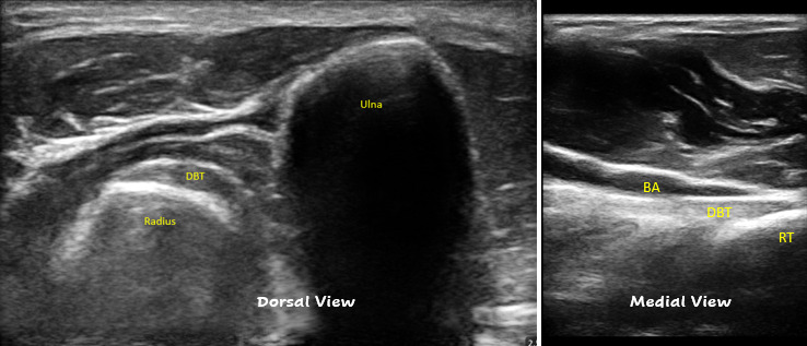
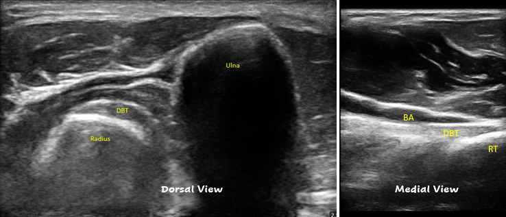
Tenderness specific to the tendon attachment, in addition to the other examination findings, prompted ultrasound imaging of the distal biceps brachii tendon insertion. Subjects were placed in a sitting position with their affected extremity on a nearby stand at approximately 90 degrees of elbow flexion and end-range forearm pronation. The ultrasound transducer (L7HD3, Clarius Mobile Health, Vancouver, Canada) was placed in the long axis to the tendon and transverse to the radius on the posterolateral aspect of the proximal antebrachium at the level of the radial tuberosity; this posterior approach allows for effective visualization of the distal tendon and tendon enthesis, as described by Al-Ani and Lauder,69 Moore,70 and Griffith.71 Depth was adjusted so that the tendon presented within the focal zone, allowing for improved resolution of tissues perpendicular to the ultrasound beam.72 Because the biceps tendon wraps around the radius in this approach, dynamic movement (e.g., supination and pronation) and rocking of the probe are necessary to ensure that different segments of the tendon are perpendicular to the ultrasound beam for optimal visualization to reduce anisotropy (ultrasound waves reflecting away from the transducer).69–72 A medial view was used to further confirm tendon pathology in Subject A. Normal distal biceps brachii tendon sonoanatomy comprises a hyperechoic, unidirectional fibrillar pattern with a tapering contour into the insertion at the radial tuberosity; this fibrillar pattern can be seen in Figure 1.70,71
Interventions
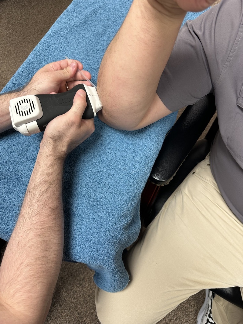
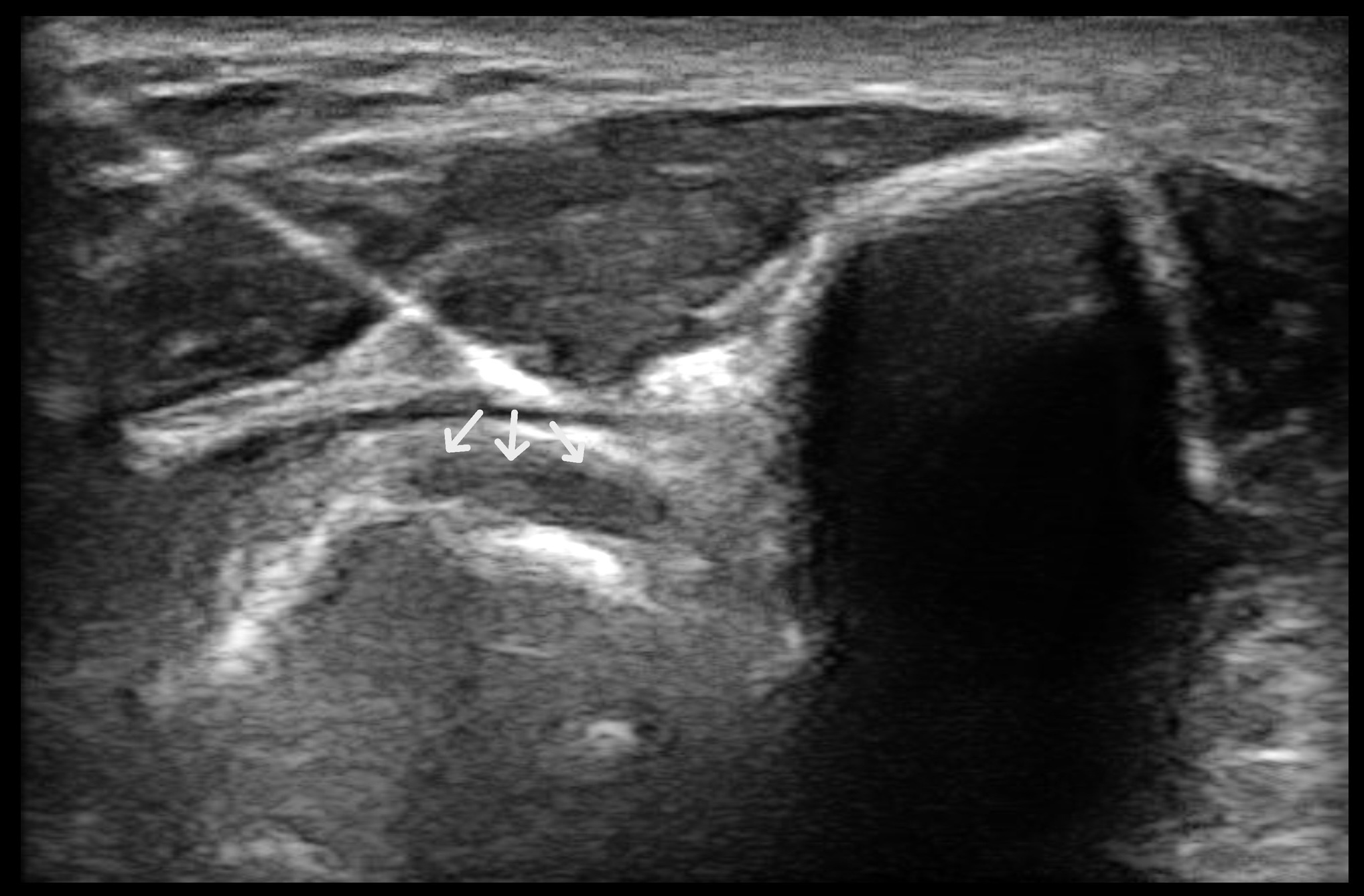
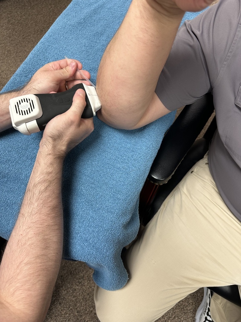
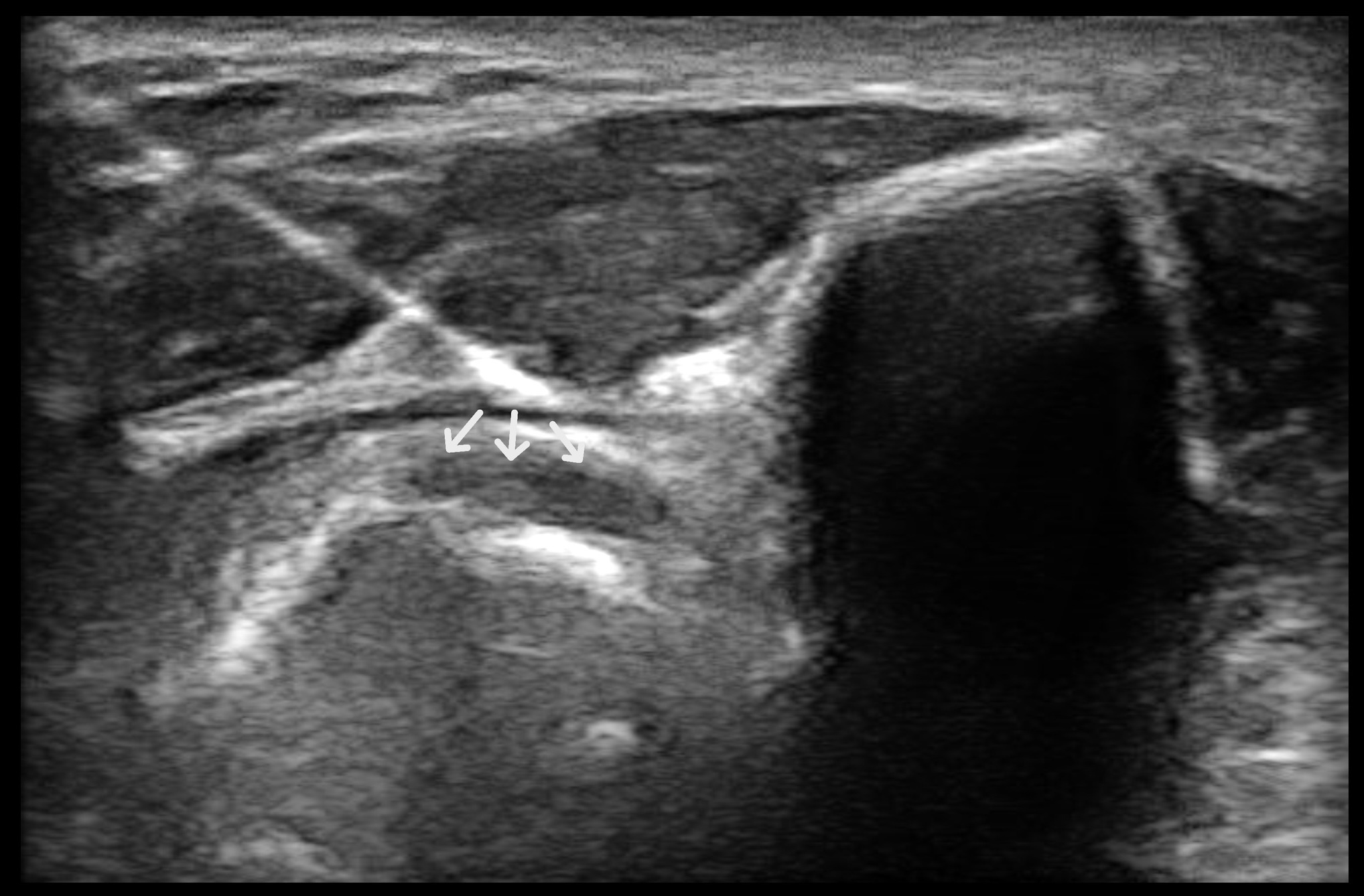
Subjects first underwent screening for contraindications to dry needling, including needle aversion, cognitive impairment, dyskinesia, bleeding disorders, vascular disease, local or systemic infection, and recent surgery. In the absence of contraindications, ultrasound-guided dry needling with tendon fenestration of the distal biceps tendon, combined with graded tendon loading, was implemented to ameliorate pain and disability associated with chronic distal biceps tendinopathy. Subjects were seen once weekly for five weeks, with ultrasound-guided tendon dry needling performed at the first three visits. After education on the purposes of incorporating dry needling, the role of mechanical loading on tendon healing, and graded loading strategies for a home-exercise program, the subject was positioned in a sitting position with their elbow on a nearby stand in approximately 90 degrees of flexion and full pronation (Figure 2). Under ultrasound guidance, a single 50 mm length by 0.30 mm diameter needle was used to fenestrate the distal biceps tendon for 10 seconds (approximately 15-20 times) with subtle positional changes within the tendon (Figure 3).
After dry needling, interventions that emphasized isotonic loading of the biceps were performed by each subject, including coupled eccentric elbow flexion and supination (i.e., 3 to 5 second eccentric phase), unilateral rows, resisted supination, and resisted elbow flexion with neutral and supinated forearm positions (Supplemental File 1). Subjects were initially educated on graded loading principles consistent with some of the other load strategies for tendinopathy.73,74 First, because initial activity tolerance was limited by pain to a greater extent than muscular fatigue, subjects were instructed to perform the highest tolerable resistance that did not provoke discomfort greater than 3/10 on a NPRS scale (minimal pain) during or after isotonic loading with a duration of greater than 24 hours for 3-4 sets of 15 repetitions.73 This guideline stayed in place until pain was abolished in both subjects. They were then advised to lift with 2 reps-in-reserve at 3-4 sets of 6 repetitions, allowing for dynamic intensity regulation that can facilitate self-progression.75 Both subjects performed these exercises 3 times per week. Baseline and 6-month resistances can be seen in Table 2. Most of the activity performed by the subjects was instruction-based and performed outside of the clinical environment. Each participant verbally reported compliance with this home exercise program at five weeks, three months, and six months.
RESULTS
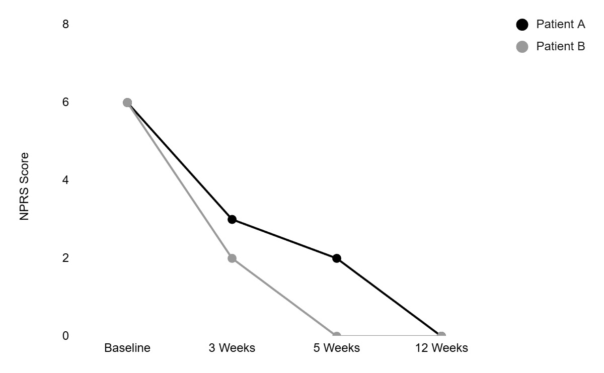
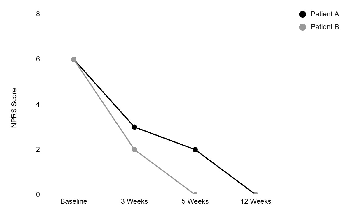
The same clinician treated each subject with a similar treatment approach that incorporated ultrasound-guided tendon dry needling with fenestration (included in the first 3 weeks) and a graded loading program (6 months). Both subjects noted a reduction in pain and had NPRS scores that surpassed the MCID from baseline versus three, five, and 12 weeks, with symptom relief persisting at 6 months (Table 3, Figure 4). The reduced NPRS scores correspond with perceived improvements greater than the +/- 5 MCID on the 15-point GROC scale from baseline to five weeks and onward in both subjects (Table 3).
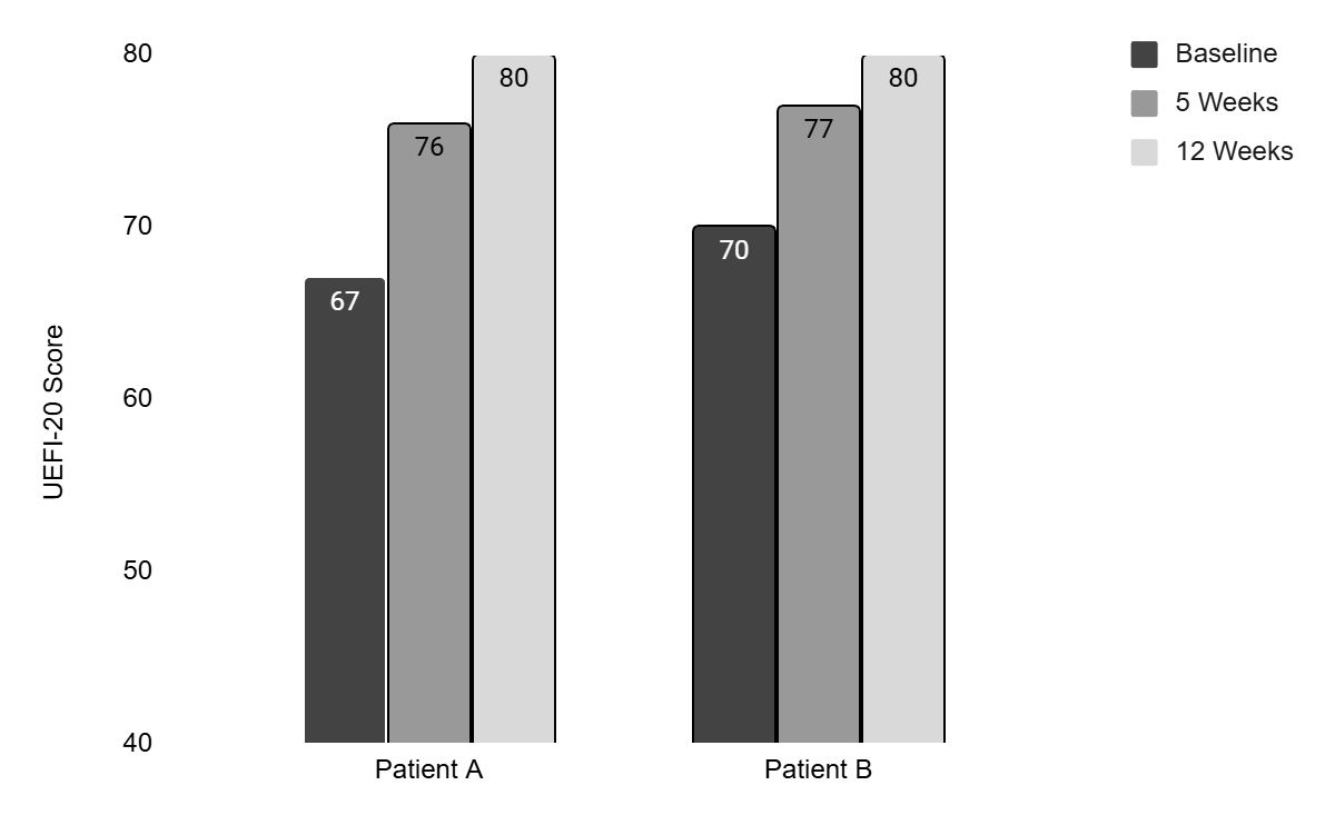
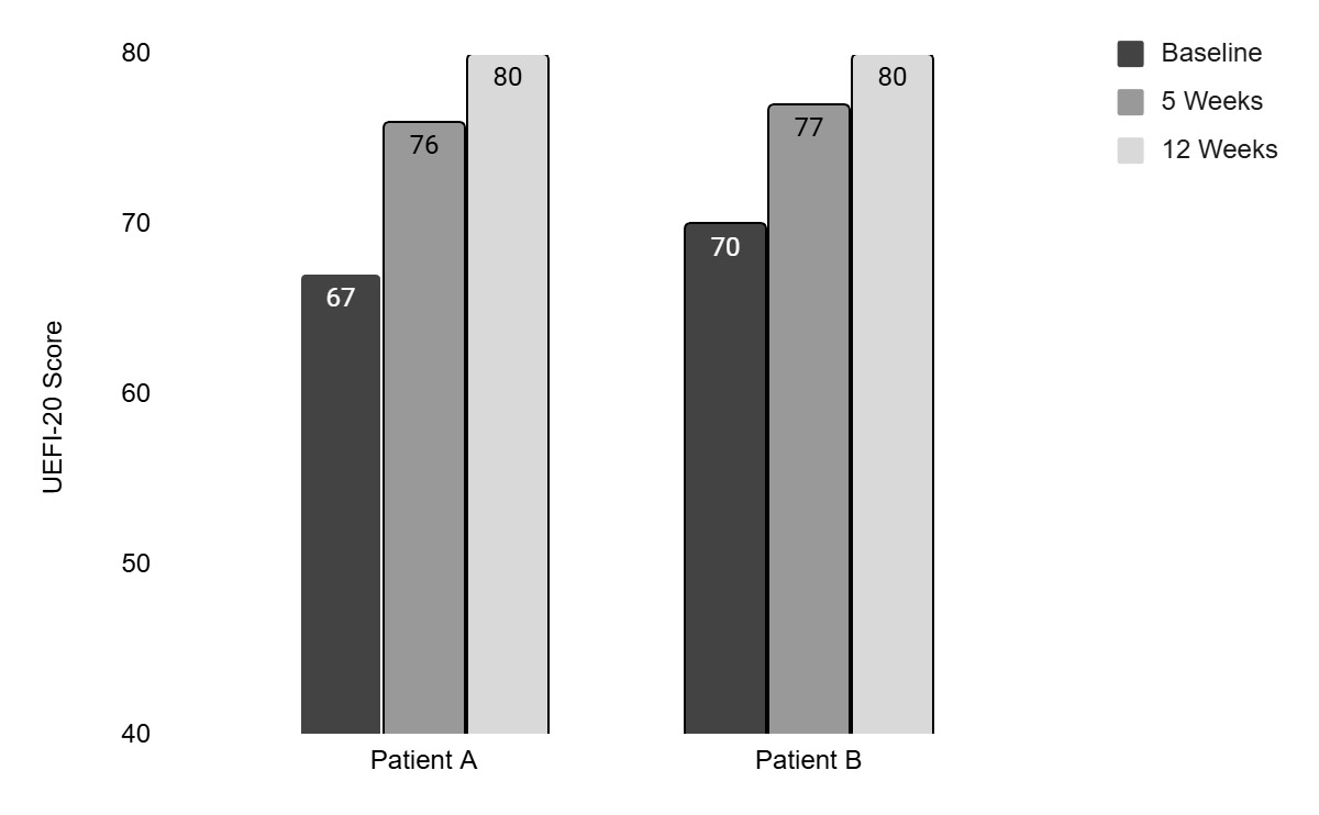
Similarly, each subject noted a reduction in upper extremity disability with UEFI-20 scores that met or exceeded the MCID of 7 points; furthermore, subject A and subject B reported no difficulty with all UEFI-20 domains at 12 weeks, which remained consistent at six months (Table 3, Figure 5). Therefore, each subject reported clinically meaningful improvements in pain and disability.
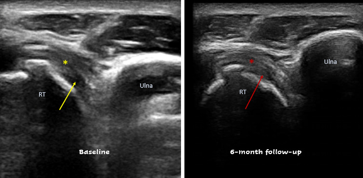
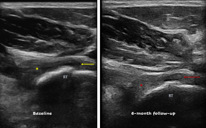
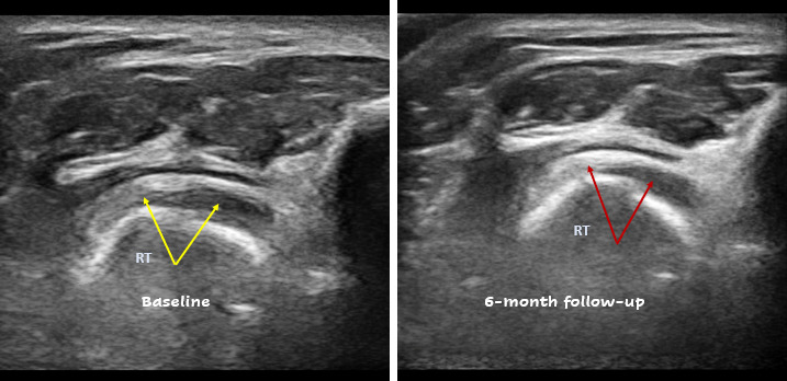
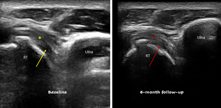
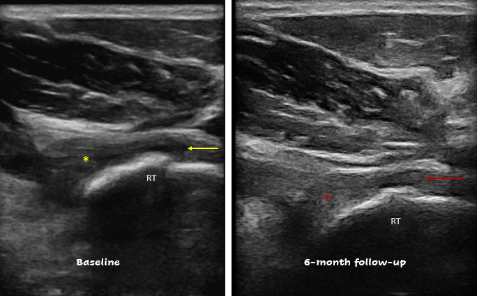
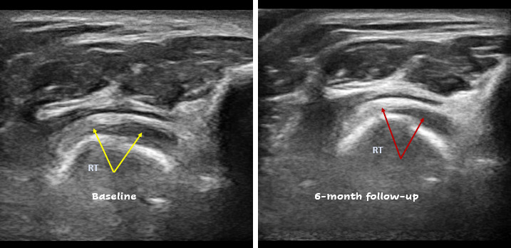
Notable healing characteristics were visualized in both subjects at 6-month follow-up imaging and reviewed by clinicians registered in musculoskeletal sonography (RMSK). At baseline, Subject A presented with cortical irregularity at the radial tuberosity with a focal anechoic region of the distal biceps tendon that was tender to sonopalpation and end-range passive pronation, suggestive of a partial-thickness tear. Six-month follow-up images demonstrate early regeneration at the tear site with improved fiber visualization (Figures 6 and 7). Subject B presented with milder cortical changes with thickening of the distal biceps tendon (0.25cm) and areas of hypoechogenicity but with preservation of tendon continuity. 6-month rescan demonstrates improvements in tendon echogenicity, a reduction in tendon width (0.25cm to 0.19cm), and normalization of mechanosensitivity with absent tenderness to sonopalpation and absent discomfort at end-range passive pronation (Figure 8).
Treatment Adverse Events
Adverse events were defined as distressing or intolerable events (e.g., pain) that lasted for more than one week.76 Neither subject reported any adverse events other than post-needling soreness and mild ecchymosis, which resolved by the next visit. Additionally, no subjects required load regression secondary to activity intolerance.
DISCUSSION
Two subjects participated in a care plan that incorporated ultrasound-guided dry needling of the distal biceps brachii tendon with tendon fenestration, alongside a graded loading program, resulting in clinically relevant reductions in pain and disability. Tendon fenestration is thought to produce a microtrauma that is beneficial for healing secondary to numerous biochemical and neurophysiological factors. This microtrauma may disrupt the maladaptive healing response seen in tendon disrepair, upregulate inflammatory cells to produce a controlled inflammatory response, and upregulate satellite cells to assist with renormalization of collagen phenotype (i.e., Type III to Type I) and matrix alignment.10,11,32,77 Pathologic tendons often have increased neuronal ingrowth and angiogenesis, which is likely a contributing factor to pain severity and chronification.9,25,34 Studies investigating lateral epicondylalgia have demonstrated that ultrasonic percutaneous tenotomy and electrolysis of the common extensor tendon results in a marked reduction in Doppler activity, suggesting reduced tendon hypervascularity.6,9 However, subjects in the study by Ang and colleagues6 also appeared to have benefited from relatively quick reductions in pain, suggesting the possibility of early symptom modulation using the technique. With tendon pathology often necessitating longer healing timeframes, early symptom modulation may be invaluable for subject buy-in and improving their participation in personally meaningful activities.4,34 A predominant theme highlighted in a systematic review of qualitative studies investigating subject experiences with tendinopathy suggests that a significant subject concern is their ability to fully return to participating in daily activities, suggesting a reduction in quality of life and potential psychosocial strain (e.g., less participation in social activities with friends, mental strain from the financial ramifications of reduced occupational participation).4 Thus, interventions that provide early symptom relief may facilitate continued engagement in rehabilitation for pathologies that often have longer-term recovery processes.
At six months from the initial evaluation, each subject presented with favorable ultrasonographic features, accompanied by pain and disability resolution. These structural changes included improvements in tendon echogenicity, fibrillar organization, and thickness, and findings were accompanied by marked reductions in sonopalpation tenderness and by improvements in NPRS, UEFI-20, and GROC beyond the MCID in both subjects. These changes may support structural change that should continue to demonstrate beneficial tendon remodeling when combined with a progressive loading protocol.78–80 Tendon healing and adaptation often occur along prolonged timelines, potentially beyond symptomatic resolution. Tendon fenestration is postulated to disrupt a maladaptive repair process with chronic inflammatory infiltration and create an environment conducive to improved remodeling versus rest alone.10,11,32,77,80 The inferior tensile tolerance that accompanies glycosaminoglycan and immature collagen deposition may be ameliorated through activity that specifically addresses the tensile capacity of the affected tendon, emphasized clinically through end-range eccentric loading.41,80–82 Among the two subjects, the combination of education on tendon management strategies and response to load, tendon fenestration to facilitate an optimal healing response, and implementation of a graded loading program appeared to improve pain and disability.
The microtrauma induced by tendon fenestration was accompanied by a graded loading program to optimize load tolerance of the affected biceps brachii for improved upper extremity functional capacity among the two subjects. The localized microtrauma induced by fenestration may initiate a controlled inflammatory response that is thought to facilitate tendon healing.10,11,32,77 The subsequent application of progressive mechanical loading may then provide an appropriate stimulus for tendon adaptation secondary to tenocyte sensitivity to load.81,83 Mechanical load stimulates insulin-like growth factor-1 and transforming growth factor-β secretion to promote tendon remodeling, and long-term compliance with an exercise program that emphasizes tendon tensile load tolerance appears to ameliorate abnormalities seen in tendon structure (e.g., disruption in fibrillar alignment) through a variety of mechanisms, including increased collagen synthesis.83–88 Continued participation in a loading protocol is important as it allows for epigenetic changes of the tendon, allowing it to adapt specifically to the loads the individual must endure (e.g., occupational lifting demands).86 However, some studies suggest that changes in tendon structure and pain may not always be well correlated.89,90 The intervention incorporated pain as the progression strategy for isotonic load management, avoiding pain levels above a 3/10 on NPRS.73
To the authors’ knowledge, this is the first study to report ultrasound-guided dry needling with fenestration of the distal biceps tendon at the radial tuberosity from a posterior approach as an adjunct to a physical therapy care plan. The dorsal approach used in this study is beneficial as it avoids the neurovascular structures anteriorly.12,13 A limitation of this retrospective case series is that Doppler ultrasound images were not included, which would have added additional information regarding hyperemia. Additionally, both subjects had minimal comorbidities and were physically active; one may expect varying subject responses in the presence of extensive comorbidities or chronic nicotine use.24–27 Lastly, this is a case series without the ability to discern cause and effect or the ability to determine the contribution of improvements secondary to the passage of time. Future research would benefit from incorporating a multi-site, randomized controlled trial comparing fenestration of the distal biceps tendon and a graded loading program to a graded loading program alone or no intervention controls to quantify the value of tendon dry needling with fenestration for short-term analgesia and long-term clinical outcomes in those with chronic distal biceps tendinopathy.
CONCLUSION
Two subjects with chronic distal biceps brachii tendinopathy achieved clinically meaningful improvements in pain and disability with ultrasound-guided tendon dry needling with fenestration and graded resistance exercise. Further, improved tendon sonographic characteristics were observed at six months versus baseline in both subjects. Randomized controlled trials investigating ultrasound-guided needle fenestration for distal biceps tendinopathy are necessary to establish causal inference and discern generalizability.
Conflicts of Interest
The authors report no conflicts of interest.