Loading [Contrib]/a11y/accessibility-menu.js
1.
Manske RC, Wolfe C, Page P, Voight M. Diagnostic Musculoskeletal Ultrasound in the Evaluation of the Extensor Digitorum Longus. IJSPT. 2026;21(11). doi:10.26603/001c.172997
Download all (5)
  • Extensor Digitorum Longus
  • NORMAL VIEW IN LONG AXIS (LAX)
  • NORMAL VIEW IN SHORT AXIS (SAX)
  • EXTENSOR DIGITORUM LONGUS TENOSYNOVITIS
  • PARTIAL TEAR OF THE EXTENSOR DIGITORUM LONGUS TENDON

Abstract

The extensor digitorum longus muscle and tendons are among the longer muscle-tendon units in the lower leg and foot. This feather-like, unipennate muscle is part of the anterior ankle group, attaching proximally along the lateral side of the lateral condyle of the tibia and the upper three-quarters of the fibula’s inner surface. Near the ankle, the tendon narrows as it passes beneath the extensor retinacula and divides into four smaller tendons that connect to the toes. Since it crosses the ankle joint and attaches to the second through fifth toes, it facilitates dorsiflexion of the foot and extension of the toe joints.
An accurate diagnosis of extensor digitorum longus tendonitis or rupture is crucial for appropriate treatment planning and optimal patient outcomes. Diagnostic musculoskeletal ultrasound (MSKUS) has become a widely used imaging modality for assessing many ankle and foot pathologies. It offers a portable, real-time, and cost-effective alternative that is gaining traction in rehabilitation and sports medicine settings. MSKUS has emerged as a valuable, non-invasive imaging modality for evaluating ankle and foot injuries. This manuscript reviews the utility of MSKUS in evaluating the quality, size, sonographic techniques, and clinical implications for rehabilitation professionals who need to visualize the extensor digitorum longus. By integrating MSKUS into clinical practice, providers can improve diagnostic accuracy, increase diagnostic confidence, monitor healing progression, and guide rehabilitation strategies to optimize outcomes for patients with ankle and foot injuries.

Introduction

Diagnostic musculoskeletal ultrasound (MSKUS) has become an essential tool for evaluating musculoskeletal structures. Foot and ankle pain are very common injuries in orthopedic and sports physical therapy clinical practice. Fortunately for therapists, MSKUS can visualize many potential pain generators superficially in the foot and ankle. One pain generator in the lower leg, ankle, and foot is the extensor digitorum longus. This paper provides a comprehensive review of diagnostic MSKUS of the extensor digitorum longus, including relevant anatomy, scanning technique, normal and abnormal imaging characteristics, and common pathological findings.

Anatomy of the Extensor Digitorum Longus Muscle and Tendon

The extensor digitorum longus muscle and tendons are one of the longer muscle-tendon units in the lower leg and foot. The muscle is a feather-like unipennate muscle that is part of the anterior ankle complex with a proximal attachment site along the lateral side of the lateral condyle of the proximal tibia, and the upper 3/4ths of the inner surface of the fibula.1 Distally, just above the ankle, the tendon narrows as it passes beneath the extensor retinacula, where it then divides into four smaller tendons attaching to each of the four lateral toes. Because it crosses the ankle joint and attaches to the 2nd through 5th toes, it dorsiflexes the foot and extends the metatarsophalangeal joints.2,3

However, although textbooks describe the most common anatomical origins and insertions, variation of the extensor digitorum longus muscle and tendon is very common, especially near its distal parts, and includes duplication or absence of its tendinous slips.2 Newton documented the absence of the muscle unilaterally,4 while Sakuma found the absence of the tendon slip to the 4th toe.5 These are extremely important to note, as these absences could obviously cause concern to the casual sonographer unfamiliar with these unique variations.

The etiology of injury to the extensor digitorum longus is diverse and includes rupture due to its superficial location on the extensor side of the foot with little soft tissue coverage.6 Tendon ruptures or partial tears are relatively rare but can occur as a result of sport-related trauma from inversion and plantar flexion injuries.7,8

The Role of MSK Ultrasound in Evaluation of the Extensor Digitorum Longus Muscle and Tendon

Because these extensor digitorum longus tendons are superficial, MSKUS visualizes these injuries well. Additionally, because MSKUS allows dynamic examination while viewing, it may be better than static imaging such as magnetic resonance imaging (MRI) or computed tomography (CT). Rockett et al. reported that MSKUS was superior to MRI in detecting ankle tendon abnormalities.9 MSKUS showed a much higher sensitivity (100% vs 23.4%), nearly identical specificity (89.9% vs 100%), and much higher accuracy (94.4% vs 65.75%) when compared to MRI.

Advantages

  • Real-Time Imaging: MSKUS allows dynamic evaluation of muscle and tendon while the lower leg, ankle, and foot can be moved through the available range of motion. It provides real-time assessment of soft tissues and their motion, offering information beyond the static morphology seen on radiographs, MRI, and CT.

  • High-Resolution Visualization: MSKUS provides detailed images of the muscle and tendon structure.

  • Accessibility and Cost-Effectiveness: MSKUS is portable, widely available, and less expensive than MRI.

Limitations

  • Operator Dependency: MSKUS requires skill and experience for accurate interpretation of findings. With practice, visualizing muscle and tendon tissue with MSKUS is relatively easy and largely depends on operator skill, the availability and technical considerations of state-of-the-art equipment, and the positioning of the lower leg, ankle, and foot during the assessment.

  • Depth Limitations: Visualization is usually not a problem as the extensor digitorum muscle and tendons are superficial and well within the appropriate depth limitations of MSKUS.

  • Artifacts and Shadows: Because of the superficial location, deeper shadowing from the ankle or tarsal bones is rarely an issue during image interpretation.

Sonographic Technique for Evaluating the Extensor Digitorum Muscle and Tendon

  • Probe Type: The depth of the extensor digitorum muscle and tendons allows for a standard high-frequency 3-13 MHz linear array probe.

  • Patient Position: The optimal position for imaging of the extensor digitorum longus tendon is with the patient in a supine position with the knee flexed so that the sole of the foot is resting flat on the examination table.

  • Dynamic Assessment: Passive or active movement of the ankle and toes can create dynamic movements that allow stress to be applied to muscles and tendons, helping to accentuate issues seen during ultrasonography.

Normal Sonographic Appearance of the Extensor Digitorum Longus Muscle and Tendon

Normal, healthy extensor digitorum longus tendons will appear as a continuous, echogenic, fibrillar structure with minimal to no fluid in its tendon sheath. During dynamic movements, they should glide smoothly without interruption. Note that the bursa of Gruberi may be seen between the extensor digitorum tendons and the talus.10

Pathologic Findings of the Extensor Digitorum Longus Muscle and Tendon

  • In the muscle of the extensor digitorum longus, a herniation of a fascial defect may be seen as a bulge through the fascia that may be seen in both short axis (SAX) and long axis (LAX) views.

  • A partial tear may be seen as disruption of continuity of the fiber arrangement. A hypoechoic gap may be seen with tendon retraction in a complete tendon rupture.

  • Tendinosis may be seen as loss of normal fibrillar echogenicity, with replacement by disorganized hypoechoic tissue. Additionally, thickening of the tendon edges or margins may be seen, as well as muscle swelling.

  • In the case of an avulsion fracture, a subtle cortical irregularity may be seen and is not always clearly visible on radiograph.

Clinical Implications for Rehabilitation Providers

MSKUS provides real-time feedback for rehabilitation professionals, facilitating early diagnosis and intervention. Key applications include:

  • Early Detection of Injury / Accurate Injury Grading: MSKUS can determine normal tendon and muscle tissue from pathologic, inflamed, swollen, torn, or ruptured tissue.

  • Dynamic Functional Testing: Rehabilitation professionals can use MSKUS during physical therapy sessions to monitor recovery and assess tendon tissue thickness and consistency in both static and dynamic assessments. Serial MSKUS imaging helps assess decreasing thickness and tissue remodeling to support readiness for rehabilitation progression.

  • Guided Interventions: Ultrasound imaging assists in precision-guided interventions such as dry needling or injections, such as corticosteroids for inflammation.

  • Patient Education: Real-time imaging serves as a visual aid to explain the nature of the injury and set realistic expectations for recovery. Dynamic imaging during lower leg, ankle, and foot movements can show the tendon’s actual movement.

Limitations and Challenges

Despite its advantages, MSKUS has limitations and challenges. Like many other areas of the body examined with MSKUS, ultrasound accuracy depends on several factors, including probe placement, applied pressure (small changes in probe angle or pressure can compress the tendon and alter its appearance), operator experience, and ultrasound quality. MSKUS can show anatomical structure but cannot differentiate cellular inflammation from other potential findings, such as acute inflammatory reactions or chronic fibrotic thickening. However, it allows for detailed anatomical examination and outperforms MRI in spatial resolution of small superficial structures in the foot and ankle.7

Conclusion

In summary, diagnostic MSKUS offers rehabilitation professionals a powerful, clinically relevant extension of the physical examination when evaluating the extensor digitorum longus muscle and tendon. Its ability to provide high-resolution, real-time, and dynamic visualization of the tendon architecture enhances diagnostic precision, supports early clinical decision-making, and allows serial monitoring of tissue healing and load tolerance. When integrated with a thorough understanding of anatomy, injury mechanisms, and functional biomechanics, MSKUS becomes more than an imaging modality—it becomes a performance-informed clinical tool. While operator skill and certain anatomic limitations must be acknowledged, thoughtfully incorporating MSKUS into sports and orthopedic practice can elevate assessment accuracy, refine rehabilitation progression, and ultimately improve return-to-play outcomes for athletes with foot and ankle injuries.
References

1.
Kamaşak B, Kırındı MH, Ulcay T, Uzun A, Aycan K. An unique unilateral tendon variation of the extensor digitorum longus muscle. Surg Radiol Anat. 2022;44(11):1427-1430. doi:10.1007/​s00276-022-03040-4
Google Scholar
2.
Banerje A, Singh S, Raza K, Rani N, Kaler S. Unilateral Variation in Extensor digitorum longus muscle. Clin Ter. 2016;167(5):150-151. doi:10.7417/​CT.2016.1946
Google Scholar
3.
Standring S, Borley N, Collins P, et al. Gray’s Anatomy: The Anatomical Basis of Clinical Practice. 40th ed. Elsevier, Churchill Livingstone; 2008.
Google Scholar
4.
Newton B. Unilateral absence of an extensor digitorum longus muscle and variations of toe tendons. Int J Anat Var. 2014;7:42-45.
Google Scholar
5.
Sakuma E, Kato H, Honda N, Mabuchi Y, Soji T. A rare anomaly of the extensor digitorum longus. Anat Sci Int. 2004;79(4):235-238. doi:10.1111/​j.1447-073x.2004.00082.x
Google Scholar
6.
Yeo ED, Han JK, Lee HS, et al. Direct reconstruction of chronic extensor digitorum longus tendon rupture using interposed scar tissue in the foot: A case report. Medicine (Baltimore). 2020;99(40):e22506. doi:10.1097/​MD.0000000000022506
Google Scholar
7.
Deka JB, Deka NK, Shah MV, Bortolotto C, Draghi F, Jimenez F. Isolated partial tear of extensor digitorum longus tendon with overlying muscle herniation in acute ankle sports injury: role of high resolution musculoskeletal ultrasound. J Ultrasound. 2022;25(2):369-377. doi:10.1007/​s40477-021-00572-0
Google Scholar
8.
Pointinger H, Munk P, Poeschl G. Rupture of the extensor digitorum longus muscle: additional finding in an ankle sprain. Eur J Trauma. 2003;29(3):161-163. doi:10.1007/​s00068-003-1270-z
Google Scholar
9.
Rockett MS, Waitches G, Sudakoff G, Brage M. Use of ultrasonography versus magnetic resonance imaging for tendon abnormalities around the ankle. Foot Ankle Int. 1998;19(9):604-612. doi:10.1177/​107110079801900907
Google Scholar
10.
Gaetke-Udager K, Jacobson JA, Bhatti ZS, Smith J, Parameswaran A, Fessell DP. Ultrasound of the gruberi bursa with cadaveric and MRI correlation. Am J Roentgenol. 2016;207(2):386-391. doi:10.2214/​AJR.15.15955
Google Scholar


Extensor Digitorum Longus

Patient Position
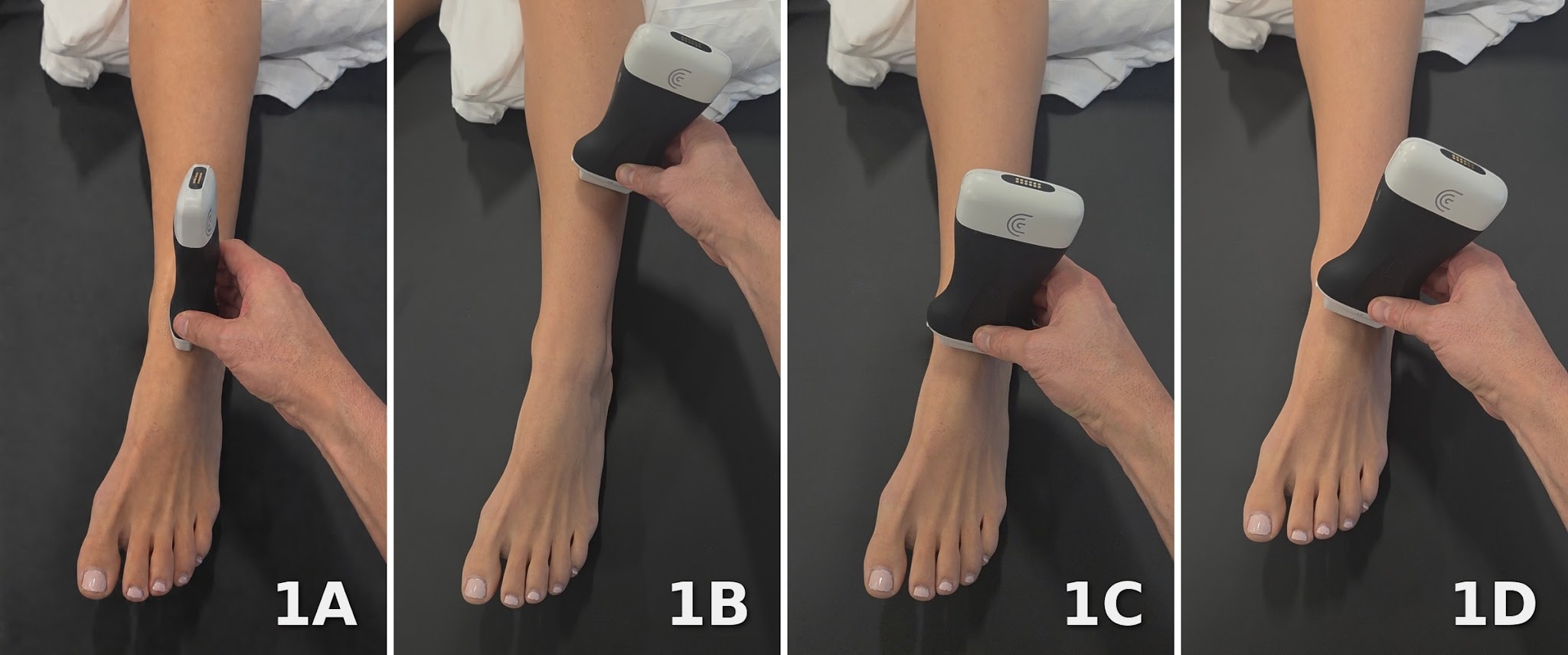
The patient is positioned supine or in supported long sitting with the knee slightly flexed and the anterior ankle exposed. The foot is supported in a comfortable position with slight plantar flexion as tolerated. This position provides access to the superficial extensor tendons while allowing movement of the ankle and toes during dynamic assessment. Sufficient ultrasound gel and light transducer pressure help maintain contact without compressing the superficial tendon sheath.

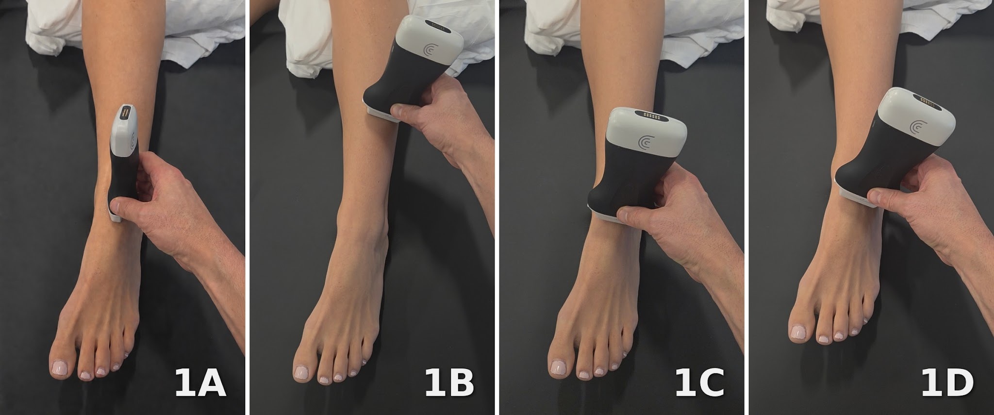
Figure 1A: Long Axis (LAX) Transducer Placement
The LAX view is obtained by positioning the transducer over the anterior ankle, parallel to the extensor digitorum longus tendon fibers. First identify the tendon in a transverse survey, lateral to the extensor hallucis longus, and then rotate the transducer to follow its longitudinal course. The distal tibia and talus provide deep bony landmarks as the tendon crosses the ankle. Trace the tendon proximally toward the musculotendinous junction and distally toward its divisions on the dorsum of the foot. Active extension of the second through fifth toes helps confirm extensor digitorum longus identification by demonstrating tendon excursion.

Figures 1B–1D: Short Axis (SAX) Transducer Placement (Muscle Belly, Anterior Ankle, Dorsal Foot)
The SAX view is obtained by rotating the transducer 90 degrees from the LAX position, placing it perpendicular to the tendon fibers. At the anterior ankle, identify the tibialis anterior tendon medially, the extensor hallucis longus tendon centrally, and the extensor digitorum longus tendons laterally. Translate the transducer proximally and distally to assess tendon size, internal echogenicity, and the surrounding sheath. Extension of the second through fifth toes helps distinguish extensor digitorum longus excursion from that of the neighboring extensor hallucis longus during great-toe extension.

NORMAL VIEW IN LONG AXIS (LAX)

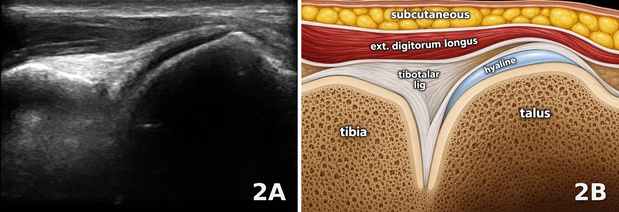
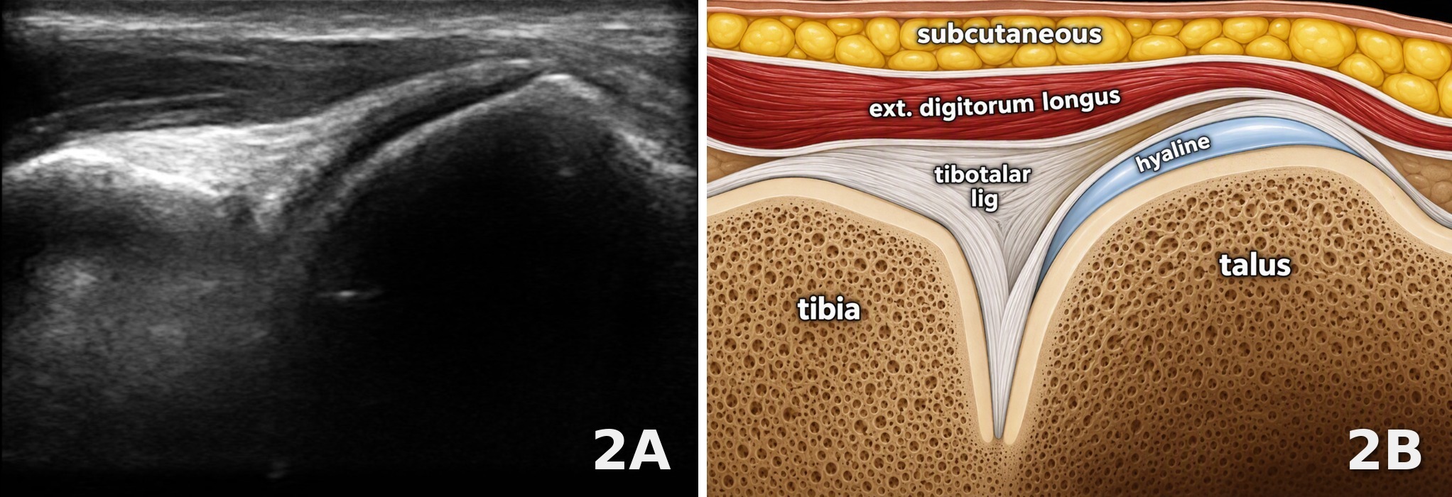
Figures 2A and 2B: Long Axis View
In the LAX view, the extensor digitorum longus tendon appears as a continuous, hyperechoic fibrillar structure crossing superficial to the anterior ankle joint. The distal tibial and talar cortices form bright interfaces deep to the tendon and joint capsule. Normal tendon fibers maintain a uniform, parallel arrangement without focal thickening or disruption. Minimal to no fluid should be visible within the tendon sheath. Small heel-to-toe transducer adjustments may be necessary to reduce anisotropy, particularly where the tendon changes direction across the ankle. Dynamic extension of the second through fifth toes should demonstrate smooth tendon excursion.

NORMAL VIEW IN SHORT AXIS (SAX)

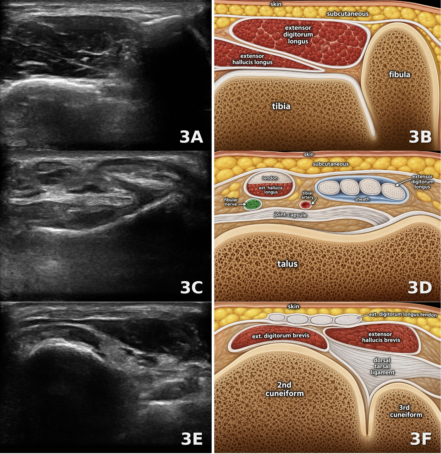
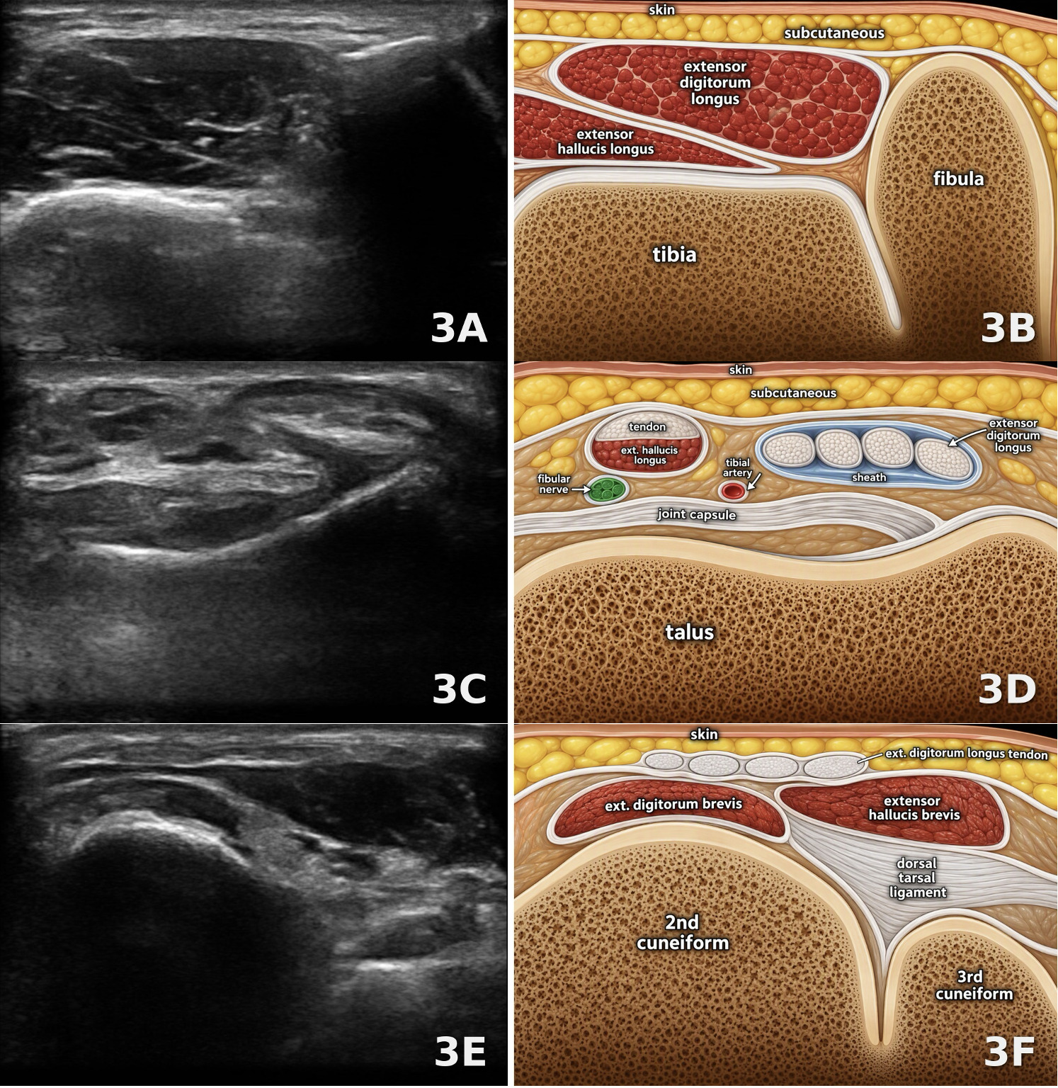
Figures 3A–3F: SAX View (3A/3B Muscle Belly, 3C/3D Talus, 3E/3F Midfoot)
In the SAX, the extensor digitorum longus can be followed from its muscle belly in the anterior compartment of the leg across the anterior ankle and onto the dorsum of the foot. Proximally, the muscle belly lies adjacent to the fibula and superficial to the extensor hallucis longus, displaying hypoechoic muscle fascicles separated by echogenic connective tissue. At the anterior ankle, the extensor digitorum longus tendons appear as echogenic oval or rounded structures with a finely stippled internal pattern within a common tendon sheath. They lie lateral to the extensor hallucis longus and tibialis anterior tendons and superficial to the anterior joint capsule, with the bright cortical reflection of the talus providing a deep bony landmark. Further distally over the cuneiforms, the individual tendon slips become more distinctly separated, coursing superficial to the intrinsic extensor musculature and dorsal capsuloligamentous structures toward the second through fifth toes. Throughout the examination, tendon contours should remain well defined, without focal enlargement or surrounding sheath distention. Adjust the transducer angle as the tendons change course to maintain perpendicular insonation and minimize anisotropy.

EXTENSOR DIGITORUM LONGUS TENOSYNOVITIS

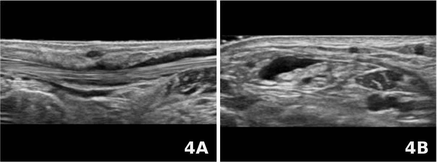
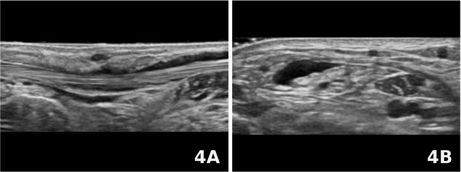
Figures 4A (LAX) and 4B (SAX): Extensor Digitorum Longus Tenosynovitis
The supplied images demonstrate fluid distending the extensor digitorum tendon sheath in both LAX and SAX. In the LAX view, hypoechoic to anechoic fluid extends alongside the echogenic tendon fibers. In the SAX view, fluid outlines portions of the tendon cross sections, making the expanded sheath more apparent. These findings are compatible with tenosynovitis in the appropriate clinical setting. Tendon fiber continuity should be assessed separately to determine whether associated tendon injury is present.

PARTIAL TEAR OF THE EXTENSOR DIGITORUM LONGUS TENDON

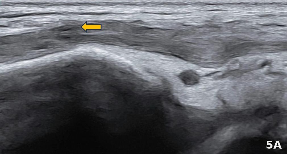
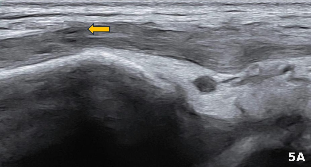
Figure 5A (LAX): Partial Tear of the Extensor Digitorum Longus Tendon
A partial tear demonstrates focal disruption of a portion of the tendon fibers, with some fibers remaining continuous across the involved region. In the LAX view, a focal hypoechoic defect interrupts the normal fibrillar arrangement shown at the orange arrow. Associated tendon thickening or surrounding fluid may be present. Gentle dynamic assessment may help demonstrate preserved continuity of the remaining fibers.