INTRODUCTION
Low back pain (LBP), a prevalent issue in athletic and physically active populations, is estimated to affect 10-30% of the adult population and is the fifth most frequently reported issue to clinicians by patients.1–4 Low back pain is often categorized as specific or non-specific.5 Specific LBP is characterized by pain and symptoms caused by non-spinal or spinal origin, while non-specific LBP is frequently classified as back pain with or without leg pain and without a specific underlying cause.5 However, differentiating between specific and non-specific LBP presents a distinct clinical challenge in sports physical therapy, particularly when referred pain from the sacroiliac joint (SIJ) compromises kinetic chain force transfer and athletic participation.5,6 Referred pain from the SIJ can cause radicular pain symptoms in various body parts, such as the buttocks, anterior thigh, groin, posterior thigh, and lower back, accounting for 15% to 30% of non-specific LBP cases, and creates a primary obstacle to high-load stabilization training in athletes.3,4,6
Medical professionals commonly treat patients experiencing non-specific LBP originating from the SIJ with muscle relaxers, non-steroidal anti-inflammatory drugs (NSAIDs), high-velocity, low-amplitude manipulations (HVLA), or surgery.1,3,4,7,8 Physicians frequently use minimally invasive intra-articular (IA) corticosteroid injections to alleviate SIJ pain by reducing joint inflammation.3,4 However, these injections can pose risks such as increased susceptibility to osteoporosis, fragility fractures, and weakened immune function, which may be problematic for naturally immunocompromised patients.3,4 HVLAs, while uncommon, could have adverse effects, such as localized tenderness, weakness, soreness, and rigidity, which could be contraindicated in those with hypermobility.9 While surgical interventions have shown promise in treating SIJ dysfunction, it is important to note that not all patients experience significant improvement following these procedures.4 Additionally, some patients may be hesitant to undergo invasive surgery that requires fixation of the pelvis.
Physical therapy that targets the surrounding muscles to improve stability in the SIJ can help reduce pain caused by hypermobility.4,7 Clinicians use pelvic stabilization exercises, primarily engaging the gluteus maximus and pelvic floor muscles, to reduce SIJ motion, thereby decreasing pain and disability.1,2,10 The Sabrina et al. meta-analysis explored physical therapy interventions to treat SIJ dysfunction, either as a standalone treatment or an adjunct to other methods.10 Their results indicated that physical therapy approaches were more effective, either independently or in conjunction with electrotherapy, mobilization treatments, and pelvic floor exercises, including transcutaneous electrical nerve stimulation, than treatments that did not include physical therapy.10
Blood Flow Restriction (BFR), also known as occlusion or hypoxic training, has become increasingly popular over the last 15 years for its ability to enhance strength gains in both healthy and injured individuals.11 Healthcare professionals commonly use this technique to counteract muscle weakness and atrophy after surgery and during rehabilitation following injury.11–13 Jessee et al. and Rohde et al. found that low-load BFR can produce results similar to, or even better than, those of high-load resistance training for muscle adaptations including strength, hypertrophy, and power.12,13
Athletes suffering from SIJ hypermobility often experience severe local and referred pain that prevents them from tolerating the high-load stabilization exercises typically required for return-to-play.1,2 Low-load BFR provides a potential unique clinical bridge, enabling athletes to engage the pelvic and gluteal musculature necessary for joint stabilization without aggravating hypermobile joint structures. In sports physical therapy, tracking patient-reported outcomes such as pain intensity, functional capacity, and perceived disability serves as an essential benchmark for evaluating an athlete’s readiness to advance through rehabilitation and return to competition. Therefore, the purpose of this case series was to describe the preliminary clinical outcomes specifically regarding pain, function, and disability in a small series of subjects with SIJ dysfunction following a single session of BFR training.
METHODS
Study Participants
All subjects provided informed consent and voluntarily participated in this case series, which was approved by a university IRB. All subjects also consented to the findings being submitted for publication. A convenience sample of four consecutive subjects who presented with non-specific LBP and were diagnosed with SIJ dysfunction participated in the case series. This case series took place in an outpatient athletic training clinic to replicate a typical patient-clinician interaction.
Patient inclusion required a presentation of SIJ dysfunction with pain in the buttock or lower back, with or without any leg symptomology; patient assessment for inclusion was performed with a thorough orthopedic examination and positive special test (i.e., Cluster of Laslett) findings.14,15 Further, subjects had to be over the age of 18 and had to self-report that pharmaceutical intervention (e.g., NSAIDs) for pain was not occurring. Exclusion criteria included pregnancy, recent lower extremity surgery, other current pain treatments (e.g., nerve blocks), radiculopathy symptoms, the use of blood thinners, or the presence of other medical conditions (i.e., hypertension, deep vein thrombosis (DVT), cancer, osteoporosis, or a true leg length discrepancy) in the health history or physical exam.
General Exam
Clinicians conducted a general orthopedic exam to assess the presence of SIJ dysfunction in response to the LBP complaint. Clinicians performed the exam based on the patient’s provided medical history and objective findings, including inspection, palpation, active and passive range of motion testing, manual muscle testing, and special tests to confirm or exclude various pathologies. Potential pathologies include, but are not limited to, SIJ dysfunction, facet syndrome, piriformis syndrome, inflammatory arthropathies, ankylosing spondylitis, and lumbar disc pathologies.
SIJ Dysfunction Diagnosis
The investigating clinicians classified SIJ dysfunction using the Cluster of Laslett, a previously studied battery of pain-provocation tests.14 Clinicians can achieve a more accurate diagnosis of SIJ pathology by using the pain provocation test battery than by using a single test, with high sensitivity (88%) and specificity (78%) for the battery.14 The Cluster of Laslett includes four pain provocation tests: the Distraction test, the Thigh Thrust test, the Compression test, and the Sacral Thrust test. Positive cluster findings and SIJ dysfunction classification required at least two individual tests of the battery to be positive.14 Just as the Cluster of Laslett was used in the diagnosis at initial evaluation, the cluster was also used as a discharge criterion at the discharge evaluation to assess for any ongoing SIJ involvement.
Blood Flow Restriction Protocols
Participants engaged in the conventional BFR protocol utilizing the Delfi Personal Tourniquet System (PTS) (Vancouver, BC). The clinicians supervising the interventions were certified in BFR protocols developed by Owens Recovery Science, and all obtained their Delfi PTS certification from Owens Recovery Science at the same time. Clinicians intended that subjects receive BFR treatment weekly, with reassessment occurring every seven days. In cases where pain resolution was achieved, subjects were promptly discharged.
Clinicians placed a four-inch-wide tourniquet on the upper thigh of the affected leg, which was chosen based upon on the side where the SIJ pain was experienced. The required pressure for achieving 80% arterial occlusion was calibrated using the Delfi PTS (Personal Tourniquet System) in accordance with the recommendations from its developers. This system enables clinicians to consistently apply the same level of pressure to the extremity throughout the various ranges of motion during exercise. Pressure settings for attaining the 80% occlusion were established prior to the treatment session.
Strength exercises were performed using bodyweight squats to a minimum depth of 90 degrees with their arms in front of their body throughout the exercise in a series of sets: Set 1 consisted of 30 repetitions followed by a 30-second rest; Set 2 included 15 repetitions with another 30-second rest; Set 3 also involved 15 repetitions and a 30-second rest; and Set 4 concluded the session with 15 repetitions. Bodyweight squats were chosen for these exercises due to their complexity, as they engage multiple muscle groups in the lower extremities. Furthermore, these muscles originate in the pelvis and contribute to both movement and stabilization of the pelvis.
Clinicians intended to treat subjects until symptoms resolved or another treatment modality was indicated. Although the subjects received only a single BFR treatment, they remained under the continuous care of these case series clinicians.
Patient-Reported Outcome Measures
Patient-Reported Outcome Measures (PROMs) were assessed at baseline and post-treatment at the end of each BFR session and at one-week follow-up. The outcome measures collected before and after each session consisted of the numerical pain rating scale (NPRS), patient-specific functional scale (PSFS), and the Disablement in the Physically Active Scale Short Form 8 (DPA SF-8).
The NPRS is a validated tool used to measure a patient’s perceived pain intensity on a scale from 0 to 10, where 0 indicates no pain and 10 represents the worst pain imaginable for the patient.15–18 According to research by Childs et al., the minimal clinically important difference (MCID) for patients with non-specific low back pain ranges from 1.7 to 2.2 points on the NPRS.19 Additionally, the minimal detectable change (MDC), which a patient can distinguish, is two points.19 Both the MCID and MDC serve as important metrics for analyzing subjective outcome measures. Clinicians rely on these metrics to determine whether observed changes in pain levels are statistically significant rather than due to measurement error or individual patient variability. For this case series, clinicians focused on a minimum 2-point change: the MCID and MDC. Subjects were asked to report their pain levels before treatment, after treatment, and at a one-week follow-up.
The PSFS is a patient-driven scale measuring a patient’s perceived physical function and activity level.20,21 Subjects selected two specific tasks, including but not limited to running, walking, jumping, or picking up something from the ground, and then rated their perceived function performing the task from 0-10: Zero indicated that the task could not be performed, while ten was equated with performing the task at a preinjury level.20,21 The MCID for the PSFS has been reported to be between two and three points.20 The MDC has multiple values based on the patient’s age and the specific condition. For example, Stratford reported an MDC for chronic pain of two points, while Maughan and Lewis reported an MDC of 1.4 points for low back pain.22,23 For this case series, an MCID and MDC of two points were used based on the previous findings.20–23 The PSFS was administered verbally before and at a one-week follow-up.
The assessment of disablement is increasingly recognized as a vital dimension of health care.24 Given that disablement comprises both physical and mental health components, clinicians have emphasized the necessity of incorporating a PROM that utilizes a multi-dimensional model, especially for chronic pathologies such as LBP.24 The Disablement in the Physically Active Short Form-8 (DPA SF-8) has been developed in accordance with a multi-dimensional framework specifically for physically active individuals who suffer from musculoskeletal injuries.24 This instrument consists of a 16-item scale that evaluates impairment, functional limitations, disability, and quality of life.24–26 The MCID has been evaluated to be two points for persistent injuries, while the MDC was shown to be 5.83 points.24 For this case series, the MCID and MDC were valued at two and six points, respectively, as the patient can only report whole numbers. The DPA SF-8 was used at the initial examination and one-week follow-up.
CASE DESCRIPTIONS
Subject one was a 52-year-old male tennis player actively competing in USA-ranked tennis tournaments. He presented with an acute episode of LBP and sought treatment on the same day. His symptoms were characterized by sharp pain localized over the left SIJ despite a lack of prior similar symptoms. The patient reported an inability to identify a specific mechanism of injury (MOI). Positive findings from special tests included the long sit, compression, and thigh thrust tests (Table 1). For the PSFS assessment, the patient selected two meaningful tasks: touching his toes and donning socks. A BFR cuff was affixed to his left limb based on the symptomatology observed.
Subject two was a 17-year-old male goalkeeper for a Major League Soccer (MLS) team. He felt a sharp pain in the left Posterior Superior Iliac Spine (PSIS) area after kicking the ball with his left leg during practice. He sought treatment at the clinic the same day. Previously, he had been treated by a chiropractor for an unrelated injury to the left PSIS and had received care at the clinic for right hip flexor pain. The patient tested positive for thigh thrust, compression, distraction, sacral thrust, and long sit tests (Table 1). However, during a follow-up, he tested negative for all the special tests (Table 1). The important activities he selected for the PSFS were running and moving from sitting to standing. Given the patient’s current symptoms, a cuff was applied to the left limb.
Subject three was an 18-year-old male MLS soccer athlete who had an acute onset during practice several weeks before his involvement in the case series. His MOI was kicking a ball with his left leg, and he felt a sharp pain over his left PSIS. The patient has a previous history of hip flexor pain in his right limb and previous right ankle injuries. The positive special tests included the long sit, compression, distraction, and sacral thrust tests (Table 1). Post-treatment, the subject tested negative for all the previously positive special tests (Table 1). The patient’s two meaningful tasks for the PSFS were jogging and transitioning from sitting to standing. Based on his current symptoms, the cuff was placed on the patient’s left limb.
Subject four was a 64-year-old female recreational athlete participating in walking, strength training, and conditioning. She had an acute onset while carrying heavy boxes out of her garage into her truck. She felt a sharp pain in her right PSIS region and reported to the clinic the next day. She had a previous history of hip flexor pain as well as previous episodes of pain over both her left and right PSIS. The positive special tests included the long sit test, the thigh thrust test, the compression test, and Sacral thrust tests (Table 1). Post-treatment, the subject tested negative for all the previously positive special tests (Table 1). The PSFS score was averaged between two meaningful tasks: sitting in a chair and walking up the stairs. For the treatment of this episode, the cuff was placed on her right limb, given her symptomology.
RESULTS
Clinical Presentation and Special Tests
All four subjects completed the single BFR session early in the course of their treatment, on the same day as the initial examination, and then were seen for the one-week follow-up. Subsequently, they were discharged from the therapy due to symptom resolution and did not require any additional interventions. At the initial examination, all subjects presented with at least two positive pain-provocation tests from the Cluster of Laslett, confirming the diagnosis of SIJ dysfunction. Following the BFR intervention, all subjects tested negative for all previously positive special tests. These negative findings were maintained at the one-week discharge follow-up, indicating a complete resolution of mechanically provoked SIJ signs.
Pain Outcomes (NPRS)
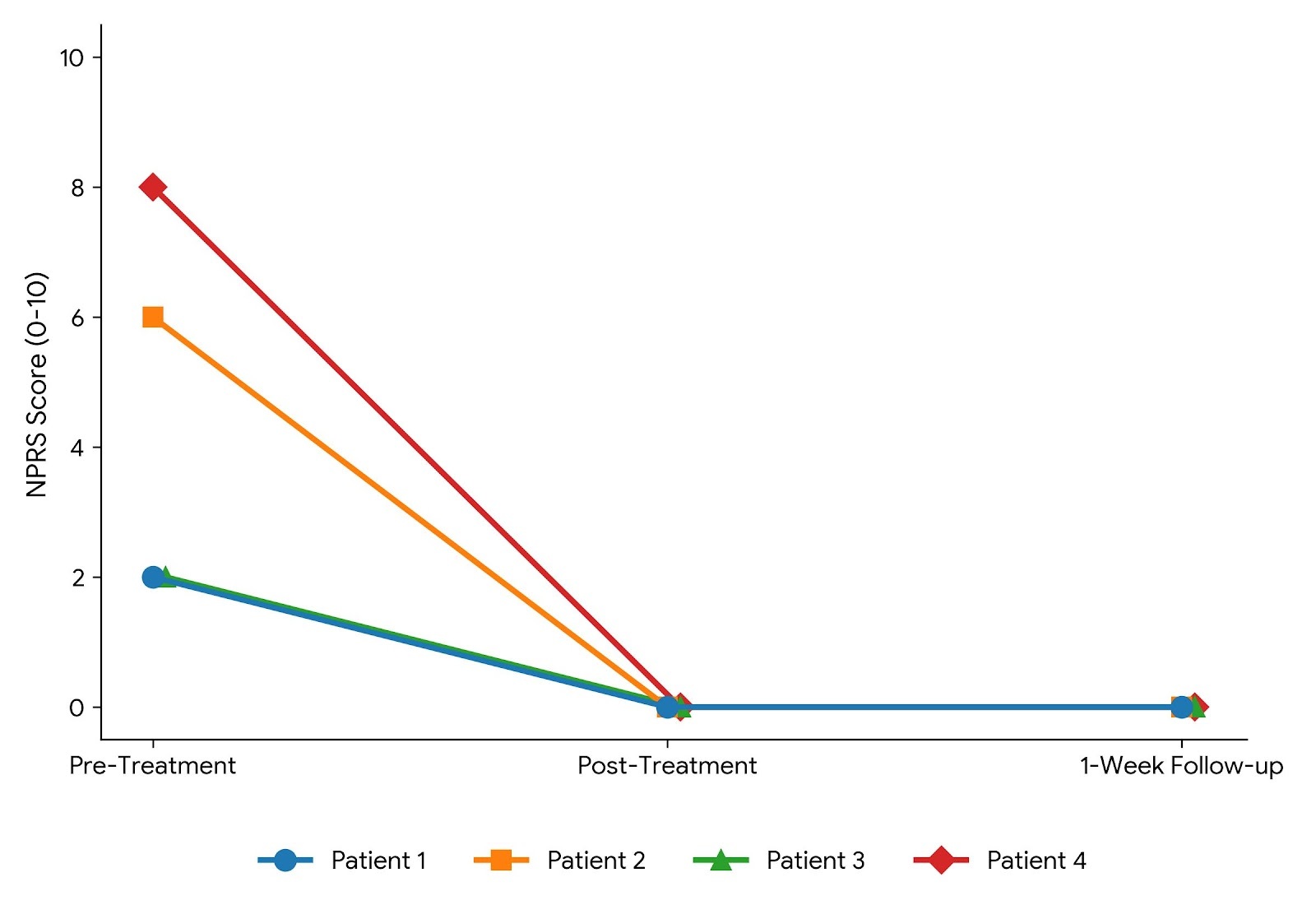
Subjects experienced rapid and clinically significant pain relief following the intervention. At baseline, before the BFR intervention, the mean current pain score on the NPRS was 4.5 ± 2.55, and the mean worst pain score was 7.5 ± 1.10 (Figure 1). Immediately post-treatment, all subjects reported a current NPRS score of 0, reflecting complete pain resolution. This pain-free state (NPRS = 0) was maintained by all four subjects at the one-week follow-up. For all subjects, this reduction met or exceeded the established MCID and MDC threshold of 2 points, signifying a clinically meaningful change (Figure 1).
Functional Outcomes (PSFS)
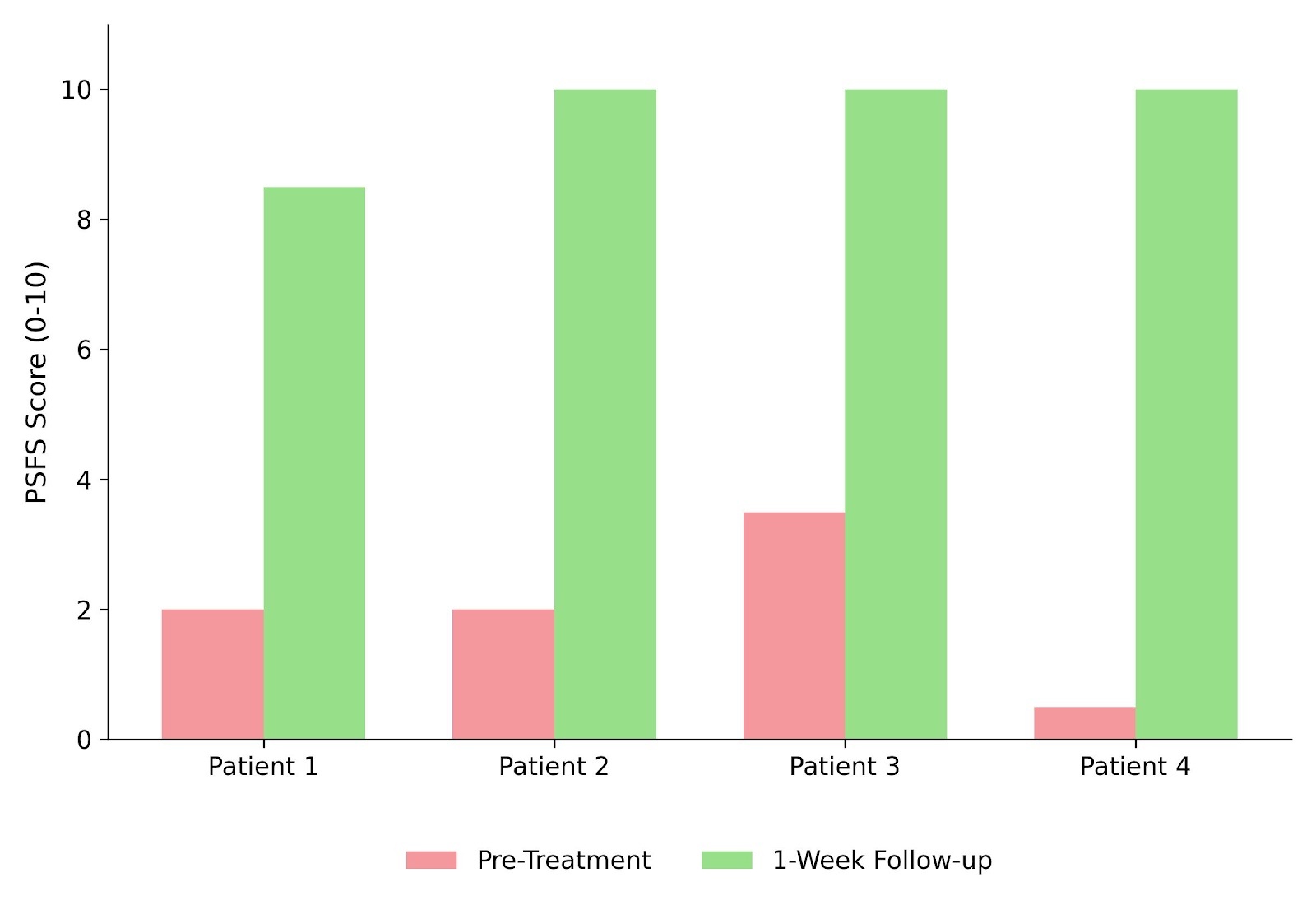
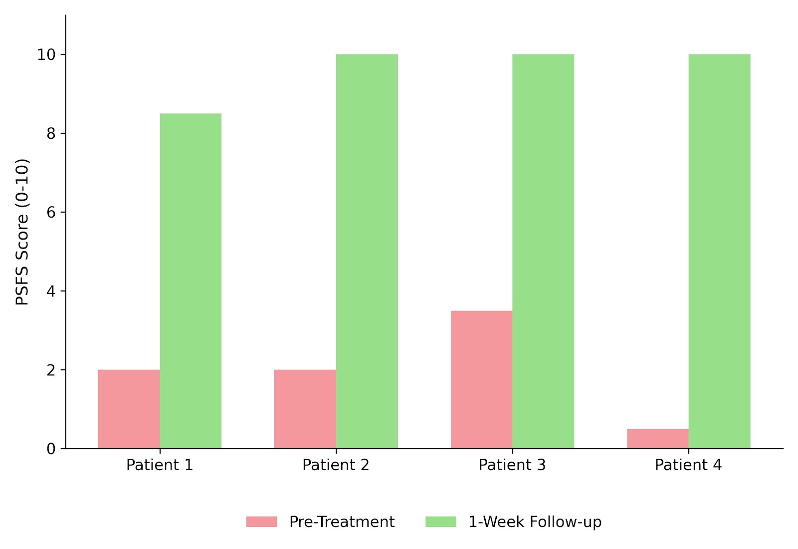
Perceived physical function, as measured by the PSFS, demonstrated substantial improvement following intervention (Figure 2). For each patient, scores from their two self-selected functional tasks were averaged to generate a single composite PSFS score (0–10 scale) at initial examination and at the one-week follow-up. Individually, three of the four patients (Patients 2, 3, and 4) achieved a maximum composite score of 10.0, indicating a full perceived return to pre-injury function across both selected tasks, while Patient 1 improved from 2.0 to 8.5. All four patients exceeded the established MCID and MDC thresholds of 2.0 points for the PSFS.
Disablement and Quality of Life (DPA SF-8)
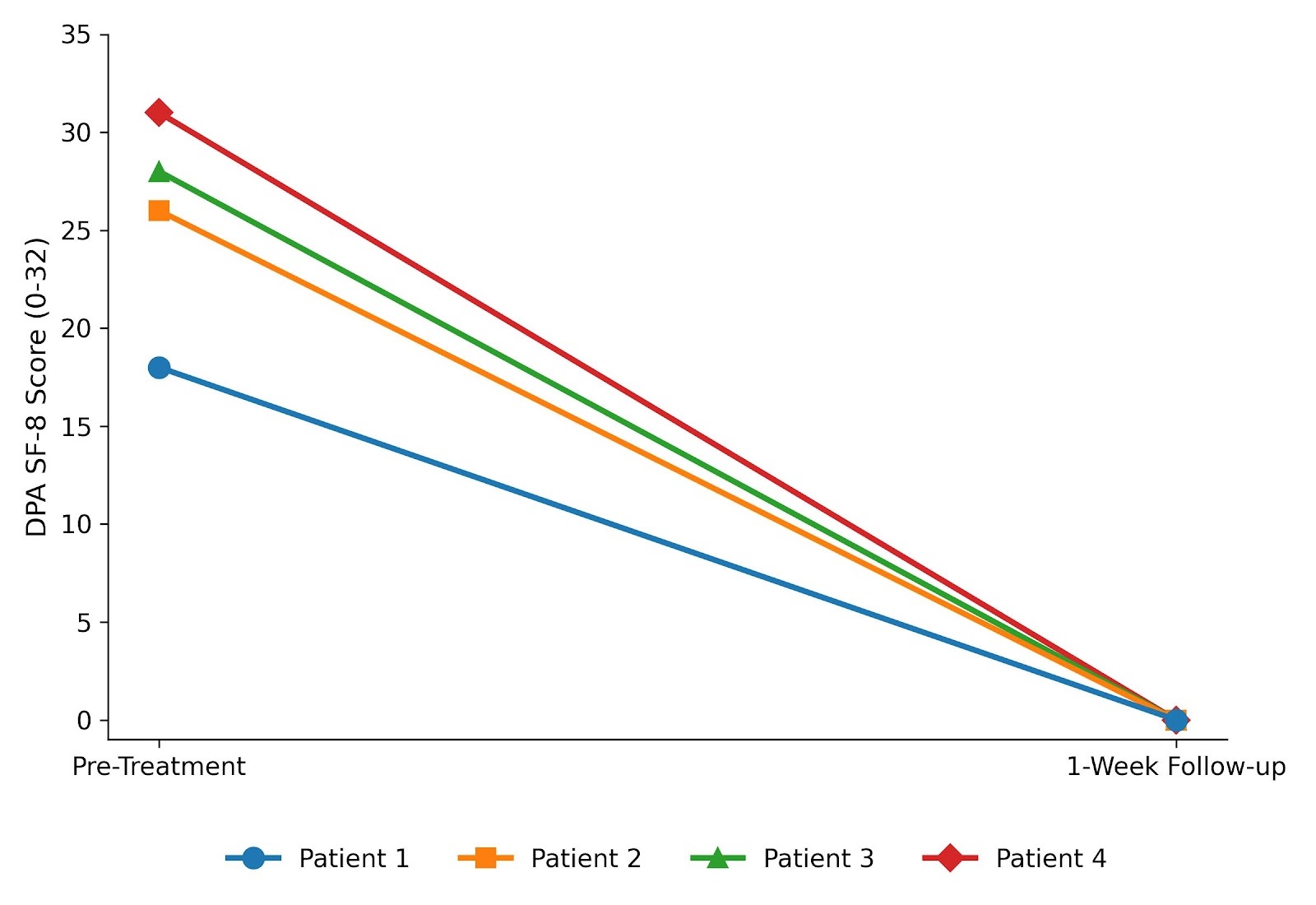
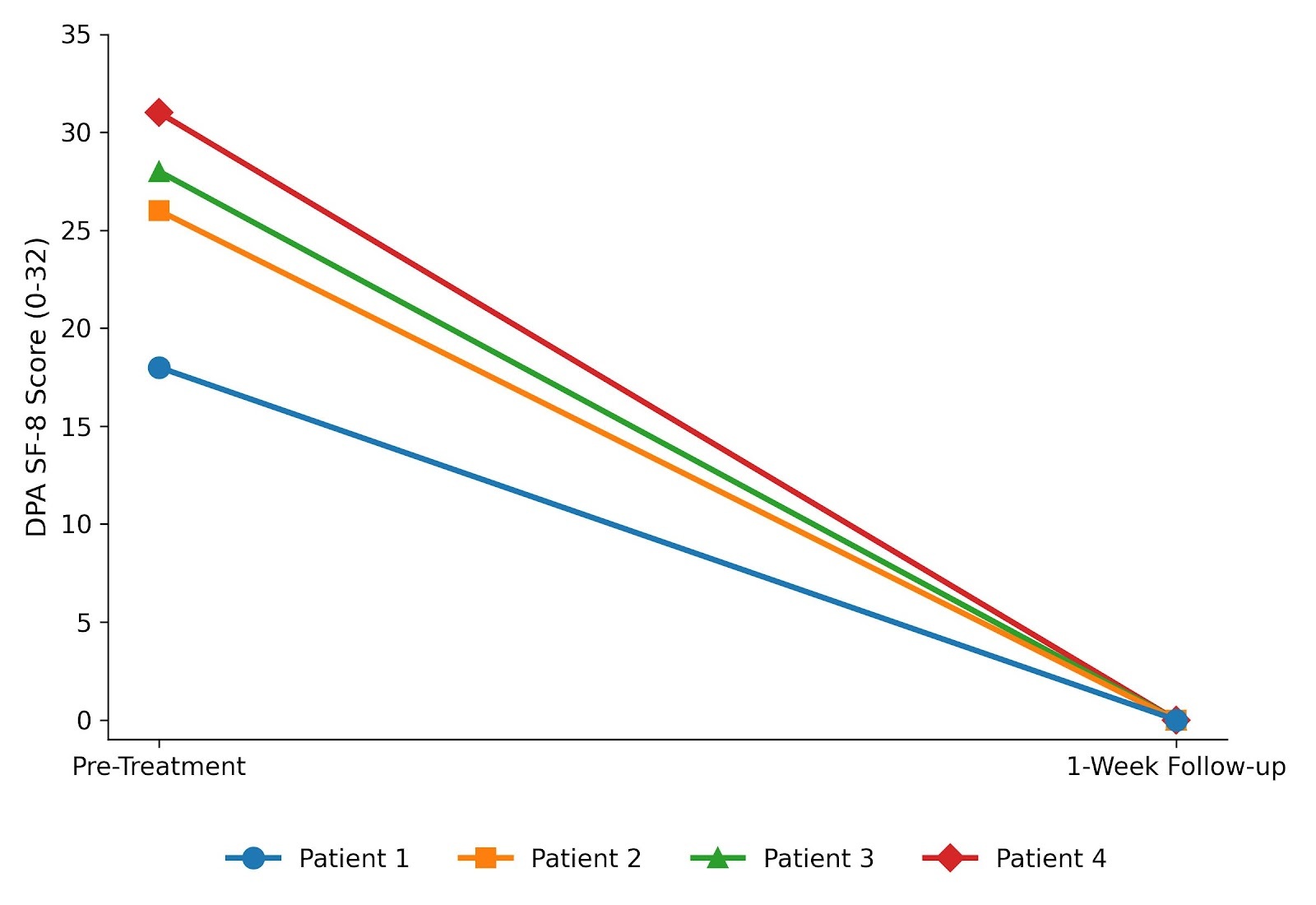
The DPA SF-8 was utilized to assess the subjects’ quality of life and pain-related disablement. At baseline, the subjects reported a high degree of disablement, with an average score of 25.75 ± 4.72 out of 32 points (Figure 3). Following the one-week post-treatment period, all four subjects reported a DPA SF-8 score of 0. This uniform drop to zero indicates a remarkable enhancement in quality of life and a complete resolution of perceived disablement. Furthermore, the average reduction of nearly 26 points (Figure 3) far exceeded both the MCID of 2 points and the MDC of 6 points established for this scale.
DISCUSSION
All participants in this case series achieved complete pain resolution following the single treatment and sustained this resolution at a one-week follow-up, demonstrating both that the intervention provided not only immediate relief but also prolonged benefits over the subsequent seven days. Each participant attained a functional score of nine or higher, reflecting high levels of functional capacity. Additionally, all subjects reported an enhancement in quality of life and a reduction in disablement after seven days, with all reaching a score of zero. The PSFS and DPA SF-8 were used to assess function and disablement, administered at baseline and at one-week follow-up. This approach allowed subjects sufficient time for self-assessment while enabling the evaluation of treatment sustainability.
Numerous therapeutic modalities have been proposed to manage SIJ pain from a physiotherapeutic perspective, including targeted resistance training for the gluteal muscles, core musculature, hip flexors, and transverse abdominis.4,27 SIJ pain is primarily linked to hypermobility and axial loads applied to the joint, exacerbated by deficiencies in stabilizing musculature, particularly the gluteus maximus and pelvic floor muscles.2,28,29 By enhancing the strength and endurance of these muscular groups, clinicians can improve the joint’s capacity to resist pathological axial loading.2 The anatomical architecture of the SIJ is also critical in alleviating pain and counteracting shearing forces.2 The optimal functioning of compressive forces within the ligaments and muscular structures of the joint facilitates stability/friction, thereby providing substantial stability. Conversely, the inability of muscular structures to produce adequate compressive forces can lead to excessive joint mobility, rendering the joint susceptible to hypermobility, which may increase strain on the ligamentous structures and the surrounding joint capsule.2
In this case series, all subjects attained a post-treatment NPRS score of 0/10 immediately following the treatment session. This is clinically significant as it indicates a complete resolution of pain. However, since clinicians only monitored the effects of the intervention on pain over a period of seven days, they cannot definitively ascertain that this pain relief was not temporary in nature. Moreover, the heterogeneity among subjects regarding age, activity levels, pain intensity, functional status, and quality of life metrics enhances the importance of this outcome. The PSFS scores reached full perceived function for three participants, while one individual approached but did not fully attain this level. Although the specific patient-selected tasks differed among subjects, all involved pelvic rotation alongside activation of the gluteal muscles and core stabilizers. Despite subject one not realizing full perceived function, each participant achieved a PSFS value indicative of eligibility for discharge from the case series. The DPA SF-8 scores averaged 25.75 points at baseline; however, at the one-week follow-up, all subjects recorded a score of 0/32, thereby meeting the discharge criteria and illustrating substantial clinical improvement.
The estimated recovery duration for newly diagnosed acute cases of low back pain ranges from approximately six weeks to twelve months.5 This prolonged recovery period may be attributed to the degree of disability experienced by individuals suffering from non-specific LBP. Elevated levels of disablement resulting from the injury can restrict the patient’s capacity to engage in specific rehabilitative exercises, thus extending the recovery timeline.13 The rapid clinical improvements observed in these four patients suggest that low-load BFR training served as a tolerable and effective rehabilitation modality for this specific cohort. Based on established BFR literature, these improvements could theoretically be driven by enhanced muscle recruitment and early hypertrophic signaling in the adjacent musculature, though these specific adaptations were not directly measured in these four subjects.13 A number of studies have investigated the potential strength gains associated with BFR training, demonstrating that low-load BFR at 20-30% of one-repetition maximum (1RM) can yield significant enhancements in strength.11,13,30 Conversely, aerobic training conducted without the application of BFR under standard conditions typically does not produce notable hypertrophy or strength improvements.13
The widely accepted theory behind BFR training is that hypertrophy and strength gains result from muscular stress caused by reduced oxygen and metabolite accumulation.11 This leads to muscular adaptations such as increased muscle fiber recruitment, fatigue, and anabolic signaling.11,13,30 Typically, achieving similar results would require multiple sets at 70% of 1RM; however, with BFR, performing to failure at 20-30% of 1RM, known as low-load BFR, can yield the same benefits.11,13,30 These findings can be valuable for clinicians working with injured subjects, as they promote hypertrophy with low volume and load. An additional finding in the literature is a theory with mixed evidence regarding the effect of proximal musculature on the cuff used in BFR.30–35
In a randomized controlled trial, Bowman et al. demonstrated a systemic and crossover effect when evaluating strength gains in the contralateral leg and proximal musculature (chest, trunk, and gluteal muscles). In their study, the limb subjected to BFR showed an increase in thigh and leg girth, total work for knee flexion, hip abduction, extension, and plantar flexion strength compared to the control group.30 Conversely, the limb not subjected to BFR also exhibited increased thigh girth, quadriceps peak torque, and single-leg heel raises.30 These results suggest that BFR has a systemic effect on the body, with the ability to impact the proximal musculature (in this case the gluteal muscles), which offers a potential physiological rationale for the rapid pain relief and functional improvements observed in these four subjects.
While structural adaptations may explain long-term improvements, the rapid pain relief observed in this case series may be linked to exercise-induced hypoalgesia (EIH), a phenomenon where pain sensitivity is acutely reduced following exercise.36,37 EIH is commonly hypothesized to be mediated by the release of endogenous opioids following the activation of group III and IV primary afferent muscle fibers.37,38 A systematic review by Karanasios et al. demonstrated that low-intensity BFR at 80% arterial occlusion pressure (AOP) significantly increases pain pressure thresholds compared to high-intensity exercise, BFR at 40% AOP, or low-intensity exercise alone.38 These findings align with a recent randomized controlled trial by Proppe et al., which observed that low-load BFR at 80% AOP produced a repeatable, systemic EIH response that lasted up to 60 minutes post-exercise.36 Although Karanasios et al. and Proppe et al. evaluated these thresholds in healthy populations, their recommendation to utilize BFR as a pain management strategy provides a strong physiological rationale for these findings.36,38 The profound, acute EIH response likely presents a potential rationale for the immediate, complete resolution of pain (NPRS = 0) and the improved functional capacity these four subjects experienced.
LIMITATIONS
Given the clinical setting, this case series is limited to a small, heterogeneous athletic cohort presenting with SIJ dysfunction, which restricts the generalizability of these findings to broader or non-active populations. Furthermore, as an uncontrolled case series lacking a control or comparison group, a cause-and-effect relationship between low-load BFR training and rapid symptom resolution cannot be established. Without controls, observed improvements may be influenced by the natural history of acute SIJ pain, which can improve spontaneously over time, as well as regression to the mean, given that patients typically seek clinical intervention when symptom severity is at its peak.
Additionally, because neither the treating clinicians nor the subjects were blinded to the intervention, the exclusive reliance on self-reported outcome measures introduces susceptibility to placebo effects and clinician expectation bias, both of which could artificially elevate patient-reported outcomes. While PROMs provide critical insight into patient perception and functional readiness, the absence of objective biomechanical, force-plate, strength, or neuromuscular performance metrics limits the ability to quantify physical adaptations, in contrast to traditional BFR literature that primarily evaluates objective muscle strength gains. Finally, although the clinical protocol was designed for weekly treatments until complete recovery, all participants achieved full symptom resolution after their initial session; consequently, the effects of repeated BFR treatments over extended periods could not be evaluated. Future research should use randomized controlled trials incorporating a sham-BFR and a standard exercise control group, incorporate objective physical and neuromuscular assessments, and follow for longer-term follow-up periods to isolate the specific therapeutic efficacy of BFR in active populations with SIJ dysfunction.
CONCLUSION
The results of this case series indicate that a single session of low-load BFR training produced immediate, complete pain resolution and substantial short-term improvements in perceived function and disability in four athletically active subjects with SIJ dysfunction. Future studies should explore the potential benefits of BFR for individuals with SIJ dysfunction to determine how BFR influences muscular adaptations in the proximal musculature, and additional patient reported outcome measures. While these findings only represent individual responses in four subjects, they highlight BFR as a potential adjunct to current treatments for SIJ dysfunction. By enabling low-load exercise without provoking symptoms, BFR may serve as a tolerable early-stage bridge in rehabilitation, particularly for individuals who are too restricted by pain to tolerate traditional, higher-load protocols.