Introduction
Diagnostic musculoskeletal ultrasound (MSKUS) has become an essential tool for evaluating musculoskeletal structures. Shoulder pain and disability are very common in physical therapy clinical practice. Luckily for therapists, most pain generators can be visualized with MSKUS. One of the more common pain generators in the shoulder can be the subacromial-subdeltoid (SA-SD) bursa. This paper aims to provide a comprehensive review of diagnostic MSKUS of the SA-SD bursa, including relevant anatomy, scanning technique, normal and abnormal imaging characteristics, and common pathological findings.
Anatomy of the Subacromial – Subdeltoid Bursa
The SA-SD bursa is the largest bursa in the body.1 The subacromial bursa lies within the subacromial space, sitting above the supraspinatus yet below the acromion. The subdeltoid bursa is a lateral extension of the subacromial bursa. They both limit frictional forces between the humeral head, the supraspinatus, the acromion, and the deltoid.1–3 These two structures communicate with each other and are contiguous about 95% of the time.1 The distalmost aspect of the bursa sits on the superior surface of the supraspinatus muscle and tendon, which sits deep to the inferior surface of the deltoid muscle. The bursa is separated from the joint cavity by the rotator cuff muscles and tendons. Despite its large size, the bursa’s two sides are normally separated by no more than 2 mm in the normal shoulder.
The etiology of bursal injury or inflammation (bursitis) in the shoulder may be due to a variety of factors and is usually due to overuse caused by repetitive overhead movements, but can also include pathology including subacromial impingement, acute shoulder trauma causing bursal inflammation, rotator cuff injury, infections, gout, rheumatoid arthritis, and occasional crystal deposition in the form of calcium hydroxyapatite deposition in bursa of the shoulder.1 The bursa itself contains free nerve endings and nociceptors, which may be a large source of shoulder pain.
The Role of MSK Ultrasound in Evaluation of the SA-SD Bursa
Advantages
-
Real-Time Imaging: MSKUS allows dynamic evaluation of the bursa while the shoulder can be moved through the available range of motion. It provides real-time assessments of soft tissue and its motion, offering information beyond what static morphology provides.
-
High-Resolution Visualization: MSKUS provides detailed images of the bursa.
-
Accessibility and Cost-Effectiveness: MSKUS is portable, widely available, and less expensive than magnetic resonance imaging (MRI).
Limitations
-
Operator Dependency: MSKUS requires skill and experience for accurate interpretation of findings. The ability to sonograph bursal tissue and surrounding structures is relatively easy and, to a large extent, influenced by the operator, the availability and technical considerations of state-of-the-art equipment, and the position of the shoulder during the assessment.
-
Depth Limitations: Visualization is usually not a problem as the bursa are superficial compared to other shoulder structures and are easily within the appropriate depth limitations of MSKUS.
-
Artifacts and Shadows: Bone shadowing from the acromion and humerus may create image artifacts, requiring adjustments in probe positioning and frequency.
Sonographic Technique for Evaluating the SA-SD Bursa
Equipment Setup
-
Probe Type: The depth of the bursa allows for a standard high-frequency 3-13 MHz linear array probe.
-
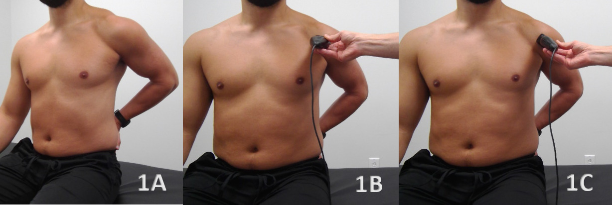
Patient Position: The optimal position for imaging of the SA-SD bursa is one that exposes the supraspinatus footprint and clears the acromion enough to reach the bursa without acoustic shadowing of the acromion. The modified Crass position is probably the most commonly used position and is performed by placing the seated patient with their hand on the ipsilateral hip or with their arm behind their back. This position internally rotates and extends the shoulder, bringing the bursa out from under the acromion for very clear visualization on MSKUS examination.
-
Dynamic Assessment: A passive or active movement of the shoulder can create dynamic movements of the humerus that allow stress to be applied to bursal tissue, helping to accentuate issues seen during ultrasonography.
Examination Protocol
Normal Sonographic Appearance of the SA-SD Bursa
A normal healthy bursa is seen as an anechoic, fluid-filled, thin structure with a hypoechoic layer of synovial fluid surrounded by a small hyperechoic bursal tissue wall surrounded by peribursal fatty tissue.1,4
Pathologic Findings in the SA-SD Bursa
-
A hyperechoic wall may be seen with synovial thickening.
-
In the case of swelling and inflammation, a swollen, distended bursa may be seen and appear hypoechoic or hyperechoic due to complex fluid or synovial hypertrophy. In cases of hemorrhage within the bursa, hyperechoic blood may be seen.
-
Doppler can be used to differentiate complex fluid from plain synovium, as blood flow suggests synovial hypertrophy.5
-
It is not uncommon in older patients to also see degenerative changes to the rotator cuff supraspinatus tendon, degenerative changes to the humeral head, and possibly calcifications in the bursa and adjacent rotator cuff tendons.
Clinical Implications for Rehabilitation Providers
MSKUS provides real-time feedback for rehabilitation professionals, facilitating early diagnosis and intervention. Key applications include:
-
Early Detection of Injury / Accurate Injury Grading: MSKUS can determine normal bursal tissue from pathologic, inflamed, and swollen tissue.
-
Dynamic Functional Testing: Rehabilitation professionals can use MSKUS during physical therapy sessions to monitor recovery and assess bursal tissue thickness and consistency in both static and dynamic assessments. Serial MSKUS imaging aids in assessing bursal thinning and tissue remodeling to support readiness for rehabilitation progression.
- Guided Interventions: Ultrasound imaging assists in interventions such as dry needling or precision-guided injections, such as corticosteroids for inflammation.
- Patient Education: Real-time imaging serves as a visual aid to explain the nature of the injury and set realistic expectations for recovery. Dynamic imaging during shoulder elevation into flexion and abduction can be used to see actual compression of bursal tissue.
Limitations and Challenges
Despite its advantages, MSKUS is not without limitations and challenges. MSKUS has low specificity with fluid and thickening or non-specific signs and symptoms. MSKUS can show bursal distension or hypoechoic fluid, but these findings can occur in many shoulder conditions. A systematic review by Mackie and colleagues found sensitivity around 80%, and specificity only 68% for detecting SA-SD bursitis, meaning that false positives are very common.6
As with many other areas of the body examined with MSKUS, ultrasound accuracy depends on several factors, including probe placement, pressure applied during sonography (small changes in probe angle or pressure can compress the bursa and alter its appearance), operator experience, and ultrasound quality. MSKUS can show anatomical structure but cannot differentiate cellular inflammation from other potential findings, such as acute inflammatory bursitis, chronic fibrotic thickening, or reactive fluid.
Conclusion
In summary, diagnostic MSKUS offers rehabilitation professionals a powerful, clinically relevant extension of the physical examination when evaluating the bursal tissue. Its ability to provide high-resolution, real-time, and dynamic visualization of the bursal tissue architecture enhances diagnostic precision, supports early clinical decision-making, and allows serial monitoring of tissue healing and load tolerance. When integrated with a thorough understanding of anatomy, injury mechanisms, and functional biomechanics, MSKUS becomes more than an imaging modality—it becomes a performance-informed clinical tool. While operator skill and certain anatomic limitations must be acknowledged, the thoughtful incorporation of MSKUS into sports and orthopedic practice has the potential to elevate assessment accuracy, refine rehabilitation progression, and ultimately improve return-to-play outcomes for athletes with shoulder bursal injuries.
Patient Positioning for Subacromial-Subdeltoid (SA-SD) Bursa
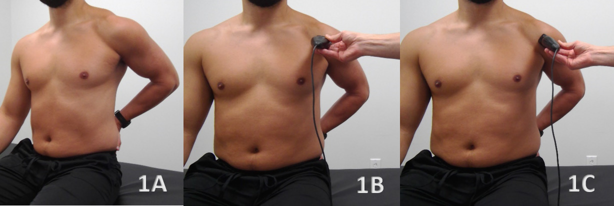
Figure 1A: Patient Position
The patient is seated with the upper extremity placed in the modified Crass position, with the hand positioned as high as comfortably tolerated along the ipsilateral posterior hip and the elbow directed posteriorly. This position brings the distal supraspinatus tendon and overlying SA-SD bursa anterior to the acromion, improving sonographic access to the bursa. The position is maintained for evaluation of the bursa in both short axis (SAX) and long axis (LAX) orientations.
Figure 1B: Transducer Placement in SAX
For SAX imaging, the transducer is placed over the anterolateral shoulder, parallel to the clavicle and perpendicular to the supraspinatus tendon as it courses over the humeral head toward its insertion on the greater tuberosity. The transducer should be positioned to visualize the humeral head, supraspinatus tendon, overlying SA-SD bursa, and deltoid muscle within the same field of view.
Figure 1C: Transducer Placement in LAX
For LAX imaging, the transducer is rotated into an oblique orientation and aligned parallel with the supraspinatus tendon fibers as they approach the greater tuberosity. This orientation allows visualization of the SA-SD bursa along the superficial surface of the supraspinatus tendon and facilitates assessment of the bursa as it extends laterally toward the greater tuberosity.
SA-SD Bursa Normal Sonographic Anatomy View
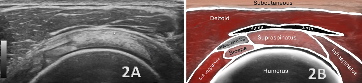
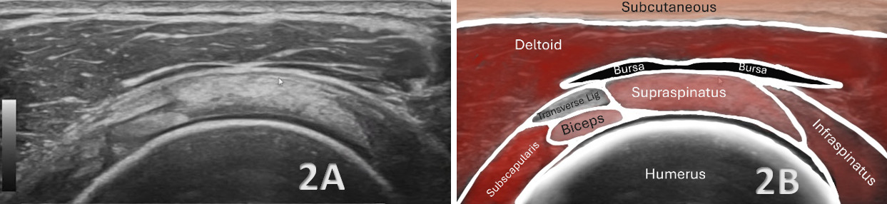
Figures 2A and 2B: Normal SAX View
In SAX imaging (Figure 1B), first identify the bony cortex of the humeral head and the overlying supraspinatus tendon, which demonstrates the characteristic “tire on the rim” appearance as the tendon wraps uniformly over the humeral head. Deep to the tendon, the hyaline cartilage is visualized as an anechoic interface immediately superficial to the hyperechoic cortical surface. The SA-SD bursa is located superficial to the supraspinatus tendon and deep to the deltoid muscle, where it normally appears as a thin potential space bordered by hyperechoic interfaces. A thin layer of peribursal fat may also be visualized between the tendon and deltoid. In the normal, non-distended state, the bursa should remain thin without focal fluid accumulation, distention, or synovial thickening, while the underlying supraspinatus tendon maintains uniform thickness and echotexture.
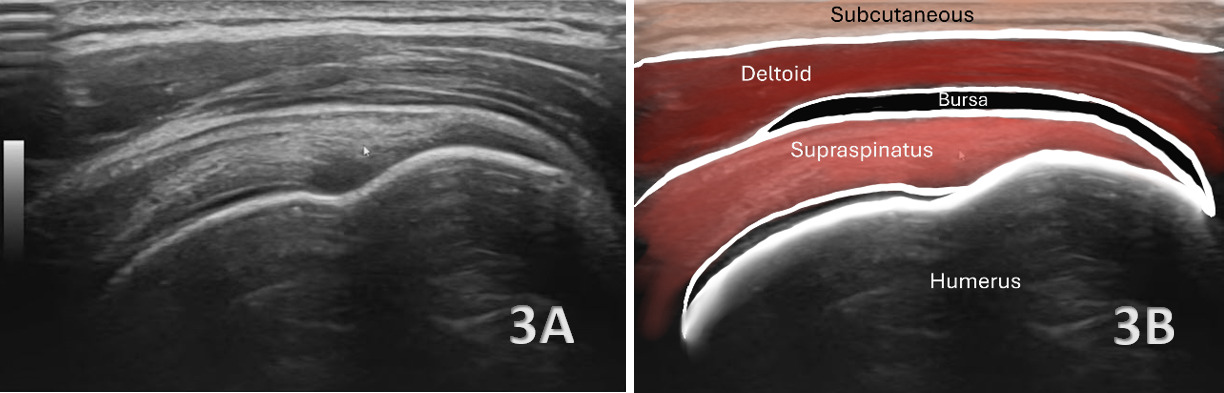
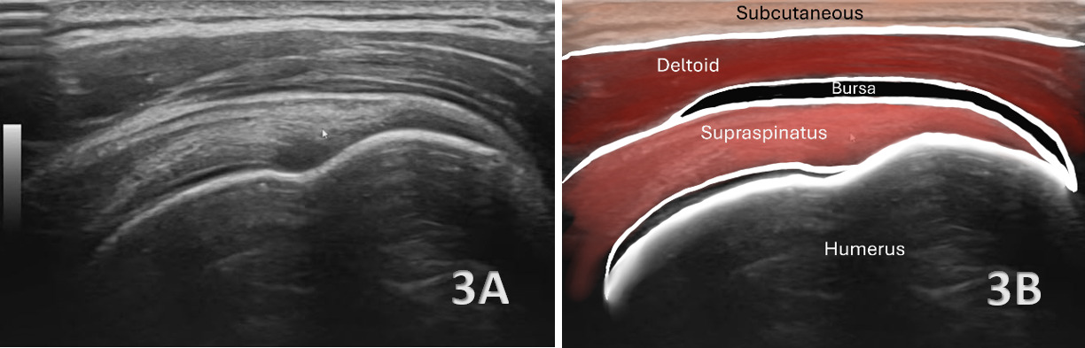
Figures 3A and 3B: Normal LAX View
In LAX imaging (Figure 1C), the transducer is oriented slightly obliquely to remain parallel with the supraspinatus tendon fibers as they approach their insertion on the greater tuberosity. The bony landmarks should first be identified, including the sloping contour of the greater tuberosity, the adjacent concavity of the humeral head, and the anatomical neck. The normal supraspinatus tendon demonstrates an organized fibrillar appearance and progressively tapers toward its distal attachment at the greater tuberosity. Superficial to the tendon, the SA-SD bursa is visualized between the supraspinatus and the overlying deltoid muscle as a thin potential space with hyperechoic margins. In the normal state, the bursa should remain thin and uniform along the course of the tendon without focal distention or abnormal fluid accumulation. Sweeping laterally toward the greater tuberosity can further assess the bursa for subtle fluid that may collect within its lateral recess.
SA-SD Bursa Pathology
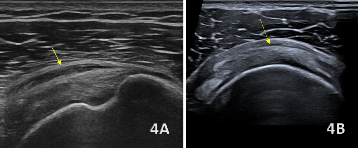
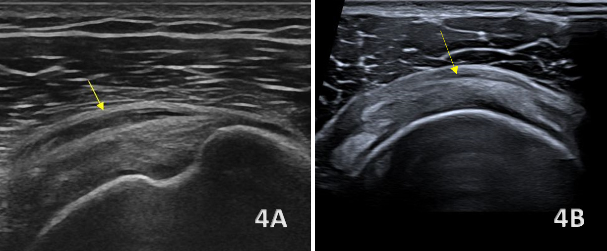
Figures 4A (LAX) and 4B (SAX): SA-SD Bursitis
SA-SD bursitis, as shown above in Figures 4A and 4B, is characterized sonographically by distention of the normally thin bursal space with hypoechoic or anechoic fluid, which may be accompanied by thickening of the bursal walls. Fluid may become particularly conspicuous as the transducer is swept laterally toward the greater tuberosity or anteriorly toward the long head of the biceps tendon. Dynamic imaging can demonstrate abnormal movement or redistribution of bursal fluid during shoulder motion and may reveal compression of the distended bursa as it passes beneath the acromion. When present, these findings should be evaluated in conjunction with the underlying supraspinatus tendon for associated rotator cuff pathology.