Introduction
Anterior cruciate ligament (ACL) injury remains a pivotal problem in sport and orthopaedics. Despite advances in screening and rehabilitation, the annual incidence in pivoting sports has plateaued, and up to one-third of athletes never regain their pre-injury performance. Addressing this challenge requires an integrative approach combining biomechanics, neurophysiology, and molecular muscle biophysics. High-speed video reconstructions and in-vivo strain recordings show that non-contact ACL ruptures occur within the first 0–70 ms of stance, when peak ground-reaction forces (GRF) and anterior tibial shear converge on a knee that is often near extension.1 At that instant, the neuromuscular system is caught mid-stream: quadriceps are resisting forced lengthening, hamstrings have only begun to fire, and the fastest spinal stretch reflexes are still travelling to the spinal cord.
Traditional risk-factor models explain this vulnerability in macroscopic terms valgus knee alignment, limited hip flexion, or excessive quadriceps dominance increase the external moment arm and overload the ligament during cutting or landing.2 Yet such frameworks leave open a fundamental question: what happens inside the muscle–tendon complex during those critical milliseconds, and how do molecular events dictate whether the ACL survives the load?
Recent biophysical studies offer important clues. Calcium influx before touchdown not only triggers actin–myosin cross-bridge cycling but also locks titin’s N2A segment onto actin, instantly tripling the protein’s spring stiffness. As the foot strikes, this pre-loaded titin and the population of strongly bound cross-bridges create a short-range stiffness (SRS) regime that can resist the first 1–2 % of fascicle stretch with remarkable rigidity. If the external impulse exceeds that buffer, residual force enhancement (RFE) maintains tension with minimal metabolic cost yet even RFE saturates rapidly under combined anterior shear and valgus torque. Crucially, the ACL–hamstring reflex arc, often invoked as nature’s failsafe, activates only after 35-40 ms and therefore is too late to avert the initial peak strain.3
These insights motivate the central hypothesis of this clinical commentary: pre-contact modulation of hamstring-titin mechanics constitutes the first and perhaps only line of defense capable of acting within the injury-relevant timeframe. To examine this proposition, the authors synthesize data spanning whole-body kinematics, joint kinetics, electromyography, single-fiber mechanics and titin nanobiology. The 150 ms surrounding foot strike is partitioned into four phases preactivation, initial contact, early post-contact and late post-contact and trace how forces propagate from GRF vectors down to sarcomeric springs. By linking cortical preactivation, spinal reflex latency, and titin-based stiffness within a single time-resolved framework, this model provides a novel explanation for why ACL ruptures occur before feedback arrives and how targeted eccentric and neuromuscular training can enhance early protection. The purpose of this clinical commentary is to provide a theoretical model intended to guide future empirical investigations.
Preactivation Phase (≈ –150 to 0 ms) before Initial Contact.
Before foot strike, the athlete decelerates and prepares to absorb impact. The entire lower limb and trunk shift to absorb load: hip extensors and knee flexors brace, and the contralateral leg may adjust. There is no ground contact yet, so GRF = 0, but the body’s center of mass is lowering.4 Hip and trunk position influence loading: e.g. forward-leaning posture can reduce knee extension moment. Key risk factors during flight emerge here – video/simulational studies show that landing with low knee flexion and high knee valgus angle/moment predisposes to higher ACL strain.5 In practice, athletes use visual and vestibular cues to orient the body and pre-plan the landing strategy.
The ankle is dorsiflexed or plantarflexed depending on landing style, the knee is partially flexed (often ~20–30° in injury cases), and the hip flexion is moderate. Musculoskeletal geometry determines how GRF will translate into joint moments on touchdown.6 For example, an extended knee at contact places the tibial plateau more anterior relative to the GRF, increasing anterior shear and knee extension moment (which quadriceps must resist).7 Conversely, a flexed knee spreads the force but can increase quadriceps demand.
The central nervous system issues feed-forward commands, hamstrings, quadriceps, gluteals, soleus/gastrocnemius, and dorsiflexors ramp up activity before contact.8 This co-activation stiffens joints (increasing joint impedance) and engages short-range muscle stiffness. At the molecular level, Ca²⁺-triggered cross-bridge cycling begins, so many myosin heads attach to actin.9 These attached cross-bridges behave like elastic springs: an infinitesimal perturbation will be met by a stiff response. In effect, muscle fibers gain an immediate elastic component even before stretch (often called preflex).10 A fraction of titin filaments also begin to stiffen: calcium can induce binding of titin’s N2A region to actin, preventing the slackest immunoglobulin (Ig) domains from unfolding.11 Thus, even before impact, muscle exhibits viscoelastic properties that will shape the initial response to stretch.
Proprioceptors (muscle spindles in quads/hamstrings/calf, joint receptors in ankle/knee) signal limb position and tension during the flight phase, refining the anticipatory set.12 Importantly, mechanoreceptors in the unloaded ACL (Ruffini endings, Pacinian corpuscles) sense joint angle but are minimally loaded.13 Pre-activation EMG is seen in all lower-extremity muscles ~20–50 ms before contact in trained athletes; this anticipatory activity is crucial because reflexive feedback lags behind the impending impact.14
Initial Contact (0 ms)
At foot strike, the ground generates a rapid force impulse on the body. The GRF vector typically has a large vertical component (often on the order of 2–3 times body weight in jumping/landing tasks) and a horizontal (posterior) component that decelerates forward motion.15 For example, Cerulli et al. measured a ~5.5% peak ACL strain during a rapid stop jump, with strain rising sharply in concert with GRF16 Increases in vertical and braking GRF correlate with higher ACL load. The direction of the GRF (relative to foot placement) creates knee moments: an anteriorly placed CoP produces a knee flexion moment, while a medial offset can induce a valgus moment.17 Early in stance, the ankle dorsiflexes under the load and the knee flexes slightly as momentum is absorbed.
Immediately at contact, the ankle, knee, and hip bear axial compressive load.18 At the knee, the tibiofemoral joint experiences high compression (from the vertical GRF) and rapidly rising anterior shear (from any horizontal GRF component and quadriceps tension). The hip similarly takes compressive load and an extension moment (the pelvis resists collapse).19 Knee valgus torque is often observed in injury cases – small misalignment here means the lateral knee stresses the ACL and medial structures. In general, extended or near-extended knee positions increase the proportion of shear vs. pure compression, placing more demand on ligamentous restraint.20 Typical knee flexion angles at impact in ACL injuries are shallow (often <30°), so the quadriceps insertion has a strong anterior pull on the tibia.21 Joint moments at this instant set the stage for muscle response: for example, a posterior GRF with an extended knee produces a large knee extensor moment (quadriceps must counter it) and an internal hip flexor moment (requiring gluteal response).22 Ankle plantarflexors (soleus/gastrocnemius) engage to control dorsiflexion. Muscles transition from feed-forward preactivation into immediate eccentric response to absorb impact energy and stabilize the knee.
The already-activated muscles now begin to contract eccentrically.23 Quadriceps resist knee buckling, hamstrings decelerate anterior tibial motion, and calf muscles brake ankle dorsiflexion.24 The short-range stiffness of each muscle (due to preattached cross-bridges) generates an immediate force jump: in muscle fiber experiments, an active stretch yields a steep initial force rise, then a brief “give” as cross-bridges break.25 At ~0–1% fiber strain (just fractions of sarcomere length), cross-bridges behave elastically; beyond ~11–12 nm per sarcomere displacement, many cross-bridges detach and force plateaus.26 Thus, at impact the limb’s muscles provide a stiff elastic response, partly buffering the load. However, force is only partly maintained by cross-bridges – additional viscoelastic elements (notably titin) also stretch and store kinetic energy.27
At the instant of contact, no new reflexive signals have yet influenced muscle.28 All joint stiffness is from preactivation. ACL mechanoreceptors begin sensing any load change right away, but any reflex path has a latency of tens of ms.29 Thus, in this first moment, the knee’s stability is purely feed-forward controlled and utilizes passive resistance.30 Joint protection during initial contact therefore depends entirely on the pre-established viscoelastic properties of active muscle fibers and the intrinsic short-range stiffness of the titin-based spring system.
Early Post-Contact (≈ 20–40 ms)
After impact, the GRF continues to rise (often peaking around 20–30 ms) as the leg decelerates the body’s center of mass.31 During this early stance, the knee typically flexes further (up to 30–45°) and hip flexes to absorb energy. Peak vertical force occurs; simultaneously the posterior shear and any medial/lateral shear press on the knee.32 Because knee flexion is still relatively small, the geometry keeps the quadriceps moment arm large, the anterior tibial shear (and thus ACL load) remains high. Hip extension torque increases as the body brakes.33 This period defines the peak of impact-phase loading, where the combination of vertical and posterior GRF components maximizes deceleration forces transmitted through the lower kinetic chain.
Knee compressive force may reach ~3× body weight in a hard landing.34 Anterior shear from the posterior GRF and the continued quadriceps pull builds, loading the ACL and posterior knee capsule. If a valgus moment is present (due to lateral foot placement or weak hip abductors), tensile stress on the anteromedial ACL region rises.35 At the ankle, dorsiflexors (tibialis anterior) and plantarflexors (soleus) co-contract to stabilize the foot under load, transferring force up the chain.36 The hip undergoes large extension moment (gluteals and hamstrings active) as the body’s momentum is arrested.
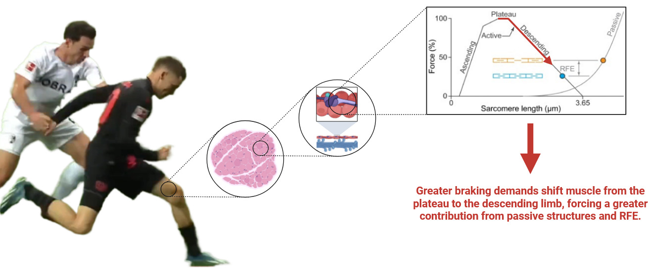
Muscle fibers undergo rapid lengthening.37 Quadriceps fibers (especially the vasti) and soleus fibers are eccentrically stretched while activated. Cross-bridge dynamics now govern force development: the initial short-range stiffness transition is over, and many cross-bridges have detached.38 The muscle enters a steady stretch phase where residual force enhancement (RFE) can arise39 (Figure 1).
__the_more_the_musc.png)
Empirically, active fibers show higher force post-stretch than in a purely isometric contraction at that new length.40 This is attributed to titin: as thin filaments slide by (driven by cross-bridge pulling), titin’s PEVK region (an elastic, highly extensible domain named after the four amino acids that make up most of its sequence: Proline [P], Glutamate [E], Valine [V], and Lysine [K]) may wind onto actin, storing elastic energy.41 Thus titin carries part of the load. Measurements show forces up to ~2.5× the isometric Fmax on the descending limb, far above what cross-bridges alone predict.42 Calcium-dependent stiffening of titin (via N2A-actin binding) also increases resistance at these lengths (Table 1).
By ~20 ms after impact, the very fastest reflex arcs fire.43 Muscle spindles in the quadriceps and calf, stretched by the sudden lengthening, produce a short-latency reflex (SLR) in those muscles (~20–30 ms latency).44 Friemert et al. report hamstring SLR onset at ~20.3 ± 3.5 ms after a rapid anterior tibial displacement.45 This SLR is largely spinal and stiffness-regulating. Concurrently, ACL mechanoreceptors (primarily Ruffini endings sensing tension) begin firing due to rising ACL load, but any resulting reflex is slightly longer.46 A medium-latency reflex (MLR) in the hamstrings (often attributed to the ACL–hamstring reflex arc) occurs around ~38–40 ms. At ~30–50 ms, hamstrings receive both direct muscle spindle input and some input via the stretched ACL.47 These reflexive hamstring contractions tend to counteract anterior tibial translation and knee extension, opposing ACL strain.48 However, note the timing: even the MLR (~39 ms) trails well behind the initial ligament loading. EMG studies classify these as the medium and long-latency responses; the long-latency reflex (LLR) involving transcortical pathways typically emerges at ~80–100 ms post-perturbation, likely too late to protect the ligament in a rapid injury scenario.49
Mid-Early Post-Contact (≈ 40–60 ms)
In this interval, the knee continues to flex and muscle forces evolve.50 Ground reaction forces begin to decline after peak, but knee joint load can still increase due to ongoing inertial effects. The combination of forces – the (now somewhat lower) GRF vector, high quadriceps tension, and any valgus/rotational torque – produces maximal strain on the ACL.51 Cadaveric simulations suggest that at this stage the ligament strain ramps to its maximum.52 Video-based injury analysis infers that “ACL injuries are generally believed to occur within 0–67 ms of initial ground contact”. Thus, the ligament may fail around ~50–60 ms post-contact (consistent with Cerulli’s ~5.5% strain peak).53 In practical terms, the knee is highly loaded: at ~40–50 ms the knee may still bear ~2× body weight in shear, and valgus/internal-rotation moments (if present) peak.54
Knee extension moment from GRF shrinks as flexion increases, but the quadriceps is still working eccentrically (and increasingly concentric as flexion slows).55 Hip extension/horizontal abduction moments are high, driven by hamstrings/gluteals countering forward momentum.56 Ankle plantarflexion moment reduces as dorsiflexors catch the foot. If the foot is not flat (e.g. heel-strike), forefoot rocker may abruptly load the midfoot and knee differently.57 At this stage, the large extension moments on knee tend to push the tibia forward; the hamstrings strive to pull it back but are only beginning to catch up reflexively.58
The activation of the hamstrings becomes significant around 35–50 ms.59 Some studies show that hamstrings EMG may peak ~60–80 ms post-contact, by which time the ACL may already have failed.60 Quadriceps EMG, conversely, tends to peak earlier (often at contact or within 20–30 ms) due to the larger initial stretch (this bursts their SLR).61 The interplay of quadriceps vs hamstring forces critically affects ACL load: a strong late hamstring contraction would protect the ACL, but here it likely comes too late.62
The muscle-tendon unit continues its eccentric-shortening cycle.63 Cross-bridge reattachment rates cannot build force instantaneously; at these velocities many cross-bridges may enter a non-force-bearing (slipped) state.64 Meanwhile, titin’s engagement peaks: according to the winding-filament hypothesis, the continuing cross-bridge activity has wound additional titin onto actin, storing elastic energy.65 When the stretch ends (foot lift-off), this stored energy contributes to force maintenance. The viscoelastic nature of muscle is evident: force does not instantaneously fall to zero when stimulation ceases, but decays over ~100–200 ms due to internal drag (titin and surrounding matrix).66 Notably, Hessel et al. showed that “Ca²⁺-sensitive viscoelastic elements” (i.e. titin) account for much of the force and work in dynamic stretch-shortening cycles, underscoring that the muscle force at high stretch is not purely cross-bridge-driven.67
By ~50 ms the ACL–hamstring reflex should be underway.68 If the ACL is still intact, mechanoreceptor signals arriving at the spinal cord produce additional hamstring drive. However, clinical and animal studies indicate that ACL-driven hamstring reflex amplitudes are modest, and in ACL-deficient knees this reflex latency is prolonged and weakened.69 Long-latency reflexes (like the stretch reflex modulated by cortical circuits) can modulate the muscle activation pattern, but typically have a latency >70 ms – beyond the usual injury window.70 In summary, neuromuscular feedback is active by 40–60 ms, but it is barely in time to influence the outcome of an ACL tear that likely happens around this time.
Late Post-Contact (≈ 60–95 ms)
If the ACL is going to rupture, it usually does so by ~60 ms after contact.2 Video and experimental data agree that nearly all non-contact ACL tears occur within ~0–70 ms after ground contact.71 After ligament failure, the remaining soft tissues (menisci, collateral ligaments, capsule) and muscles then bear the remaining load.72 The hamstrings, now fully reflexive and under conscious control, co-contract to stabilize the knee.73 By ~80–100 ms, long-latency (LLR) neuromuscular responses kick in: these are partly transcortical reflexes that allow voluntary correction. EMG often shows a later burst in hamstrings and other stabilizers at this time.74
In a typical ACL rupture scenario, the tibia may sublux anteriorly once the ligament snaps.75 The knee often flexes rapidly thereafter, as muscle contraction and inertial forces drive motion. The hip flexes and trunk may move out of typical alignment.53 With no ACL, the quadriceps contraction now causes more anterior tibial shift, emphasizing why aggressive quadriceps use immediately after an ACL tear (as in some training) is avoided.76
Muscles are now in their late stance pattern. Titin still contributes to passive tension; in fact, after ~95 ms most active cross-bridges have completed their pull, so residual force is mostly passive (titin and connective tissue).77 Short-range elastic recoil of muscle can assist joint repositioning.78 Note that at this late stage, ground forces may be declining and the foot may be starting to lift; thus, net loading on the knee falls. The principal stabilizers become hamstrings and hip extensors.79
After the acute injury event, higher-level sensorimotor control responds.80 Body sense (via remaining proprioceptors) triggers reflexive muscle patterns to protect the knee: for example, gamma motor neurons may increase spindle sensitivity.81 However, if the ACL is torn, its own mechanoreceptors no longer function normally, disrupting normal feedback.82 In the following hundreds of ms to seconds, arthrogenic muscle inhibition often sets in – a protective reduction in quadriceps drive due to pain and swelling – but that is beyond the first 100 ms window discussed here.83
Integration and Injury Mechanism
Putting it all together, an ACL rupture typically falls into the first ~50–70 ms after foot contact.84 At impact the combined effects of whole-body deceleration (via GRF), joint alignment (valgus/internal rotation), and muscle force orientation (eccentric quadriceps vs hamstrings balance) produce a high anterior shear and valgus torque at the knee.85 Muscles are caught in mid-response: hamstrings have begun to contract but are not yet maximally engaged, while quadriceps may still be resisting stretch.22 The molecular spring–damper system of muscle (cross-bridges and titin) helps absorb energy, but if force exceeds ACL strength (which can happen under high-load, near-extension conditions), the ligament tears. Reflexively, nothing much can be done: the ACL–hamstring reflex latency (~35–40 ms) lags behind the loading, and even the fastest SLR (~20 ms) only acts on spindles (not the ACL).86 In fact, it is widely noted that “the ACL may tear in <70 ms but reflex muscle tension takes ≥35 ms to develop”. Thus, prevention relies on pre-contact strategies (flexed knee, balanced co-contraction, neuromuscular training) rather than rapid post-impact reflexes.87
Hypothetical Molecular Events Prior to ACL Rupture.
During the last 150 ms that precede an anterior cruciate ligament (ACL) rupture, a tightly choreographed series of neuromuscular and molecular events unfolds inside the posterior thigh musculature, particularly the hamstrings, which act as the primary dynamic restraint to anterior tibial translation.88 From approximately –150 ms to –40 ms, a feed-forward motor command originating in the primary motor cortex and cerebellum anticipates ground contact and depolarizes α-motoneurons innervating the hamstring motor units while the limb is still airborne.89 The descending volleys reach the sarcolemma, traverse the transverse-tubule network and trigger conformational opening of the dihydropyridine receptors mechanically linked to ryanodine receptor 1 in the terminal cisternae of the sarcoplasmic reticulum.90 The consequent Ca²⁺ surge saturates troponin-C, displacing the tropomyosin strands and permitting actin–myosin interaction; in parallel, Ca²⁺-dependent myosin light-chain kinase phosphorylates the regulatory light chains on the S1 neck region, which increases the stiffness of each cross-bridge and biases them toward strong-binding states.91 Concomitantly, elevated Ca²⁺ strengthens the electrostatic association between titin’s N2A element and actin filaments and induces folding of the PEVK-spring segments, producing an immediate two- to three-fold rise in titin-based stiffness.92 This molecular stiffening primes the muscle–tendon unit for the eccentric loading that begins roughly 40 ms before foot strike.93
Between –40 ms and –5 ms the hamstring muscle–tendon complex lengthens under a rapidly growing external moment, and sarcomeres experience forced-lengthening while still activated.94 Under these conditions residual force enhancement (RFE) develops: stretched but still cycling cross-bridges remain in high-force states, passive titin segments partially unfold yet stay latched to actin, and the overall sarcomeric tension exceeds isometric levels by an additional 15–30 %.95 Because this augmented tension is maintained largely without further ATP hydrolysis, the hamstrings can sustain high force despite decreasing neural drive as touchdown approaches.96 The mechanical consequence is an increased posterior shear at the tibiofemoral joint that counteracts anterior tibial translation generated by the quadriceps.97
At the instant of impact (0 ms) and during the first ~30 ms of stance, the system enters the short-range stiffness (SRS) paradigm.98 Here, the first one to two percent of fascicle stretch elicits an almost step-like rise in resistance on the order of 20–30 N mm⁻¹ before “yielding” occurs and stiffness falls.99 Two molecular contributors underpin this behavior. First, titin’s Ca²⁺-dependent stiffness, amplified by the prior N2A-actin binding, resists elongation like a pre-loaded spring.100 Second, strongly bound myosin heads act as transient pulleys that wrap additional titin segments onto actin, further shortening and stiffening the spring element.101 The stored elastic energy and the rapid torque development decelerate anterior tibial shift well within the 20 ms window that precedes the monosynaptic stretch reflex, a period during which ligamentous structures are most vulnerable because reflexive muscular support has not yet arrived.16
When any component of this multiscale protective cascade is inadequate delayed cortical preactivation, insufficient Ca²⁺ release, reduced titin phosphorylation, constrained cross-bridge recruitment, or premature yielding of the SRS the posterior shear generated by the hamstrings falls below the threshold needed to offset the quadriceps-driven anterior tibial impulse.102 In such cases the ACL is left to absorb a sudden spike in tensile load, and if this load exceeds its material capacity before reflexive hamstring activity can ramp up, rupture ensues.103 Thus, the millisecond-by-millisecond coordination of neural drive, excitation-contraction coupling, titin-based spring modulation, and cross-bridge mechanics constitutes a critical molecular buffer that buys time for slower feedback mechanisms and, when intact, markedly reduces the probability of catastrophic ACL failure.
In the integrated model that spans cortical pre-activation down to sarcomeric nanomechanics, the hamstrings’ feed-forward tension emerges as the first and virtually only line of defense that arrives early enough to matter.104 Because even the fastest spinal stretch reflexes lag tens of milliseconds behind the explosive force peaks generated when the limb decelerates, any protection that depends on feedback arrives after the critical load has already threatened the ACL.105 If the quadriceps fires in relative isolation while the hamstrings and soleus remain under-recruited, anterior shear on the tibia rises sharply and shifts the burden onto the ligament.106 Although short-range stiffness supplied by Ca²⁺-sensitized titin and strongly bound cross-bridges can momentarily buffer the joint, this visco-elastic capacity is finite; once the external impulse exceeds that threshold, the excess force is shunted into passive tissues, most notably the ACL.107 The cascade therefore underscores that real-time joint safeguarding in high-load scenarios relies chiefly on the intrinsic, milliseconds-fast mechanical properties established during pre-activation rather than on the ACL’s own reflex arc, which intervenes only after the decisive moment has passed.108
By integrating current neurophysiological and molecular biology insights, we can outline a hypothetical adaptation cascade through which rehabilitation and ACL-injury prevention programs gradually restore and ultimately amplify the muscles’ capacity for fast pre-activation before intial foot contact (Table 2).
Eccentric Training as a Vaccine?
During a rapid deceleration or a high-speed landing, the working sarcomeres are abruptly driven from the plateau region of the force-length curve toward the descending limb, sometimes overshooting the 3.6 µm region where passive tension begins to rise steeply.109 This transition illustrates the unique load-bearing behaviour of skeletal muscle during eccentric braking, where sarcomeres operate near their mechanical stability limit. In the first instants, cross-bridges that were already cycling at the plateau are forcibly lengthened; because detachment kinetics cannot keep pace with the imposed stretch, their strain increases and the instantaneous active force spikes above the isometric reference.110 Almost immediately, however, cross-bridge contribution starts to fall because the overlap between actin and myosin declines on the descending limb.111 What prevents a catastrophic drop in torque is the engagement of titin. Activation-dependent stiffening of titin through Ca²⁺ binding to its N2A and PEVK segments and through thin-filament interaction envisaged in the winding-filament hypothesis turns the giant protein from a slack spring into a viscoelastic damper that bears load while storing elastic Energy.112 This viscoelastic damping property is essential for safe deceleration, as it allows muscles to dissipate energy while preserving structural integrity. At the high stretch velocities typical of landing, titin’s viscoelastic drag rises sharply, so the molecule resists elongation more forcefully and augments total muscle force with virtually no extra ATP cost.113 This velocity-sensitive titin behavior explains why eccentric force rises as landing speed increases and why energy absorption is metabolically inexpensive.114
When the body’s momentum has been arrested and the joint angle stabilizes, a portion of the titin strain and any non-uniform half-sarcomere overstretches are retained within the lattice.115 The muscle therefore settles into an isometric state whose steady-state force is still higher than the purely isometric reference at the same length this is residual force enhancement (RFE) (Figure 1).116 RFE is largest when the landing stretches the fibers onto the descending limb or slightly beyond the passive up-turn, because here titin stiffness is highest and sarcomere-length non-uniformity is mechanically stabilized rather than destabilized.117 The knee extensors are a good macroscopic example: after an eccentric stretch matched to landing angles, vastus lateralis maintains ~10 % extra force for several seconds with no rise in EMG, demonstrating a titin-dominated RFE that can keep the knee stiff while the center of mass rebounds.118
At the molecular scale, three complementary mechanisms underpin this landing-related RFE. First, Ca²⁺-dependent binding of titin’s N2A region to actin reduces titin’s free-spring length, so the PEVK segment bears more of the stretch load and its spring constant rises.119 Second, strong cross-bridge binding rotates the thin filament and winds the proximal titin domains around it, further shortening the titin contour length and ratcheting up tension.120 Third, mechanosensing within the thick filament promotes additional myosin head activation under high axial stress, offsetting part of the cross-bridge deficit on the descending limb.114 Together these mechanisms convert the kinetic energy of landing into stored elastic energy and elevated steady-state force without a proportional increase in actomyosin ATP turnover, which explains the paradox of high eccentric force and low metabolic cost.119
Functionally, the enhanced steady-state force that lingers after foot-ground contact increases joint stiffness and accelerates the transition into the concentric push-off phase, improving rebound efficiency in drop jumps or cuts.40 Because titin’s contribution dominates the extra force, the neuromuscular system can keep activation low, limiting fatigue and heat, yet the muscle–tendon unit can still accept or release large mechanical power swings.121 The same interplay of descending-limb mechanics and RFE underlies effective braking in sprint stops, cushioning in gymnastics landings, and the protective “stiff landing” strategy seen in athletes with strong eccentric capacity.
These adaptations reflect the synergy between neuromuscular preactivation, titin-based viscoelastic stiffness, and residual force enhancement as core elements of injury prevention.
Molecular and Neurophysiological State of Arthrogenic Muscle Inhibition After Anterior Cruciate Ligament Reconstruction (ACLR)
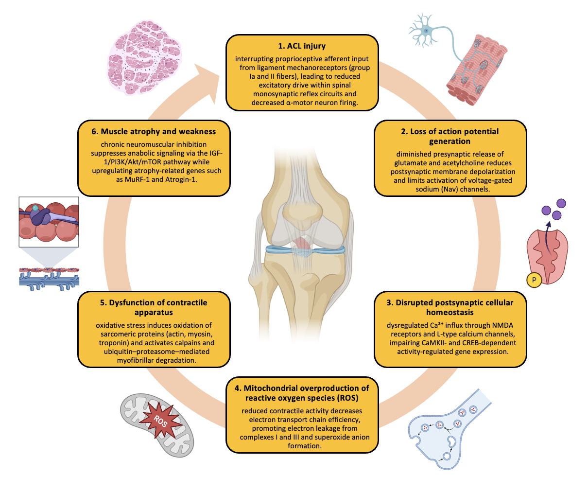
Most athletes who have injured their ACL undergo reconstruction of the ligament. Arthrogenic muscle inhibition (AMI) persists in as many as one-third of patients after anterior cruciate ligament reconstruction (ACLR) and is now understood to be driven by a cascade of molecular events that unfold from the myofiber to the cortex. In the quadriceps, early postoperative elevations of the atrophic regulators myostatin and transforming growth factor-β are detectable within the first postoperative week, coinciding with transient rises in C-reactive protein and cartilage oligomeric matrix protein; these changes normalize as gross knee function returns, yet precede long-lasting weakness.122 These early molecular alterations establish the biochemical foundation for subsequent neural inhibition and delayed strength recovery. Concurrently, gene-expression studies show sustained up-regulation of the ubiquitin-proteasome E3 ligases atrogin-1 and MuRF-1 alongside increased myostatin mRNA, indicating that protein breakdown, rather than synthesis failure alone, drives the 5–10 % cross-sectional fibre loss observed in the first month.123,124 Down-stream neuromuscular junction (NMJ) remodeling has been documented in ACL-transection models that recreate the postsurgical milieu: agrin and nicotinic acetylcholine receptor γ-subunit expression rise while ε-subunit and muscle-specific kinase (MuSK) decline, a pattern that fragments end-plate architecture and further depresses contractile efficiency; cryotherapy appears to partially rescue agrin expression and stabilise NMJ morphology, underscoring the inflammatory contribution to synaptic degradation.125,126
Beyond the muscle fiber, voluntary activation deficits reflect altered motor-unit behavior. Motor-unit decomposition demonstrates smaller recruitment thresholds and slower discharge rates in the vastus medialis at 70–100 % maximum torque, while high-threshold units fail to fire synchronously, a signature of insufficient synaptic drive rather than simple disuse.127,128 Electrophysiological evidence links these peripheral findings to spinal disfacilitation: quadriceps H-reflex amplitudes are reduced in the involved limb for up to six months, and γ-loop dysfunction marked by diminished Ia-afferent transmission persists bilaterally, implicating increased presynaptic inhibition of α-motoneurons and excessive Ib feedback from the injured joint capsule.83,129,130 Changes in inhibitory neurotransmission extend to the dorsal horn, where GABA-mediated modulation of primary afferent depolarization is heightened, plausibly dampening proprioceptive inflow and reinforcing quadriceps inhibition.131 This spinal-level inhibition forms the neurophysiological substrate of chronic quadriceps weakness observed after ACLR.
At the supraspinal level, corticomotor pathways exhibit both structural and functional plasticity. Transcranial magnetic stimulation studies reveal greater long-interval intracortical inhibition, lower corticospinal excitability and expanded cortical silent periods in the quadriceps representation; these neurophysiological deficits correlate with lower voluntary activation scores and predict delayed return-to-running milestones.132,133 Functional MRI conducted as early as six weeks after surgery shows a redistribution of BOLD signal away from primary motor cortex toward premotor, cerebellar and visual-association areas during active knee extension, suggesting compensatory visuomotor strategies when efferent drive is compromised.134 EEG and resting-state fMRI corroborate these findings with increased θ-band power and strengthened interhemispheric sensorimotor connectivity up to one year post-reconstruction, indicating durable network-level adaptations135,136
Collectively, these data position AMI after ACLR as a multi-system disorder initiated by local cytokine and atrophy-related gene activation, propagated through NMJ deterioration and spinal reflex inhibition, and ultimately consolidated in cortical reorganisation (Figure 2). Understanding this continuum highlights why rehabilitation methods (Appendix 1) are continually being investigated to target specific nodes within the muscle–spine–brain axis.
_injury.png)
Molecular aspects of ACL Injury: Eccentric Training Perspective
ACL injury is increasingly being understood as a neurophysiological injury rather than a purely mechanical one. Following ACL rupture and reconstruction, widespread alterations occur across spinal-reflexive, subcortical, and cortical pathways, including reduced spinal-reflex excitability, diminished corticospinal drive, impaired α-motor neuron recruitment, and selective inhibition of fast-twitch motor units.137–140 In parallel, neuroimaging studies show increased reliance on higher cortical regions (premotor, parietal, posterior cingulate, lingual gyrus) and reduced cerebellar activation during motor tasks, shifting the athlete toward slower, visually guided, consciously controlled movement strategies.93,141 This pattern reflects a maladaptive reorganization in which subcortical and cerebellar structures normally responsible for automatic, feed-forward motor control are underutilized, while cortical resources compensate for deficits in reflexive and predictive stabilization.
Eccentric training is uniquely positioned to counter these maladaptive neural adaptations. Whereas concentric contractions depend strongly on spinal-reflexive pathways which are suppressed post-ACLR142 eccentric contractions elicit a distinct neural strategy characterized by greater cortical and subcortical activation, higher α-motor neuron recruitment, improved motor-unit synchronization, and enhanced tolerance for high forces.143 Importantly, just twelve sessions of eccentric quadriceps training have been shown to significantly increase corticospinal excitability and reverse quadriceps activation failure after ACL reconstruction.144 In this way, eccentric loading addresses the very neural deficits that limit effective strength restoration.
Eccentrics also target cerebellar mechanisms that are impaired after ACL injury. ACLR patients typically exhibit reduced cerebellar excitability and compromised feed-forward error correction.93,141 Because eccentric actions depress muscle-spindle gain, they increase reliance on cerebellar predictive control,145 reinforcing the anticipatory mechanisms required for rapid, high-velocity joint stabilization during sport. This becomes particularly relevant in the context of fatigue: recent work shows that the cerebellum prioritizes fatigue regulation over movement precision under load,146 meaning that high-fatigue conditions common in traditional hypertrophy-based rehab may further reduce cerebellar contribution and hinder motor relearning. Thus, controlled eccentric loading may help stabilize cerebellar involvement while avoiding the fatigue-driven neural downregulation that impairs skill reacquisition.
Beyond neural adaptations, eccentric training enhances mechanical properties that are essential for knee stability during landing and cutting. One key enhancement is short-range stiffness (SRS), the rapid increase in muscle stiffness during the first 20–40 ms of stretch the precise window in which most ACL injuries occur.147 Eccentric training improves SRS by increasing cross-bridge stiffness147 and enhancing pre-activation and early-phase stiffness during dynamic tasks.148 Improved SRS enables the quadriceps–hamstring complex to stabilize the knee instantaneously, before reflexive or cortical corrections can occur.
Eccentric training also improves residual force enhancement (RFE), a property in which muscles produce more force at the same activation level following an active stretch. RFE is mediated by titin-based stiffness and cross-bridge dynamics149,150 and enhances joint stability with lower neural drive. This is particularly beneficial in ACL-injured individuals, who often compensate with excessive cortical activation during movement.151 By improving force economy and force steadiness after stretch, eccentrics support reactive stabilization in chaotic, high-speed sport scenarios where automated feed-forward control is essential.
Taken together, eccentric training directly counteracts the sensorimotor deficits that characterize ACL injury it increases corticospinal and α-motor neuron excitability, restores subcortical and cerebellar automaticity, reinforces predictive control, re-engages high-threshold motor units, and enhances the rapid mechanical properties (SRS and RFE) necessary for knee stability under real-world demands. For these reasons, eccentrics should not be viewed simply as a loading strategy but as an intervention targeting the neuro-mechanical root causes of persistent dysfunction after ACL reconstruction.
Practical Applications: Integrating Accentuated Eccentric Loading, Fast Eccentric Loading, and Horizontal Deceleration into ACL Rehabilitation
The mechanistic alterations observed after ACL injury reduced corticospinal excitability, impaired α-motor neuron recruitment, diminished subcortical automaticity, cerebellar underactivation, and loss of short-range stiffness necessitate rehabilitation strategies that directly target these neuro-mechanical deficits. Among available modalities, accentuated eccentric loading (AEL), fast eccentric loading (FEL), and structured deceleration training provide complementary and progressively integrated stimuli capable of restoring neural drive, predictive motor control, and rapid joint stabilization.
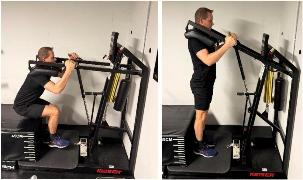
Accentuated eccentric loading (AEL), particularly through the 1–2 AEL squat, provides a safe yet potent method to overload the eccentric phase while minimizing joint stress. In this exercise, the athlete descends with control on one leg, and then rises on two legs during the concentric phase (Figure 3). This configuration allows high-quality eccentric overload. The approach directly addresses neural deficits after ACL injury, as eccentric contractions enhance corticospinal excitability and reverse quadriceps activation failure.144 AEL restores high-threshold motor-unit recruitment and fast-twitch synchronization,143 while improving short-range stiffness an essential protective mechanism in the first 20–40 ms of dynamic loading, the window in which most ACL injuries occur.147,148 By increasing reliance on cortical and subcortical structures that regulate predictive and automatic stabilization, AEL serves as a foundational intervention for re-establishing efficient neuromotor control early in rehabilitation.
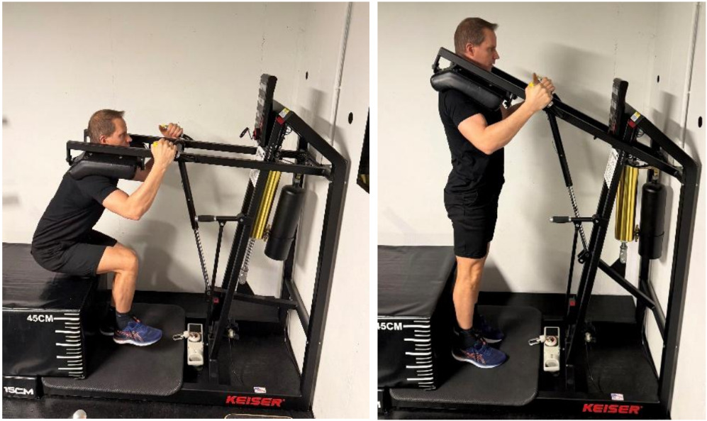
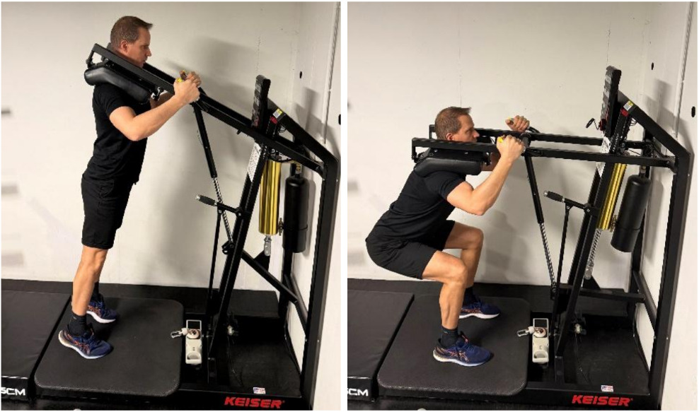
As neural drive and movement control begin to normalize, fast eccentric loading (FEL) provides the next progression by introducing high-velocity eccentric braking. In a typical FEL squat, the athlete descends as fast as possible, “catches” the load by braking forcefully in the bottom position, and then completes the concentric phase (Figure 4). This rapid braking stimulus enhances rate of force development (RFD) and fast-twitch fiber function. FEL, typically performed at 50–70% of concentric 1RM in low-volume cluster formats, produces superior increases in muscle power and type IIx fiber hypertrophy, as well as increases in fascicle length, firing frequency, and motor-unit synchronization.152,153 These adaptations reflect increases in sarcomeres in series,154 which directly improve shortening velocity and explosive capacity qualities consistently diminished after ACL injury. Although FEL has not been examined directly through neuroimaging, the neural control characteristics of rapid eccentric braking strongly suggest increased reliance on cerebellar predictive mechanisms.145 This is particularly relevant because ACL-injured individuals show impaired predictive control during movement.93,141 Under fatigue or high mechanical load, the cerebellum further shifts toward predictive stabilization,146 reinforcing the rationale that FEL increases the requirement for cerebellar engagement even if direct activation has not yet been empirically demonstrated.
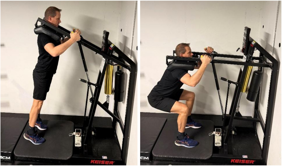
To ensure transfer of these neuromuscular adaptations to real-world movement demands, horizontal deceleration training must be incorporated as a final stage. Deceleration remains a neglected yet crucial component of injury risk reduction players perform nearly three times more high-intensity decelerations than accelerations,155 and decelerations impose substantially higher mechanical load and soft-tissue stress.156,157 Because ACL injuries frequently occur during unanticipated braking actions, rehabilitation must include both anticipated and unanticipated horizontal decelerations. In anticipated drills, athletes accelerate for a set distance (5–20 m) before braking maximally at a predetermined point, allowing controlled exposure to progressively increasing approach velocities. In unanticipated decelerations, athletes accelerate but receive a reactive cue visual, auditory, or cognitive that requires them to stop instantly at unpredictable timing. This restores the ability to decelerate under perceptual and cognitive uncertainty. Improved eccentric strength and braking capacity are strongly associated with reduced ACL injury susceptibility,158 further supporting the inclusion of deceleration as a final, context-rich component of rehabilitation.
Collectively, these three exercise types form a coherent, mechanistically grounded progression. AEL re-establishes corticospinal drive, α-motor neuron excitability, and early-phase stiffness; FEL enhances high-velocity force production, fast-twitch fiber behavior, and cerebellar predictive engagement; and horizontal deceleration training reintegrates these neuromuscular improvements into complex, context-dependent movement patterns that mirror the cognitive–motor demands of sport. This progression ensures that eccentric training is not considered merely a loading strategy but a targeted intervention addressing the neurophysiological and mechanical root causes of persistent dysfunction following ACL injury.
In-silico validation framework for AI-guided ACL prophylaxis
Finally, the authors propose an AI-assisted, open-source, in-silico framework to validate the effectiveness of an ACL prophylaxis physiotherapy protocol. In this work, we have used the framework to simulate a season with 20 games and a number of events i.e. (jog, run, cut, jump-landing). For each event, the resulting strain histories are passed to a Khatib-inspired micro-model that simulates the tendon and collagen fiber continuum, utilizing a damage parameter to account for structural degradation.159,160 A Weibull survival formulation maps the bundle-level strain exposure into a per-event injury risk index.161
R∈[0,1]R [0,1]R∈[0,1].
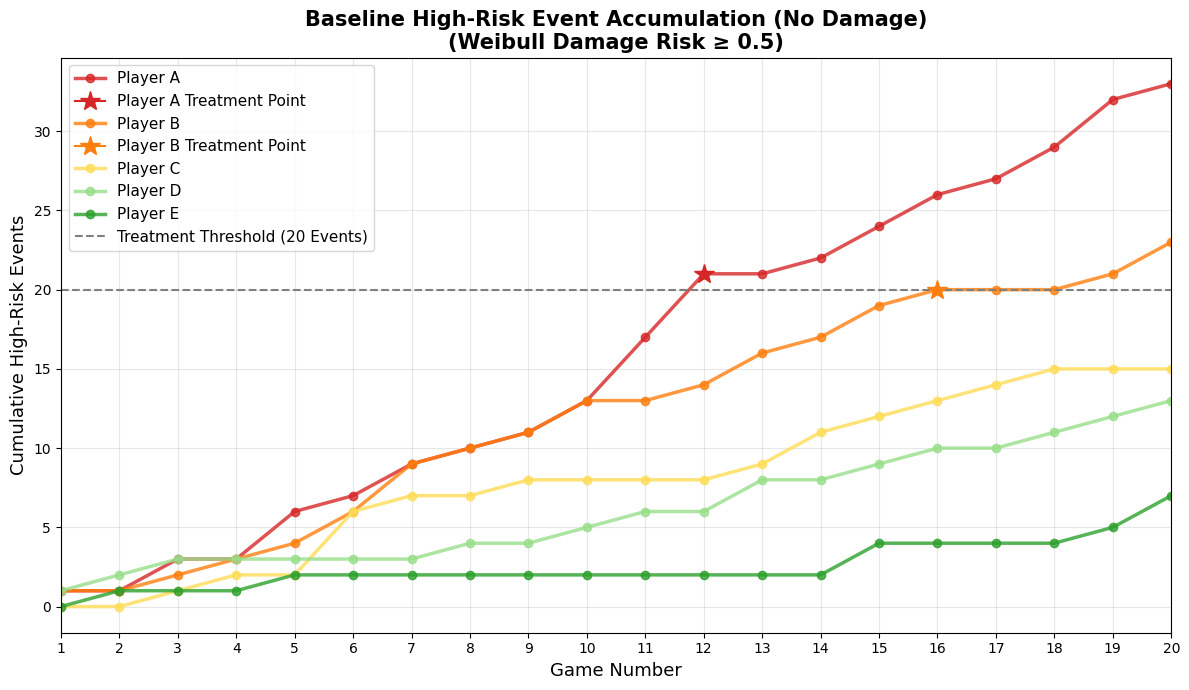
Five hypothetical ACL anatomies (Players A–E) are defined by different fibril parameters to span weak through strong ligaments.162 Season-long exposure is simulated as a sequence of match events. Events with R≥0.5R 0.5R≥0.5 are classified as “high-risk”, and the number of such events for each player are accumulating. Exceeding 20 high-risk events is treated as a trigger for intensified preventive physiotherapy proposed in this work.
The example in Figure 5 illustrates this principle: cumulative high-risk events are plotted over 20 games for five players with different ACL anatomies.
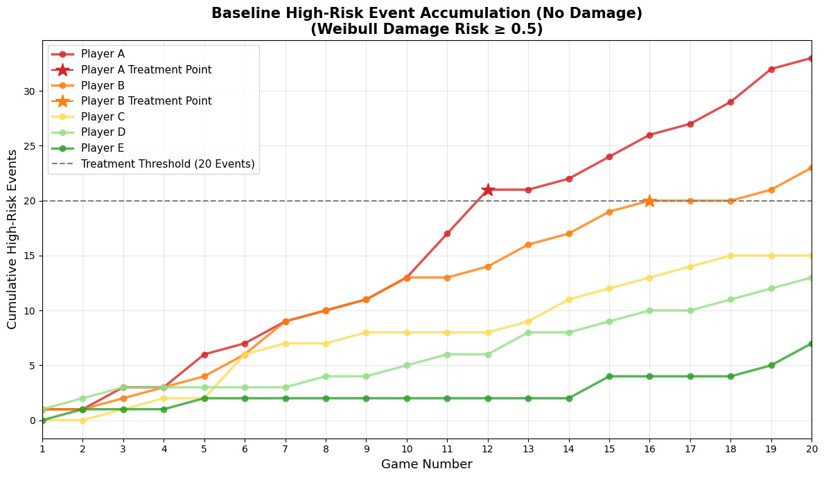
Player A (weaker/smaller ACL) exhibits the steepest trajectory and crosses the 20-event threshold by game 12, Player B crosses at game 16, whereas Players C–E (stronger ligaments) and remain below the cutoff despite comparable simulated match loads. The differing slopes show how identical external demands can translate into markedly different internal ligament risk profiles purely due to microstructural variability.
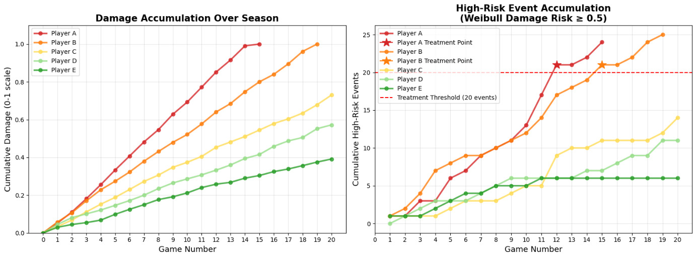
A parallel example in Figure 6 illustrates the progression of damage accumulation relative to the count of high-risk events over time.
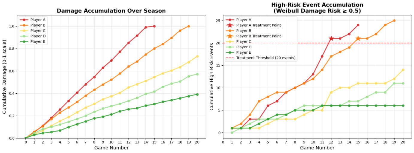
Player A, characterized by weaker/smaller ligaments, suffers catastrophic tissue failure at game 15, having breached the high-risk threshold earlier at game 12. Similarly, Player B sustains an ACL rupture at game 19 after crossing the threshold at game 15, whereas Players C–E maintain low damage levels and remain well below critical event counts. This consistent lag between the threshold breach and ultimate failure underscores the predictive value of the high-risk metric, serving as an early indicator of developing injury before macroscopic damage occurs.
The authors propose that this framework can be used to design and test prophylactic PT protocols and to evaluate across teams and seasons regarding whether implementing targeted interventions when model-derived thresholds are exceeded leads to fewer ACL injuries than expected from historical or league-wide statistics.
Future suggested work: Match video could be processed with a computer-vision pipeline or markerless motion capture to reconstruct player-specific kinematics and estimate ground reaction forces. These data could drive a simplified knee model that combines anterior shear, valgus and internal-rotation moments to obtain ACL force and strain, which are then partitioned into anteromedial and posterolateral bundles.
The following provides a link to an example of this type of work. GitLab source: https://gitlab.com/adam.kozlowski1/khatib-inspired-acl/-/tree/main?ref_type=heads
Conclusion
During sudden braking the body encounters extremely large ground-reaction forces (GRF). Vertically they reach 6–8 × body mass about 1.5–3 × higher than during acceleration while in the anterior-posterior direction they can be up to four times greater than in other movement tasks.163 The peak loads arrive almost instantaneously, within the first 30–50 ms after the foot contacts the ground, so the muscles must oppose the rising forces immediately.164
Within this critical 0–50 ms “window,” joint protection comes first and foremost from the muscles’ inherent, nonlinear elasticity.165 Classic cat-soleus experiments by Nichols and Houk showed that the muscle’s own structure through the visco-elastic properties of its contractile elements and connective tissue can stiffen instantly under stretch and slacken under unloading, stabilising the limb before sensory feedback arrives.165 Only after ~20 ms do spinal reflexes driven by muscle spindles and Golgi tendon organs join in, modulating motor-unit recruitment and matching tension to the changing load.166
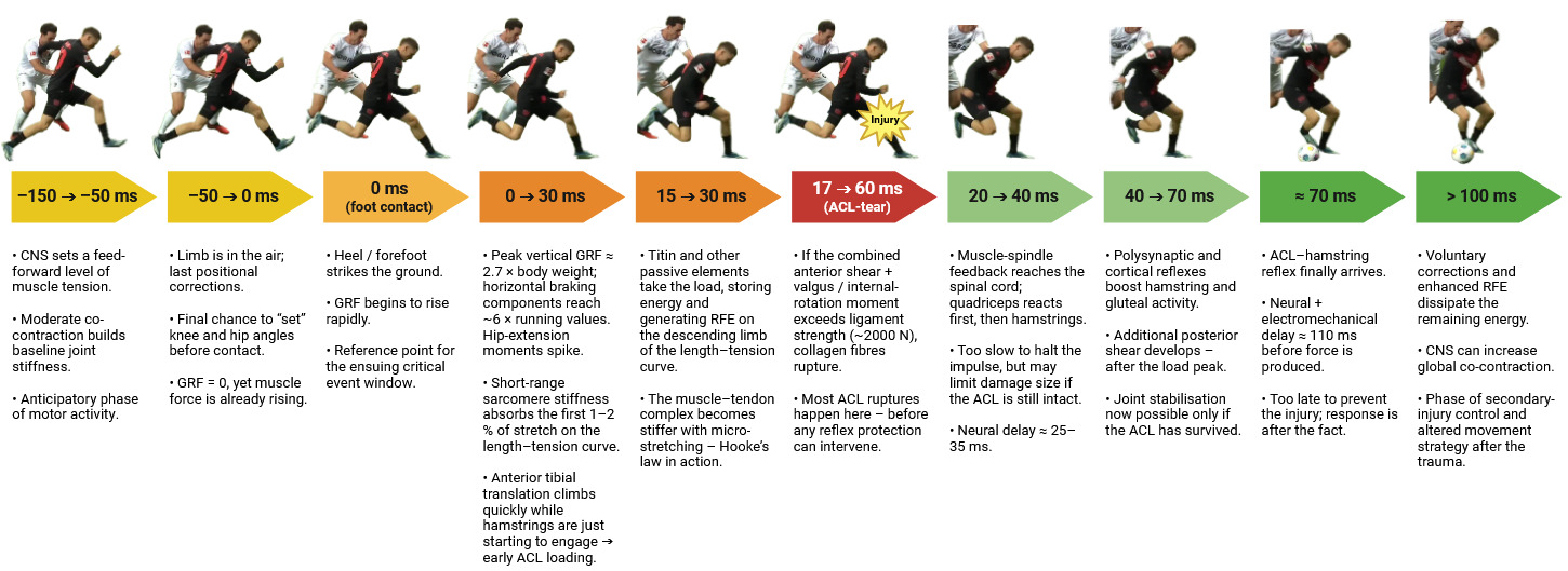
The so-called ACL reflex is much slower.167 Activation of mechanoreceptors in the anterior cruciate ligament elicits a muscular response around the knee only after ~70 ms, and with electromechanical delay at least 110 ms are needed before the muscles generate significant force.168 In practice, by the time the peak GRF has already acted, the ACL reflex is just beginning and cannot serve as an immediate “fuse” for the ligament.169 Its role is more one of updating motor programmes online.
This integrated view underscores that, in overload situations, direct joint protection hinges chiefly on the muscles’ instant, built-in mechanical properties and their rapid reflex regulation not on the ACL reflex, which comes into play only after the fact (Figure 7) .114
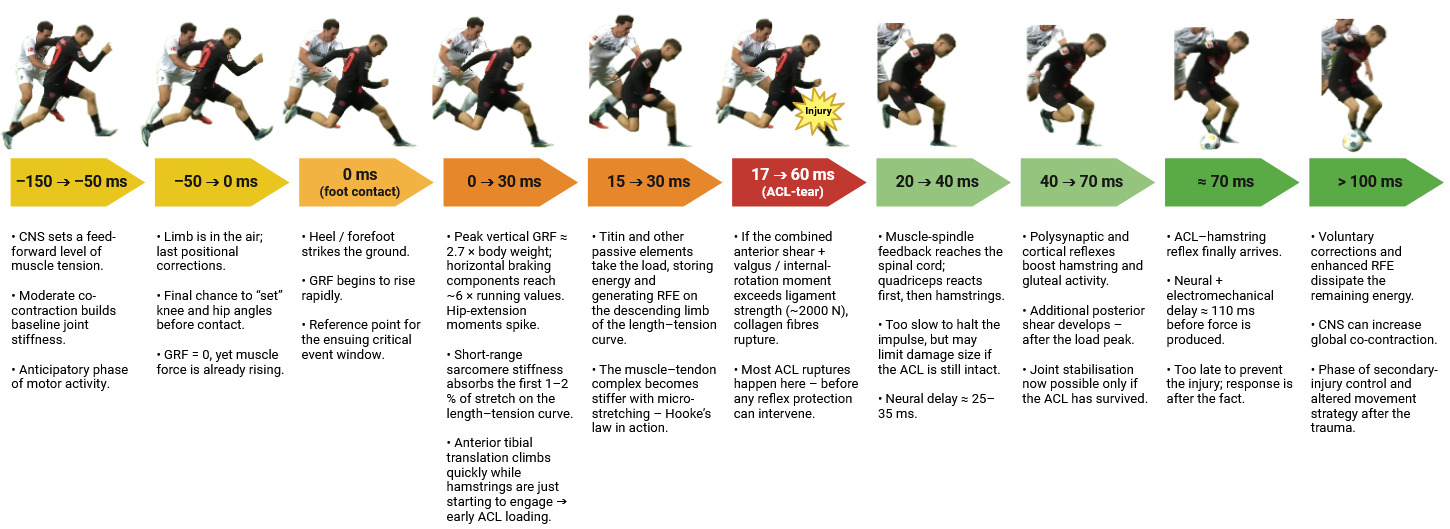
This framework highlights an essential shift in understanding injury prevention: the decisive defensive mechanisms are preprogrammed and molecular in origin, not reactive or purely biomechanical. The integration of biomechanics, neurophysiology, and muscle nanomechanics provides a unified model explaining how the ACL survives or fails under extreme deceleration.170 Nevertheless, the present synthesis also faces several limitations. Most current data originate from isolated in-vitro muscle models or simulations, which may not fully capture in-vivo neuromechanical coupling during high-speed movements. Temporal resolution in EMG and imaging studies remains insufficient to map millisecond-scale titin–actin dynamics in humans. Moreover, individual variability sex, training history, and hormonal state likely modulates both molecular stiffness and reflex latency, yet these factors are rarely controlled experimentally.171
Future research should therefore focus on real-time, multiscale measurements linking cortical preactivation, muscle-fiber mechanics, and ligament strain. Combining ultrafast ultrasound elastography, nanomechanical assays, and high-density EMG may bridge the remaining gap between cellular biophysics and macroscopic joint behavior.172 Translationally, longitudinal studies should determine whether targeted eccentric, reactive, or feed-forward training can modify titin stiffness and reflex timing to confer measurable protection in sport-specific tasks.120 Ultimately, advancing from descriptive to predictive models of ACL resilience will require an integrated approach spanning molecular biology, neuromechanics, and applied rehabilitation science.173
Conflicts of interest
The authors report no conflicts of interest.